What it is
The veins of the thoracic cavity are a system consisting of the main vena cava, which originates in the upper part of the chest and gradually connects with other smaller ones.
This vessel is formed from the confluence of the right and left brachiocephalic veins. It is not paired.
A few centimeters later, the azygos vein adjoins the superior vena cava, after which it enters the right atrium. Its function is to drain blood from the walls of the chest and its organs.
Veins have good extensibility and capacity, but weak elasticity.
Their inner surface, with the exception of the vena cava and portal system, has valves. They help move blood towards the heart and prevent it from flowing back.
The veins of the thoracic cavity contain up to 80% of all blood in the body. They constantly contain about 65% of the total volume.
Blood is pumped from areas of high pressure to areas of low pressure. When inhaling, negative intrapulmonary pressure appears. It provokes the expansion of large vena cava. This facilitates the flow of venous blood to the heart.
Treatment
When treating superior vena cava syndrome, the goal is to relieve symptoms and attempt to cure the underlying malignancy. Only a small percentage of patients with rapidly occurring superior vena cava obstruction are at risk for life-threatening complications.
Patients with clinical SVPV often experience significant symptomatic improvement with conservative treatment measures, including head elevation and supplemental oxygen. Emergency treatment is indicated in the presence of cerebral edema, decreased cardiac output, or edema of the upper respiratory tract. Corticosteroids and diuretics are often used to relieve swelling of the larynx or brain, although documentation of their effectiveness is questionable.
Radiation therapy has been recommended as standard treatment for most patients with SVPV. It is used as initial treatment if a histological diagnosis cannot be established and the patient's clinical condition worsens; however, reviews suggest that superior vena cava obstruction alone rarely represents an absolute emergency that requires treatment without a definitive diagnosis.
Fractionation regimens for radiation therapy typically include two to four large initial fractions of 3 to 4 Gy, followed by daily delivery of conventional fractions of 1.5 to 2 Gy, up to a total dose of 30 to 50 Gy. The radiation dose depends on the size of the tumor and radioactivity. The radiation portal should include a 2-cm field around the tumor.
During radiation, patients improve clinically before chest x-ray shows objective evidence of tumor shrinkage. Radiation therapy relieves superior vena cava obstruction in 70% of patients with lung carcinoma and in more than 95% of patients with lymphoma.
In patients with SVPV secondary to non-small cell lung carcinoma, radiation therapy is the primary treatment. The likelihood that patients will benefit from such therapy is high, but the overall prognosis for these patients is poor.
Chemotherapy may be preferable to radiation therapy for patients with chemosensitive tumors.
The most extensive experience in the treatment of SVPV secondary to non-Hodgkin's lymphoma is reported at MD Anderson Cancer Center. Patients received chemotherapy alone, chemotherapy plus radiation therapy, or radiation therapy alone. All patients achieved complete relief of SVEP symptoms within 2 weeks of any type of treatment. It is likely that no single treatment was superior in achieving clinical improvement.
(No Ratings Yet)
Characteristic
The thoracic vein system is quite simple.
The azygos and semi-gypsy veins originate from the lumbar vessels.
Then, rising into the chest cavity, they pass into a gap in the diaphragm. Then the unpaired one goes to the right of the spine, and the semi-unpaired one goes to the left.
At the level of 7-8 vertebrae they connect, and then enter the superior vena cava.
Unpaired
It originates in the abdominal cavity to the right of the spine. It runs through the entire thoracic cavity and bends over the right bronchus at the height of the third vertebra. It drains into the superior vena cava.
Semi-unpaired
It also collects blood from the abdominal and chest cavity.
It is located to the left of the spine and is a continuation of the ascending lumbar veins. It is a little thinner than the unpaired one.
Receives the veins of the esophagus, mediastial, posterior intercostal, subcostal and accessory hemizygos.
Brachiocephalic
It can be either right or left. Collects blood from the head, neck and arms.
The left brachiocephalic vein is twice as long as the right one.
They fuse together towards the medial end of the 1st rib.
Upper hollow
It collects blood from the veins of the chest and abdominal cavity, head, neck and upper extremities.
It is a short, valveless vessel. Its length is up to 7 centimeters and its diameter is up to 25 millimeters.
On the right, the azygos vein flows into it, the roots of which are the brachiocephalic veins.
Blood from the walls and organs of the chest cavity, as well as the spinal cord, flows into the veins of the chest.
Varicose veins on the chest
Varicose veins develop on the chest in women as a result of breastfeeding, and in men the causes of the pathology are associated with lifting heavy objects. In this case, the patient experiences an expansion of the subcutaneous vascular bed, spider veins and significant trophic disorders appear, up to the formation of long-term non-healing ulcers. To get rid of the pathology, complex treatment using medicinal and surgical methods is necessary.
Main functions
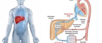
Vascular anatomy of the thoracic cavity includes arteries, veins, and the heart.
They are necessary to pump out venous blood from the lungs and for its subsequent filling with oxygen. Afterwards it goes to all internal organs.
This helps maintain proper gas exchange in the lungs and metabolic functions of body tissues.
The heart is a pump that constantly moves blood throughout the body.
It fills the lungs with venous blood, and after oxygenated arterial blood, the rest of the body tissues.
The function of the pulmonary veins of the chest is to bring carbon dioxide-free and oxygenated blood to the heart. It then travels from the left ventricle to the aorta.
The aorta, in turn, divides into three large arteries that supply oxygen to the head and arms. It, dividing into many small arteries, supplies blood to the muscles and skin of the chest.
Blood from the abdominal aorta supplies nutrients and oxygen to all abdominal organs.
The veins of the upper body complete the circulatory cycle. They carry blood with waste products and carbon dioxide from tissues to the heart. Afterwards, through the lungs it again enters the body organs.
A little more physiology
Are you an athlete (go to the rocking chair), or do you work hard and your work is physical? Well, your veins will definitely show through your skin. After all, they must intensively distill blood, removing decay products from organs and tissues. Large, convex blue wreaths are payment for physical labor.
A venous network in the chest area may appear:
- in adolescents during puberty (due to hormonal imbalance);
- in women during menopause (the reasons are the same);
- after an extreme diet, if sudden weight loss was successful.
A slight increase in the vascular pattern is noticeable in women with thin skin during the period of breast engorgement before menstruation.
Interesting, but true: prolonged exposure to the scorching rays of the sun, active cupping massage (it should not be done in the bust area), temperature changes (frequent and prolonged) and deep peeling of the skin can cause veins to become visible.
Possible pathologies
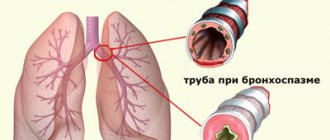
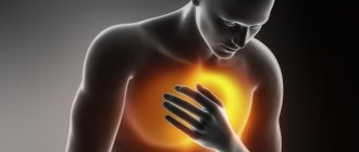
One of the most common pathologies of the chest veins is superior vena cava syndrome. Develops due to circulatory disorders.
It manifests itself as puffiness of the head and neck, dilation of the saphenous veins, shortness of breath, and cough. Similar symptoms can be observed with severe insufficiency of the cardiovascular system and various types of thrombosis.
It is also worth noting such a pathology as venous congestion. This is a violation of the outflow of blood, which leads to increased blood supply to an organ or tissue.
The main cause is acute and chronic heart failure.
Chronic venous congestion in the lungs manifests itself as multiple hemorrhages and proliferation of connective tissue.
My mirror, tell me and show all the veins...
We looked in the mirror and clearly saw the pattern of veins under the skin. If this phenomenon “haunts” you all your life, there is nothing to worry about. In people with thin skin, the venous pattern is clearly visible on the body, especially after winter, unless you use a solarium, of course. The sunny bronze fades over the winter and blue “paths” of a wreath appear under the thin white skin.
There are cases where a vascular pattern appears under the skin of the veins in men and women, without any obvious reason. And even deep research does not help to find this reason. Perhaps we are talking about a genetic predisposition, and not about a symptom of the disease.
Contact a phlebologist (a specialist who studies the condition of the venous system and treats vascular pathologies). According to “vein doctors,” physiological abnormalities are not pathology. Treatment may not be required.
Unfortunately, medicine does not deal with the elimination of this cosmetic defect. But you can try propolis preparations or other natural products. They:
- maintain skin elasticity;
- help fibroblasts (cells that produce collagen) actively create young “correct” collagen - the basis of connective tissue.
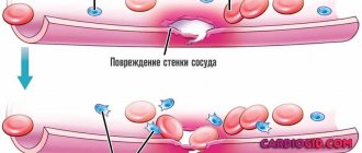
The wall of the vessel contains connective tissue, and the correct functioning of the vein in pumping blood depends on its elasticity.
Diagnostics
Modern medicine can offer simply a huge number of diagnostic methods. This allows you to identify any disease at an early stage and prevent its development.
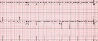
- X-ray examination. Prescribed to people who suffer from respiratory diseases. This examination helps to see the outline of the heart and major blood vessels.
- CT scan. Gives a clearer picture than an x-ray examination. Computed tomography takes several images that are analyzed by a computer.
- Magnetic resonance imaging. Prescribed for suspected diseases of the blood vessels. Unlike CT, X-rays are not used.
- Radionuclide research. Allows you to analyze blood flow in the lungs. A two-stage condition survey. In the second part of the study, a radionuclide substance is injected into a vein. This allows you to see blood clots in the lungs.
- Doppler ultrasound. Allows you to evaluate the direction and speed of blood flow in the vessels.
Based on the results of the examination, the doctor prescribes treatment procedures.
Disturbance in the blood flow in the veins of the chest cavity leads to serious consequences, including death.
Signs of impaired blood flow are:
- ulcers;
- pale skin;
- brittle nails and hair;
- constantly cold hands and feet;
- digestive problems;
- phlebeurysm.
If you notice one of the above symptoms, you should consult a doctor.
Male varicose veins on the hands
Problems with veins in the hands of men indicate a deterioration in the general condition of the body. At the initial stage, the venous pattern begins to be clearly visualized. But many do not even suspect that these are symptoms of varicose veins in men’s hands. For most representatives of the stronger sex, the presence of large nodular veins is evidence of masculinity, brutality, but not illness.
Male varicose veins on the hands
Indeed, in a physically well-developed man, the veins will be noticeable. For example, in athletes involved in bodybuilding and weightlifting, the veins in the arms protrude even in the absence of loads.
But when all the stress on the hands consists only of moving the computer mouse, the presence of protruding blood vessels on the hands should be alarming. Even people leading an active lifestyle should be concerned if the venous pattern in the upper extremities is visualized at rest.
In the future, the venous pattern may be joined by:
- heaviness and swelling of the hands;
- numbness of fingers, loss of sensitivity;
- cramps at night and after exercise;
- walking pain.
The veins are deformed, acquiring pathological tortuosity and prominence, nodes and subcutaneous lumps appear, meshes are visualized in delicate areas.
Read also: Lilac leaves have medicinal properties for varicose veins
If any sign is detected, a man should think about visiting a doctor. Delay is fraught with serious complications, which will lead to loss of ability to work.
Do you still think that it is impossible to cure varicose veins?
Judging by the fact that you are now reading these lines, victory in the fight against varicose veins and unsightly areas on the body is not yet on your side.
Have you already thought about inpatient treatment? This is understandable, because varicose veins are a very dangerous disease that, if not treated in a timely manner, can be fatal. Stars on the legs, fatigue, terrible swollen veins. All these symptoms are familiar to you firsthand.
But perhaps it would be more correct to treat not the effect, but the cause? We recommend reading the story of Ekaterina Andreeva. Read the article >>
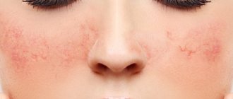
The female breast is the most unprotected and delicate part of the body, since it contains a huge number of capillaries and small vessels. Sometimes they become simply visible, but do not stick out or cause any inconvenience. This condition should not bother the representative of the fair half of humanity.
The fact is that in many women the veins can be located close to the skin, so they are clearly visible. But if the veins began to appear sharply, at the same time they became lumpy, and at the same time pain began to bother them, then this is the first signal that varicose veins are developing on the chest.
How varicose veins are treated on the female breast, and how dangerous such a pathology is, we will find out below.
Other diseases
There are other health problems that can result in prominent veins on the chest. There are many reasons: the development of a cyst or lipoma, as well as other neoplasms in the breast tissue. Sometimes blood vessels signal inflammatory processes. Vascular mesh also appears in latent cancer tumors. Be that as it may, if you notice that the veins on your chest are more visible than usual, do not panic and set yourself up for a worse diagnosis. Only a competent specialist - a mammologist or phlebologist - can reassure you and give valuable recommendations.
Symptoms of varicose veins in men
Many doctors consider varicose veins to be an insidious disease. In the initial stages of development, it causes only cosmetic defects, and there is no pain, swelling or general malaise. This significantly delays the trip to the doctor, especially in cases where the patient is a man. Later, the symptoms progress, causing an unpleasant feeling of discomfort and fatigue in the affected area.
Read also: Deep vein thrombosis ICD
Enlarged veins are manifested by the following symptoms:
- The appearance of vascular patterns and large venous patterns on the legs, arms and thighs.
- Feeling of discomfort, heaviness and fullness in the affected area.
- Night cramps and evening swelling of the legs.
- Decreased general activity of the patient, increased fatigue.
- Painful sensations and burning.
The disease progresses gradually. If you notice constant changes in your health and the regular occurrence of alarming symptoms, you should consult a doctor for timely help.
Why do varicose veins occur?
The development of varicose veins in the chest can be triggered by the following factors affecting the human body:
- excess body weight;
- errors in nutrition;
- burdened heredity;
- disruption of the structure of collagen fibers;
- atherosclerosis;
- increased blood density;
- insufficient fluid intake;
- excessive platelet production;
- hypertonic disease;
- inactive lifestyle;
- lifting weights;
- chest injury;
- increased venous blood pressure in the pelvis;
- pregnancy and childbirth;
- lactation;
- frequent constipation or diarrhea.
After feeding, women may notice that the mammary glands have become bluish, this is due to varicose veins.
This problem often occurs in women whose children were breastfed.
Often the reason that veins appear on the chest is breastfeeding. In men, pathology develops as a result of significant force loads and heavy lifting. Hypertension and abnormalities in the structure of collagen fibers located in the vessels can provoke pathology. Smoking and atherosclerotic plaques have a huge negative impact on the walls of veins.
Prevention
What should you do if the veins on your chest are more visible than usual? There are simple preventive measures that every woman can take:
- Try to lead a healthy lifestyle, give up bad habits;
- Observe basic standards of personal hygiene, adhere to the same sleep schedule, eat at a certain time;
- Avoid injury and excessive stress on the mammary glands;
- Do not burden yourself with intense physical exercise;
- Do not overcool, maintain the integrity of the skin of the chest;
- Avoid stress and sudden emotional changes;
- Drink enough fluids throughout the day;
- If necessary, take a vitamin-mineral complex;
- Eat fermented milk products, fresh vegetables and fruits - they help maintain blood vessels in tone;
- Take a walk every day, be in the fresh air;
- If you have a sedentary job, try to develop for yourself at least a minimum set of physical exercises that prevent blood stagnation in the legs.