Hemorrhagic rash can be the cause of various diseases. It occurs as a result of capillaries rupturing and red blood cells leaking out of the vessels. Most often, the rashes do not rise above the surface of the skin and cannot be felt. The exception is inflammation of the capillary walls.
Such pathology can be expressed in a variety of forms, each of which has certain distinctive features. If any rash occurs, you should immediately consult a doctor, who will determine the underlying cause and prescribe the correct treatment.
Features of the rash
A hemorrhagic rash, a photo of which is in the article, refers to the formation of small non-inflammatory rashes on the skin of different parts of the body. This is how the body reacts to the rupture of capillaries, the subsequent penetration of red blood cells into the upper layers of the skin.
When it appears, a person does not experience any discomfort at all, since the rash does not itch and does not cause pain. When pressing on the affected area, the color of the rash does not change. The number of points largely depends on the cause of the lesion and the severity.
In a child under 5 years of age, the disease can be triggered by vascular pathologies, and in a child from 5 to 15 years old - by colds and infectious diseases. In addition, the hereditary factor is important.
Features of the development of the disease with influenza
In medical practice, the number of hemorrhagic rashes on the legs has increased, and this greatly complicates the diagnosis of pathology.
A rash can appear due to hereditary and infectious diseases, which lead to vascular damage.
In medical practice, cases of vasculitis affecting children in the age group from 4 to 12 years have been recorded. Older people also tend to get sick.
The risk of contracting vasculitis increases significantly when a child has had the flu, ARVI, food allergies, or has been vaccinated.
Intensely colored bruises on the skin clearly demonstrate the presence of a serious pathology in the baby, which should be a reason for immediate contact with a hospital.
Vasculitis should not interfere with your life, which means you must immediately begin to identify the causes of the disease and their subsequent elimination.
The disease appears due to serious factors, for example, von Willebrand disease or hemophilia - a purely male pathology in which intense bleeding does not stop and blood clotting is impaired.
More often, the epidermis on the legs becomes covered with unattractive spots due to:
- Unfavorable heredity.
- Hormone abuse.
- The presence of pathologies of an infectious nature.
- Taking steroid drugs.
- Pathological changes in blood vessels.
First, a patient diagnosed with a hemorrhagic rash needs emergency medical attention.
Before the medical staff arrives, provide the patient with comfortable home conditions: give him warm sweet tea, give him an antipyretic, apply something cold to his head.
The patient spends most of his time in bed, without leaving for long. Antihistamines will ease the course of the disease; in more severe cases, take corticosteroids, the course of treatment of which can vary between 4-8 weeks.
During an exacerbation, it is advised to enrich your body with medications aimed at tightening blood vessels and giving them elasticity.
Discover skin free from itching and aesthetic defects with such preparations as “Calcium Chloride”, “Rutin”, “Ascorbic Acid”.
Classification
According to the nature of the spread, hemorrhagic rash is divided into primary and secondary. Primary is characterized by the fact that spots form on clean skin. It is divided into cavity and cavityless. The secondary form is a consequence of the development of elements of the primary rash.
According to its appearance and size, the hemorrhagic rash, a photo of which shows the features of the disorder, is divided into:
- petechiae;
- purples;
- ecchymoses.
Petechiae are small round spots. Their shade can vary from bright red to dark brown. When an insect bites, the middle of the petechia takes on a brighter shade when compared with the rest of the surface.
Purpura is a subcutaneous spot, the diameter of which is 2-5 mm, but sometimes it can be 1 cm. Often the spots merge with each other, forming one large one. May also be accompanied by burning and itching. The rash is localized on the arms, stomach, and legs. It has a purple tint, but in some cases changes to dark red or even brown.
Ecchymoses are the largest hemorrhagic rash. It consists of shapeless spots, the diameter of which ranges from 5 mm to several centimeters. Their shade can range from pink to blue-black. Elements of the rash can connect with each other, forming a large affected area, with necrotic elements in the center.
If the pathological process develops severely, then tissue necrosis can spread even further, causing rejection of a large area of skin. This can lead to gangrene.
Signs of hemorrhagic rash
It is somewhat difficult to identify the characteristic features of a pathological rash. Its manifestations are so variable. that in clinical practice several ways of differentiating them are necessary. There is a separate classification that takes into account the appearance (shape and size, as well as shades of the pathological manifestation of the disease. These are important, but not the only diagnostic criterion.
Important! Merging into spots, the elements of the rash can form entire shapeless areas, layer on visual skin manifestations of other diseases, and form stripe-like bruises or roseola. This occurs in dangerous forms of severe diseases and is considered a negative prognostic sign.
The simplest distinction between symptoms is based on their origin. This is rather a statement of the stage of development of capillary ruptures and their intensity. The primary rash appears on intact skin, the secondary rash is layered on the original elements. I differentiate rashes by etiology and age categories. In an adult, this may be a consequence of liver disease, toxic or alcoholic injuries, abuse or incorrect dosage of medications.
Attention! The child typically appears with meningitis or scarlet fever, some other dangerous diseases associated with the activity of a virus or infection. In any age group, this can be a consequence of an autoimmune disease.
There are several types of rashes based on their appearance:
- Scarlet, red, crimson, unbearably intense shades are called petechiae and are usually located on the legs. They look like tiny pinpoint rashes and are caused by damage to the capillary network. The lower extremities are not the only location of tiny rashes, but they are the most typical.
- Purpuras are larger than petechiae (can reach a diameter of 0.5 cm, and sometimes even a centimeter); they are palpable manifestations because they protrude above the skin and are easier to detect visually. If petechiae are practically not accompanied by negative sensations, then with purpura, which tends to merge into large spots, there may be accompanying itching and an unpleasant burning sensation, which cause a lot of inconvenience. The usual location of purpura is in the area of the peritoneum and chest, but the possibility of placement on the upper and lower extremities cannot be ruled out. Despite the name, purple is not the only color available, they can range in color from dark red to burgundy and dark brown.
- Ecchymoses can reach a size of 5 cm, but there are no special characteristic forms that they could acquire. The most typical thing for them is rather shapelessness and uncertainty of color, because ecchymoses, depending on the cause of their appearance, can acquire shades from pale pink to blue-black.
- Petechial-roseolous rashes are a consequence of layers of manifestations of hemorrhage on a rash that appears as a result of an illness accompanied by other rashes (for example, with typhus).
- Vibice is observed in the form of stripes. This is a characteristic sign of some dangerous infections, which is considered a negative sign and gives weak grounds for optimistic forecasts.
Interesting! Hemorrhagic spots formed as a result of the fusion of elements of the rash are not classified as inflammatory diseases.
They can be of variable sizes and take on shades from pink to black. In the photo you can see outwardly dissimilar symptomatic manifestations, but each is based on a pathological process in which hemoglobin is converted into derivatives.
For reference! The fusion of elements leads to the formation of massive areas, in the center of which necrosis can be observed. Its result is rejection of the epithelium, and then gangrenous damage to its surface. The process of transformation and the development of a negative scenario depend on the reason that caused the damage to the capillary walls and the release of red blood cells from the vessels.
Causes
The causes of hemorrhagic rash can be very different. It occurs at any age, regardless of gender. In adults, this condition is accompanied by liver damage. This may be due to:
- chemicals;
- alcoholic drinks;
- viruses;
- injuries;
- medications;
- junk food.
When the liver is damaged, the ability to clot blood is impaired. The cause of the rash may be a genetic predisposition. Hemophilia, a chronic bleeding disorder, is often identified among genetic diseases.
A hemorrhagic rash can be infectious. In this case, it is provoked by:
- meningococcus;
- scarlet fever;
- tick bites.
With an immunocomplex origin of the disorder, there are many different provoking factors. That is why a rash can occur against the background of a bacterial infection, a virus, a burn, vaccinations, hypothermia, or the use of certain medications.
The exact cause of rashes is not always determined. In this case, diagnosis and treatment should be carried out by qualified specialists.
What is it and what causes it?
Small red spots can occur from environmental factors, such as excessive friction, irritation from chemicals, and insect bites. But more often, such signs in the human body mean the presence of disorders in the body - infectious-inflammatory, immunopathological, metabolic, etc. Considering the variety of probable reasons why small red dots appeared, it is worth asking a competent specialist.
Small spots on the chest of an adult due to prickly heat
Not only young children, but also adults experience the negative effects of sweat. Chemical substances contained in the secretions of the skin glands have an irritating effect upon prolonged contact with the skin. The development of prickly heat is promoted by various environmental factors or changes in the body:
- Dry hot climate.
- Wearing clothes made of synthetic fabrics.
- Occupational hazards (work in steel foundries).
- Metabolic and endocrine diseases.
- Neurovegetative imbalance.
At first it manifests itself as mild hyperemia and itching, then small rashes appear in the form of blisters. But there are other types of prickly heat, for example, red heat, which is accompanied by the formation of bright inflammatory papules on the skin. This is observed in closed areas of the body, including the chest area, armpits, and folds under the mammary glands in women.
In conditions of physiological or pathological hyperhidrosis, signs of prickly heat appear on the skin of closed areas of the body.
Bloody color with hemorrhagic diathesis
A characteristic sign of some hemorrhagic diathesis are petechiae - small red dots that do not disappear with pressure. They occur when the permeability of the vascular wall increases, which can occur against the background of the following conditions:
- Vitamin C deficiency (ascorbic acid).
- Increased venous pressure (during coughing, physical activity, after childbirth).
- Taking certain medications (corticosteroids, allopurinol, sulfonamides, NSAIDs, methotrexate).
- Injuries and sunburn.
Petechiae on the lower extremities become the first sign of Ig-A-mediated Henoch-Schönlein vasculitis. Subsequently, they transform into typical hemorrhagic purpura - the elements are bright, slightly rising above the surface of the skin, and tend to merge into large foci, sometimes with necrosis in the center. After a few days, the rashes turn pale, acquire a brown color and gradually disappear. The disease is accompanied by other symptoms:
- Migrating pain in large joints (lower limbs, elbows, wrists).
- Abdominal pain, nausea and vomiting, gastrointestinal bleeding.
- Kidney damage (proteinuria, glomerulonephritis, nephrotic syndrome).
Congenital vasopathies deserve special consideration, in particular, Randu-Osler disease, in which multiple telangiectasias appear on the skin, mucous membranes and internal organs. Sometimes the first symptom of the disease is nosebleeds and gastrointestinal bleeding.
Petechial rash is a common sign of hemorrhagic diathesis associated with increased vascular permeability.
Rash on the arms and stomach due to infections
Many infectious diseases are accompanied by skin changes that are observed against the background of fever and signs of intoxication. A rash in the form of red dots or small spots can occur under the following conditions:
- Scarlet fever.
- Rubella.
- Meningococcemia.
- Enterovirus infection.
- Typhus and Brill's disease.
- Yersiniosis and pseudotuberculosis.
- Hemorrhagic fevers, etc.
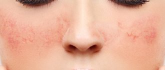
With scarlet fever, pinpoint rashes appear, thickening on the cheeks, in the area of natural folds of the skin. The infection has the following clinical features: pale nasolabial triangle, sore throat, “crimson” tongue, peeling of the skin after the rash disappears (large lamellar on the feet and palms).
Rubella is accompanied first by maculopapular and then scarlet-like rashes. They predominate on the face, lower back and buttocks, and extensor surfaces of the limbs. The disease is characterized by enlargement of the occipital and posterior cervical lymph nodes. In the prodromal period (before the rash), sore throat and conjunctivitis are bothersome.
A severe form of meningococcal infection is meningococcemia. The disease is initially accompanied by the appearance of a roseolous (pink-red, disappearing with pressure) rash throughout the body, but after a few hours the first hemorrhagic elements appear. They are localized on the buttocks and lower extremities, rise somewhat above the surface of the skin, and have a purplish-bluish color.
The rash with enteroviral exanthema may resemble other infections in nature (scarlet fever, rubella, measles). It is often combined with other symptoms - nausea and vomiting, abdominal pain, muscle aches, meningeal syndrome. Sometimes the rashes become hemorrhagic in nature, which requires differential diagnosis with meningococcal infection.
There are many infectious diseases that are accompanied by a small red rash on the skin. In typical cases, it is clinical data that becomes the basis for the diagnosis.
Specks like droplets of blood – Tuzhilin’s symptom
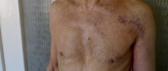
One of the symptoms of chronic pancreatitis (inflammation of the pancreas) is Tuzhilin spots. They are small hemorrhagic rashes in the form of purple droplets, mainly located on the skin of the abdomen, chest, and back. Such elements arise due to the action of excess pancreatic enzymes on the capillary wall.
Exacerbation of pancreatitis is characterized by the occurrence of intense pain in the right hypochondrium and epigastrium, which radiates under the left collarbone. Typical signs of dyspepsia:
- Nausea and vomiting.
- Flatulence (bloating).
- Loose stool.
With pancreatitis, the nature of stool changes. They become more liquid, plentiful, smelly, fatty, with undigested fibers and food particles.
Liver stars on the skin
The cause of the appearance of spider veins or “spiders” - telangiectasia - when liver function is impaired is an increase in estrogen in the blood, which provokes dilation of the walls of capillaries and arterioles. They are localized in the upper half of the body (chest, neck, face), and are dotted (1–3 mm) dark red elements, in which vascular pulsation is visible when pressed with a glass slide.
Liver diseases are accompanied by other symptoms associated with metabolic disorders, detoxification processes, and hemodynamics. A decrease in organ function can be assumed by the following signs:
- Jaundice and skin itching.
- Xanthomas and xanthelasmas.
- Palmar erythema (“liver” palms).
- Fluid retention in the abdominal cavity (ascites).
- Expansion of venous collaterals (“Medusa’s head” on the stomach).
- Hemorrhagic rash (due to decreased synthesis of clotting factors).
In severe deficiency, hepatic encephalopathy occurs due to the toxic effect of metabolic products on the brain. Due to portal hypertension, the veins of the esophagus and rectum dilate, from which bleeding often occurs.
Small hemangiomas
Hemangioma is considered one of the most common benign tumors. It is a soft formation, slightly elevated above the surface of the skin, red-cherry in color. With benign hemangiomatosis, many small millet-like spots form on the skin of a child and an adult. The same tumors can be observed in internal organs (liver, spleen, pancreas).
Main symptoms
The hemorrhagic rash itself is not the only symptom. With each type of disease, the spots have their own characteristics. One of them is hemosiderosis. The rashes merge into one common hematoma and acquire a reddish tint. Such a disorder can be provoked by the destruction of red blood cells outside the vessels, during which iron-containing pigment begins to be released.
With liver problems, toxic substances accumulate, which provokes severe skin itching. When this organ is infected, a hemorrhagic rash on the skin appears in the form of small dots. This occurs as a result of the liver not producing enzymes that help blood clot.
Another important sign is joint damage. A similar symptom is observed quite often. A similar lesion occurs in the ankle and knee joints. This condition can be painful. Sometimes an inflammatory process begins.
Hemorrhagic rash is a serious disease. It can also cause severe abdominal pain. Other symptoms include:
- diarrhea;
- nausea;
- temperature increase.
Some people experience internal bleeding. Depending on the cause that triggered the formation of the rash, its manifestations can vary significantly. In some cases, a hemorrhagic rash occurs on the legs, which is characteristic of other diseases, which greatly complicates diagnosis.
When a tick bites, the rashes are quite profuse and are accompanied by severe fever. With meningococcus, a stellate hemorrhagic rash with a necrotic lesion in the center is formed. In this situation, the patient must be urgently hospitalized. At the peak of the disease, fresh blood may appear in the area of the rash. After the patient’s well-being has stabilized, the skin gradually clears.
Why does a hemorrhagic rash appear on the legs?
A rash on the legs is a modification of the skin, accompanied by some symptoms, caused by rupture of blood capillaries. If you lightly press on the skin of your legs covered with a rash, it will not even turn pale.
In appearance, the hemorrhagic rash consists of spots of multifaceted shades: from soft lilac to dark purple.
Thin stripes of black and blue colors may occur. Small formations have a “nickname” - “petechiae”, larger spots are called “ecchymosis”.
In medical practice, various diseases of immunocomplex and infectious origin will cause hemorrhages on the legs of an adult.
The appearance of hemorrhagic rashes of immunocomplex origin provokes interaction between antibodies and antigens, which leads to the appearance of a CIC (circulating immune complex).
Provoking factors for the rapid development of hemorrhagic vasculitis:
- Genetic predisposition. Hemorrhages of red, violet, lilac, lilac colors occur on the lower extremities due to poor heredity. For example, hemophilia, accompanied by subcutaneous hematomas, the violation of which leads to external and internal bleeding, cannot be excluded.
- An infectious disease is a sure harbinger that the development of pathology has already begun. More often, hemorrhagic spots appear due to scarlet fever, meningococcal infection or tick bites.
- Pathological inflammation of the skin and blood vessels.
- Injuries of various origins.
- Malfunctions of the immune system.
- Thermal burns of varying degrees of intensity.
- Prolonged stay in the cold or under scorching sun rays.
- Intoxication of the body caused by the penetration of biological poisons into the body.
- Colds: sore throat, flu.
- An allergic reaction caused by the active influence of food agents.
Rash in a child
Among the many causes of hemorrhagic rash on children's legs, we highlight:
- hemophilia;
- vasculitis;
- purple
In some cases, the rash may cover the torso and elbows. The spots are symmetrical, do not disappear or fade during palpation. After recovery, age spots often remain. If the stage of the disease is severe, the points gradually merge with each other, forming large areas with ulcers.
Among the first signs: a sharp increase in temperature, severe headache and vomiting. In addition, there is increased weakness or anxiety, and loss of appetite. If a hemorrhagic rash forms, various types of disorders may occur on the child’s face, which is why it is important to consult a doctor in a timely manner.
The rash can also occur on internal organs; spots formed on the adrenal glands are especially dangerous. The star form is treated strictly on an inpatient or outpatient basis under the strict supervision of a doctor.
By looking at a photo of a hemorrhagic rash in children, you can determine the characteristics of the course of the disease. Especially often, such a pathology occurs in a child aged 4-12 years, if he has previously suffered from infectious diseases. With severe hemorrhagic rash in children, individual elements can merge with each other, forming ulcerative areas. After treatment, pigmentation may remain on the affected area of the skin.
FAQ
Does allergies cause fever or not?
A slight increase in temperature when an allergic component enters the body can be considered normal. Thus, a signal is given that a foreign object has penetrated the cells. A change in temperature of more than one degree should be considered a much more alarming symptom. This indicates the addition of inflammation, and therefore in this case a separate recovery course is prescribed.
What is the name of the allergy blood test?
A blood test for allergies is a test of the blood for the content of immunoglobulin E. Depending on the specific food or other allergens, different indices of this component are identified.
What to apply to an allergic rash?
To ensure effective treatment of allergic rashes, the following remedies are used:
- non-hormonal ointments, for example, Fenistil or Bepanten;
- hormonal compounds, namely Advantan or Elokom;
- Fluorocort, Afloderm and others, used for significant allergic and inflammatory reactions.
The most potent components are ointments Galcinonide, Dermovate, the use of which would be best discussed with a specialist. In addition, the treatment of allergies in an adult on the skin can be carried out using Levomekoli, Panthenol and other agents that are more likely to be preventive.
Can you be allergic to coffee?
Instant or brewed coffee is a fairly strong allergen. In the vast majority of cases, the reaction manifests itself in vomiting, severe abdominal pain and frustration. Less commonly, skin changes such as rash or itching may occur. The most rare symptoms are an increase in temperature, as well as Quincke's edema.
How to find out what an adult is allergic to?
To determine a specific allergen, specialized tests are used to identify even the rarest species. In the vast majority of cases, skin tests or blood tests for immunoglobulin E are used. It is recommended to use them all together, which will increase the accuracy of the studies, for example, if a reaction to mascara or shellac is formed.
What could you be allergic to in October?
Allergies in mid-autumn, namely in October, can develop due to three factors. When talking about this, people pay attention to pollen, fungal spores and house dust. It should be borne in mind that it is in October that quinoa, wormwood and ragweed bloom, as a result of interaction with which a rash, swelling of the throat and no less serious symptoms can occur. Some of them are directly related to how a sweet allergy manifests itself.
Allergy to milk, is it possible to have fermented milk products?
Fermented milk products can also not be consumed by everyone, because, for example, cow’s milk contains at least 20 components that have different protein natures. Lipoproteins and alpha-lactalbumin deserve special attention in this regard. This is why if an adult begins to sneeze or cough after eating dairy products, it is recommended that they undergo a special check.
Allergy to hair dye, what to do?
Tip: If women are allergic to hair dye, it is recommended to rinse their hair with a herbal decoction, such as chamomile.
If burning and itching occurs in the face or neck, the affected area of skin is lubricated with compounds such as Fenistil-gel or Psilo balm. Antihistamines are used if a person's condition quickly worsens or if they begin to itch excessively. If symptoms and fever do not go away within several hours, it is recommended to seek medical support.
Features of treatment
The main task of treating a hemorrhagic rash in an adult or child is to eliminate the main cause of such a disorder, that is, the primary disease. If the pathology was caused by meningococcus, then its treatment is carried out only in a hospital setting. If a child has been in contact with such a patient, he needs to be given immunoglobulin for prophylaxis to reduce the risk of developing an infection.
If measures are not taken in a timely manner, the patient may develop various kinds of complications. Internal bleeding, gangrene, kidney failure, and impaired functioning of the liver and heart can be dangerous to health and life.
If the first rash appears, you need to undergo a comprehensive diagnosis, as well as undergo the required tests that the doctor will prescribe. Treatment is aimed at eliminating the source of the rash, which is why therapy includes the use of certain drugs, which, depending on the diagnosis, are divided into groups:
- antibacterial agents - to eliminate infectious diseases;
- hormonal drugs - for the treatment of vascular and some congenital diseases;
- immunosuppressants - the action is aimed at eliminating cells that suppress the immune system.
As additional means, you can use drugs that saturate the blood with all the required microelements, eliminate pathogens, and also increase the level of blood clotting. In the presence of congenital pathologies, treatment is required throughout life, as irreversible changes may begin.
A hemorrhagic rash on the face requires the patient to remain in the hospital under the supervision of doctors until complete recovery. In some cases, not only the use of medications is required, but also a special diet.
Symptoms
The clinic is determined by the main diagnosis. Generally speaking, several groups of processes can be named.
- Autoimmune disorders. Inflammatory disorders. They are accompanied by both petechiae themselves and pain at the site of the lesion, changes in the laboratory picture of capillary and venous blood.
- Infectious processes. They have approximately the same manifestations. Plus focal symptoms, depending on the specific diagnosis. So, with meningitis, the brain is involved, neurological manifestations occur, mononucleosis, cytomegaly, chickenpox give, in addition to petechiae, a papular rash, the formation of small blisters filled with exudate on the skin.
- Diseases of the hematopoietic system. The main manifestations relate to vascular disorders themselves: frequent heavy bleeding, poor wound healing, decreased regenerative abilities of the body, capillary fragility, weakness, drowsiness, problems with cardiac activity, enlarged liver, spleen, bruising, etc.
As for the petechiae themselves, they are pinpoint hemorrhages on the skin, form spontaneously, cover the area of the thighs, buttocks, forearms, are found in the groin, on the face, on the mucous membranes of the oral cavity (palate, tongue), genitals, and also on the conjunctiva of the eyes ( see photo below).
They have a round, usually regular shape, and rise slightly above the skin. Pain and itching are not typical; petechiae occur without any independent symptoms.
The color varies from purple (deep red) to pink; as the pathological rashes regress, they turn yellow, lose their contours and completely disappear, leaving areas of pigmentation.
Attention:
A cosmetic defect does not go away on its own; you need the help of a specialist.
The duration of the rash's existence varies: from several days to a couple of weeks or even more. Depends on the specific diagnosis, the form of the process, the treatment provided.
Symptoms must be considered within the framework of the primary disease. The one that caused the petechial rash.
Drug therapy
Rashes necessarily require urgent consultation with a specialist. The pinpoint hemorrhagic rash itself cannot be treated, since it is only an external manifestation of another, more dangerous disease, the consequences of which may be irreversible.
Treatment of granulomatosis involves the use of cytostatic drugs in combination with glucocorticosteroids. The main cytostatic agent is Cyclophosphamide, but in some cases Chlorbutin and Methotrexate may be prescribed. They are combined with Prednisolone. If the patient has achieved stable remission, then the medications are discontinued gradually. With proper therapy, remission can last for 15 years.
Treatment of hemosiderosis involves the use of immunosuppressive drugs, but the results of therapy may not always be positive. To achieve the best effect, they are used in combination with plasmapheresis.
Additionally, symptomatic therapy is carried out with the prescription of iron supplements to replenish its reserves in the blood. For better absorption of the drug, it is not recommended to take ascorbic acid with them. Vitamin and mineral complexes are also required.
Treatment of hemorrhagic vasculitis should be carried out only in a hospital setting. The number of drugs in this case should be limited, since any auxiliary drugs can only intensify the course of the disease.
Treatment of purpura should be comprehensive. The main group of drugs are glucocorticosteroids, which are prescribed regardless of the course of the disease. The main drug is Prednisolone. In some cases, surgical intervention is indicated.
Treatment of hemorrhagic rash caused by meningitis is carried out in a hospital setting with the prescription of antibiotics to which meningococcus is sensitive.
Treatment of hemorrhagic vasculitis
Therapeutic measures for hemorrhagic vasculitis are determined by the form of its course. Protracted, acute, fulminant or recurrent forms of the disease require their own specific treatment methods. In the acute stage, treatment involves immediate hospitalization, sanitation of all existing foci of infection, anticoagulants, aspirin, thrombin receptor blockers and antagonists, and thromboxane synthetase inhibitors are used.
Important! Therapy for hemorrhagic vasculitis is carried out symptomatically. For itchy skin, antihistamines and histamine receptor blockers are prescribed. Active sorbents are used to cleanse the body of toxins.
One of the main methods of treatment is a diet from which protein-containing foods are excluded, table salt is limited, and all potential allergens are excluded. Medicines are prescribed symptomatically; cytostatics and membrane stabilizers, antacids and antispasmodics can be used, which respectively reduce the risk of gastric invasion and minimize pain.
Other treatments
In addition to prescribing medications, therapy can be carried out using other methods:
- pulse therapy;
- Acellbia;
- plasmapheresis.
Pulse therapy involves the administration of large amounts of corticosteroids. The medicine is used in critical cases when it is necessary to urgently eliminate the inflammatory process occurring in the vascular area.
Plasmapheresis is characterized by the fact that using a special device, blood is taken, which is then cleared of aggressive immune cells and returned back to the bloodstream. During the procedure, plasma is removed along with substances that provoke inflammation and some aggressive immune cells. This is one of the most effective methods of therapy, but it is short-term, which means it cannot replace taking medications. Used only in critical cases.
Acellbia involves the administration of antitumor drugs that destroy cells produced by antibodies. As a result, the activity of the immune system decreases. Such drugs have a more gentle effect compared to corticosteroids and cytotoxic agents.
Folk remedies
Instead of medications, you can use folk remedies. Garlic oil helps to cope well with any type of rash. You can use a decoction of oak bark for baths. This procedure helps speed up the healing process and also has a beneficial effect on the body.
To treat the affected area and make the skin shiny and smooth, it is recommended to use a decoction of barley grain.
Diet
If there is a hemorrhagic rash, it is imperative to adjust the patient’s diet. The principles of the dietary diet largely depend on the individual characteristics of each specific case, but there are certain general rules. The following must be observed:
- reduce consumption of protein-rich foods;
- eliminate allergens;
- limit salt intake;
- portions should be small;
- you need to eat small and regularly;
- the temperature of the food should be normal;
- diet - balanced;
- the cooking method is used to prepare dishes;
- exclude flavorings and dyes.
The patient needs to give up bad habits and adhere to a healthy lifestyle. Taking vitamins is also important.
Possible complications
Timely treatment contributes to the favorable course of the disease, and with proper therapy there are no scars left. But if you start the process of this disease, as well as in case of improper treatment, dangerous complications can arise:
- pulmonary hemorrhage;
- formation of diathesis;
- intestinal obstruction;
- disorders of the liver, heart and kidneys;
- peritonitis.
Dangerous complications can be caused by corticosteroid drugs, which are analogues of the hormone cortisol. If its level increases, pathological processes begin to occur in the body. It is possible to develop such disorders as:
- insomnia and mood swings;
- exacerbation of ulcers and gastritis;
- increased blood pressure;
- weight gain in certain parts of the body;
- swelling and fluid retention in the body;
- osteoporosis.
The greatest danger comes from damage to the bone marrow, since it is the bone marrow that is involved in the process of formation of blood cells. To monitor this process, you need to undergo tests periodically. It is important to follow preventive measures. A healthy lifestyle, proper nutrition, giving up bad habits, and maintaining a work and rest schedule help eliminate many diseases. It is also extremely important to monitor your health and the well-being of your loved ones, especially if there are small children in the family.
Diagnostics
The examination is carried out under the supervision of a hematologist. A sample list of events is:
- Patient interview. All possible complaints must be identified. A person’s task is to talk about his own well-being in as much detail as possible. The doctor draws up a complete clinical picture, analyzes the symptomatic complex and puts forward hypotheses regarding the disease. This is one of the most valuable diagnostic methods in such a case.
- Anamnesis collection. Past diseases, current pathologies and other important points, including family history, habits, and lifestyle. It makes sense to evaluate all factors to identify the likely origin of the pathological process.
- General blood analysis. The picture can be very different. Depends on the type of disorder, primary diagnosis.
- Coagulogram. An informative technique, the essence of which is to study the rate of blood clotting.
- Bone marrow sampling and biopsy. Used as a last resort. If other methods did not provide a complete answer and an accurate picture of what was happening.
There are not so many methods, however, not all diagnostic methods are named. Depending on the expected primary diagnosis, the list is expanded. It is possible to involve third-party specialists other than a hematologist.