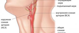
Structure of the carotid artery
This artery is a pair, the common artery is divided into two - left and right. The left carotid artery begins from the aortic arch, and the right - in the brachial trunk. They are further divided into the external carotid artery and the internal one. This place is called a bifurcation. Immediately after the branch, a certain expansion is formed in the internal carotid artery, called the carotid sinus, which is an important reflexogenic zone with numerous nerve cells. These cells take part in maintaining blood pressure, regulating heart function, blood composition and oxygen availability.
Causes
One of the main reasons influencing the appearance of vertebral artery syndrome is cervical osteochondrosis. In addition, these may be diseases that contribute to the formation of cholesterol plaques that clog blood vessels.
But the following accompanying factors can also intensify symptoms and adversely affect the course of the disease:
- Smoking.
- Head injury.
- Diabetes mellitus, both types 1 and 2.
- Ingestion of excess caffeine in the form of tea or coffee, especially if the dose is large.
- A variety of kidney diseases, which can increase swelling and poorly remove excess water from the body.
- Diseases of the cardiovascular system.
- Infections in the body.
In addition, the very appearance of the patient can indicate how the blood flow is impaired.
If the venous outflow is disrupted, then dark circles under the eyes will appear on the face. Bags may also appear.
With vertebral artery syndrome, namely when the artery is interrupted, a person’s face takes on an earthy tint.
If the arterial flow increases and the venous outflow worsens, then hyperemia of the skin will appear.
Congenital
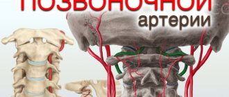
The anatomy of the vertebral artery, both left and right, is designed in such a way that they are protected by the openings of the transverse processes, starting from the 6th vertebra and up to the 2nd inclusive. The vertebral artery arises in the vast majority of cases from the subclavian artery, and only in every 20th person does it arise from the aortic arch.
The vertebral arteries then travel along the Atlantis vertebra (1st vertebra) and pass through the posterior large foramen in the skull to the brain.
Sometimes, as the fetus develops, it develops tortuosity of one or two vertebral arteries at the same time. This condition is congenital and occurs most often due to genetic inheritance. With it, there is a decrease in collagen fibers in the vessels, as a result of which they become more “soft” and pliable.
This condition requires medical supervision, since at an early stage of a child’s development, insufficient supply of parts of the brain can have negative consequences on its development, ranging from the articulatory apparatus to cognitive impairment and the formation of attention deficit hyperactivity disorder.
Purchased
Vertebral arteries can also change during life.
A decrease in blood flow can occur for two main reasons, each of which can be divided into additional ones.
The first reason is external influence, expressed in degenerative changes in the spine, in the cervical region. That is, vertebral artery syndrome most often occurs against the background of cervical osteochondrosis. Displacement of the vertebrae, the formation of protrusions and hernias, pinched nerve roots, spasms in the muscles of the neck and shoulders form either excessive pressure or displacement of the artery. This reduces the amount of blood that flows through the arteries, which causes further health problems.
The second reason is the internal effect on human arteries. As a result of various deviations in the metabolic processes of the body, cholesterol plaques appear, the walls of blood vessels become more fragile and brittle. All this leads to the formation of a small-diameter lumen in the artery. The result is a decrease in blood circulation, with all the ensuing consequences.
Development of pathological tortuosity of the artery
An important factor in the development of ICA tortuosity is heredity and congenital changes associated with the predominance of elastic fibers over collagen fibers in the tissue of blood vessels. This contributes to the wear and tear of the walls of large vessels, their thinning and deformation.
The development of pathology may be the result of excessive load on the arteries, which occurs with hypertension and atherosclerosis, when atherosclerotic plaques deposited on the walls of blood vessels reduce the lumen of the vessel and cause disruption of blood flow. It is likely that there may be other reasons that provoke this pathology, but complete clarity on this issue does not yet exist.
Meanwhile, as preventive examinations show, the manifestation of tortuosity of the carotid artery covers up to 25% of the population.
Tortuosity of the vertebral arteries. Diagnosis. Treatment
Often, patients suffering from high blood pressure and disorders of neurocirculatory functions do not know the true causes of the disease. The disease may be due to tortuosity of the vertebral or carotid arteries. This pathological problem can increase the likelihood of a stroke by 30% due to improper blood circulation in the main vessels of the body. This can even lead to disruptions in the functioning of the brain and the entire central nervous system.
Basically, this disease of the vertebral vessels is inherited and is formed when the tissue of the arteries consists more of elastic fibers than collagen. This leads to rapid wear of the walls of blood vessels, they become thin and begin to deform.
The situation can worsen with atherosclerosis, when plaques are deposited on the walls, reducing the overall patency of the arteries. And this leads to improper blood supply to the brain and other vital organs. Often, the bend may not manifest any symptoms, and only over time the patient’s blood supply to the brain will begin to be disrupted. As a result, the risk of strokes increases if diagnosis is not made in a timely manner.
Types of vascular tortuosity
The most common types of pathological tortuosity of blood vessels are:
- An S-curve is a smooth curve that can occur in one or more places. Such bends do not have a significant impact on a person’s well-being and only appear during preventive examinations. However, they can progress, leading to kinks and, accordingly, to serious disturbances in blood flow.
- Kinking is the bending of a vessel at an acute angle. In some cases, this pathology can be congenital and accompany a person with cerebral circulatory disorders from childhood. It may also develop from S-shaped tortuosity, aggravated by hypertension and atherosclerosis. A concomitant manifestation of Kinking is cerebrovascular accident and its characteristic symptoms.
- Coiling is a loop-like tortuosity of an artery. Although this type of artery pathology does not form a sharp bend, it nevertheless affects the blood flow, significantly slowing it down. The intensity of symptoms depends on the level of blood pressure, the position of the patient and other factors.
Color duplex scanning in the diagnosis of pathological tortuosity of the internal carotid arteries
Ultrasound scanner HS50
Affordable efficiency.
A versatile ultrasound scanner with compact design and innovative capabilities.
The development of ultrasound diagnostics of vascular diseases of the brain has changed the understanding of the prevalence and significance of pathological tortuosity (PI) of the carotid arteries as a cause of cerebrovascular accidents. As a cause of cerebrovascular accident in adults, pathological tortuosity of the internal carotid arteries (ICA) is second only to atherosclerotic lesions in prevalence.
A feature of the diagnosis of pathological tortuosity of the ICA is the possibility of detection during a standard ultrasound examination.
Currently, there is no generally accepted terminology to indicate violation of the straightness of the ICA. In the domestic literature, the most widely used term is “pathological tortuosity of the carotid arteries,” proposed by E.V. Schmidt in 1975 [1]. This designation of violations of the straightness of the ICA has a deep clinical and pathophysiological basis, since it characterizes various variants of ICA deformation from the standpoint of their clinical significance. The use of this term implies not only the designation of the disturbed geometry of the ICA, but also the significance of the incorrect course of the ICA in the formation of cerebral hemodynamic disorders with a certain clinical picture.
There is no generally accepted classification of pathological tortuosity of the ICA. The most widespread classification of pathological forms of ICA, proposed by J. Weibel and W. Fields [2]. The authors distinguish three types of ICA deformation, designating them as tortuosity, coiling, and kinking. Tortuosity is understood as an S- or C-shaped deformation of the ICA (Fig. 1, 2) without sharp angles and visible disturbances in blood flow. The authors consider this type of ICA deformity to be congenital and hemodynamically insignificant.
Rice. 1.
C-shaped tortuosity of the internal carotid artery (ICA) in color duplex scanning (CDS) mode.
Rice. 2.
C-shaped tortuosity of the ICA in B-mode (a) and CD (b).
Looping (“coiling”) is characterized by congenital circular deformation with the formation of a loop (Fig. 3), which can lead to impaired cerebral circulation.
Rice. 3.
Loop-shaped tortuosity of the left (a) and right (b) ICA in the CD mode.
Kinking is understood as acquired, hemodynamically significant angulation of the ICA with stenosis of its lumen (Fig. 4).
Rice. 4.
Loop-shaped tortuosity of the ICA with kinking in the CDS mode (a) and schematically (b).
Color duplex scanning (CDS) allows not only to assess the shape of the tortuous carotid artery, but also to characterize the hemodynamic state in detail. In accordance with this, all types of violations of the straightness of the ICA can be divided into “hemodynamically significant” and “hemodynamically insignificant”.
The introduction of modern ultrasound diagnostic methods into clinical practice indicates a high prevalence of pathological tortuosity of the ICA. According to F. Koskas et al. [3], the tortuous course of the carotid arteries occurs in 10-43% of cases in patients with cerebrovascular accidents. It is important that the frequency of detection of pathological tortuosity of the ICA in adults ranked second after atherosclerosis of the carotid arteries. A study conducted by V.P. Kulikov et al. [4] showed that among patients in whom pathological tortuosity of the carotid arteries was identified according to the results of CDD, there were slightly more women (56.1%) than men (43.9%), pathological tortuosity of the right ICA was more often diagnosed (42, 3%) compared to the left (25.1%), in 32.6% bilateral tortuosity was detected. Thus, it was shown that pathological tortuosity of the ICA is a common pathology not only in patients, but also in the population.
The etiology of pathological tortuosity of the ICA has not been definitively established. Today, there are two points of view on the causes of pathological tortuosity of the ICA - both congenital and acquired pathology.
Congenital genesis is supported by the detection of deformed carotid arteries in young patients in the absence of atherosclerotic process in the vessels, and frequent bilateral damage to the carotid arteries.
The acquired nature of the pathological deformation of the ICA may be supported by the high frequency of detection of this pathology with age and the dependence of the severity of the carotid artery bend on age and blood pressure level.
Pathological tortuosity of the ICA in clinical manifestations resembles the symptoms of atherosclerotic stenosis of the ICA and is manifested by signs of cerebrovascular accident. Most often, deformation of the carotid arteries is combined with arterial hypertension and atherosclerosis. In general, clinical manifestations of pathological tortuosity of the ICA are not very specific. Local signs of pathological tortuosity have some specificity, including pathological pulsation in the neck, signs of compression of the hypoglossal, accessory and vagus nerves.
However, most symptoms and syndromes are nonspecific. The most common pathological tortuosity of the ICA is motor impairment, blurred vision, headaches, speech impairment, dizziness, loss of consciousness, noise and ringing in the ears, pain in the neck, and epileptiform seizures.
The absence of specific clinical signs of pathological tortuosity of the ICA significantly complicates the identification of this pathology based on the results of a clinical examination.
Detection of pathological tortuosity of the ICA in connection with its low-specific clinical manifestations is possible only with the help of specialized instrumental diagnostic methods. Given the limitations of radiocontrast angiography and magnetic resonance angiography, the main method for detecting ICA deformity is CD. This method combines the ability to visualize vessels and blood flow in them with the study of the nature and quantitative parameters of blood flow.
Ultrasound criteria for hemodynamic disturbances in pathological tortuosity of the ICA are recorded by Doppler methods locally - in the zone of vessel tortuosity. These abnormalities can be recorded using different options for color Doppler mapping and in pulsed mode.
A typical picture of pathological tortuosity of the ICA of various shapes in the CD mode is shown in Fig. 3-6. The CDS method makes it possible to characterize in detail the shape of tortuosity, its localization and identify disorganization of blood flow. The criteria for the local hemodynamic significance of pathological tortuosity of the ICA are: gradient of peak systolic blood flow velocity between the proximal and distal sections of the vessel in relation to the site of tortuosity with a decrease in velocity in the distal direction by 20% or more; an increase in peak velocity at the site of angulation compared to the proximal portion of the ICA by 30% or more; disorganization of blood flow in the tortuosity zone, manifested by an increase in spectral expansion and disruption of the blood flow pattern in color mapping mode; the presence of asymmetry in the peak blood flow velocity in the distal section of the tortuous artery compared with the same section of the unchanged contralateral ICA. Thus, from the data presented above it follows that one of the links in the pathogenesis of cerebral circulatory disorders in pathological tortuosity of the ICA is a local disturbance of hemodynamics in the tortuosity zone with a decrease in blood flow in the distal direction.
Rice. 5.
Loop-shaped tortuosity with kinking from the mouth and high-lying S-shaped tortuosity of the ICA in the CDS mode.
Rice. 6.
S-shaped pathological tortuosity of the ICA in the CD mode (a, b).
A retrospective analysis of the medical records of 36 patients in the clinic with subsequently diagnosed pathological tortuosity of the ICA according to the CDS showed that neurologists on an outpatient basis made a clinical diagnosis of chronic cerebrovascular accident in 18.9% of cases, acute cerebrovascular accident in 6.7% of cases. , cerebral angiodystonia - in 16.4%, cervical osteochondrosis - in 10.1%, consequences of traumatic brain injury, intracranial hypertension, cephalalgia, hypertension - in 18.2% of cases. Patients referred for examination without a clinical diagnosis, in whom pathological tortuosity of the carotid arteries was discovered during CDS, amounted to 29.7%.
Moreover, to assess the informativeness of CD in diagnosing pathological tortuosity of the ICA, we analyzed the medical records of 9 patients who underwent angiography and surgical treatment. Concordance of the results regarding the presence of pathological tortuosity of the ICA was noted in 100% of cases. However, discrepancies were also found in the assessment of the shape and location of tortuosity. Obviously, the reason for the discrepancies is the lack of a generally accepted classification of pathological tortuosity of the ICA and clear assessment criteria for both the form and localization of tortuosity.
Currently, pathological tortuosity of the ICA can be the cause of transient and persistent cerebrovascular accidents. With modern ultrasound equipment, diagnosing pathological tortuosity of the ICA is not technically difficult and the main method is primarily CD.
Literature
- Vascular diseases of the nervous system / Ed. E.V. Schmidt. M.: Medicine, 1975. 663 p.
- Weibel J., Fields WS Tortuosity, coiling and kinking of the internal carotid artery. I Etiology and radiographic anatomy // Neurology. Minneap, 1965. V. 15. P. 7-18.
- Koskas F., Kieffer A., Kieffer E. et al. Loops and folds of the carotid and vertebral arteries: indication for surgery // J. Malad. Vascul. 19 Supl. A. 1994. P. 51-54
- Kulikov V.P., Khoreyev N.G., Gerasimenko I.N. and others. Color duplex scanning of vessels in the diagnosis of pathological tortuosity of the carotid arteries // Echography. 2000. N 2. S. 147-154.
Ultrasound scanner HS50
Affordable efficiency.
A versatile ultrasound scanner with compact design and innovative capabilities.
Symptoms
Pathological tortuosity of the internal carotid arteries is most often asymptomatic, however, as the patient’s pathological tortuosity progresses, he begins to be bothered by phenomena caused by cerebrovascular accidents, which manifest themselves in the following symptoms:
- frequent dizziness;
- headaches of different localization;
- violation of movement coordination;
- noise in ears;
- periodic speech impairment;
- short-term loss of consciousness;
- memory impairment;
- decreased ability to work.
Cerebral arteries
This tortuosity of the arteries is observed on a par with those described above and can develop in both small and large cerebral vessels. Its etiology is also genetic. In most cases, the arteries at the base of the brain are subject to deformation, which includes, among other things, the circle of velisium and neighboring areas.
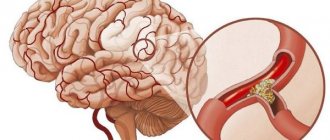
Often, tortuosity of the cerebral arteries is combined with their stenting. Thrombosis and occlusions are often observed in kinks. This leads to the development of stroke in the segment supplied by the affected artery.
In the brain, tortuosity of not only arteries, but also venous vessels is possible. Due to the deterioration of blood outflow from the brain, venous congestion occurs, the root cause of which can be difficult to find due to the fact that the clinical manifestations of many vascular pathologies are very similar to each other.
Symptoms
The clinical manifestations of symptoms of congestion in the veins of the brain may differ with different locations of the affected veins, but there are also common features:
- Intense headache, which is accompanied by neurological symptoms, impaired consciousness, nausea, vomiting, patients may become agitated. Then focal symptoms appear: convulsions, paralysis and paresis and other symptoms.
- If the affected veins become thrombosed, they can become inflamed, causing thrombophlebitis. This is indicated by hyperthermia. In advanced cases, this condition leads to stroke of the hemorrhagic type, which can cause coma and lead to death.
Pathological tortuosity of cerebral vessels can be hereditary or acquired as a result of long-term essential hypertension. The symptoms of this type are identical to tortuosity of vessels of other localization.
Venous congestion is characterized by persistent arterial hypertension that does not respond to treatment.
It also causes persistent headaches of varying intensity. The pain is usually located in the crown area and is combined with convulsive attacks and loss of consciousness. In such patients, nosebleeds may begin, after which the patient’s well-being improves.
Stagnation is also accompanied by eye symptoms: the eyes hurt, the veins in them dilate, the whites swell, the veins on the eyelids, in the temporal region and on the crown of the head become tortuous.
Treatment
Timely diagnosis significantly improves treatment results. Patients with venous stasis should be started on anticoagulants to reduce blood viscosity and reduce the risk of thrombosis. For this purpose, heparin is often used, the dosage of which is selected individually. At the same time, doctors try to prevent the possibility of a hemorrhagic stroke and, for this purpose, stabilize blood pressure.
Heparin
If inflammation occurs, the patient must be prescribed antibiotics, and if pain occurs, painkillers are needed. Treatment of pathological tortuosity of blood vessels requires great responsibility not only from doctors, but also from patients.
In order to prevent relapse of the disease, they must strictly follow all the recommendations of the attending physician even after discharge. When emergency conditions are resolved, the doctor, after additional diagnostics, must decide on the need and possibility of surgical intervention. If this is not possible, the patient is prescribed treatment to relieve symptoms. He must forget about bad habits, switch to a healthy lifestyle, closely monitor his blood pressure levels and receive courses of blood thinning medications on time.
There are special neurological sanatoriums in which such patients must undergo a course of rehabilitation at least once a year.
Diagnostics
Diagnosis of pathological tortuosity of the carotid arteries cannot be carried out only on the basis of symptomatic indicators, since these manifestations are also characteristic of other types of vascular diseases. Therefore, the results of studies conducted in the clinic are a mandatory addition.
These include:
- echo scanning with ultrasound Dopplerography and spectral analysis of the received signal;
- spiral computed tomography;
- X-ray contrast angiography.
These diagnostic tools make it possible to assess hemodynamic parameters of blood flow in the area of tortuosity, identify the presence of dysplasia of the internal carotid artery distal to the tortuosity, and determine the nature of the deformation of the vessel. Significant pathological changes identified on the basis of complex diagnostic tools serve as an indication for treatment through surgical correction.
Treatment
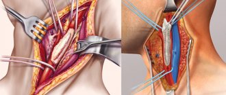
Treatment of pathological tortuosity of the carotid arteries is possible only with surgery. This type of surgery is performed in special vascular surgery centers. The section of the vessel subjected to stenosis or affected by atherosclerosis is shortened, which allows the patency of the artery to be restored. In case of extensive damage to the carotid artery, they resort to prosthetics of a section of the vessel.
If the tortuosity of the carotid arteries is accompanied by stenosis, balloon catheters and metal stents are used to widen the section of the artery, which are left inside the vessel to prevent its re-narrowing. The recovery period after surgery takes no more than 7 days. After treatment, the symptoms of oxygen starvation of the brain completely disappear.
Prevention
The following measures can be used to prevent carotid artery diseases:
- maintaining blood cholesterol levels within the required level, excluding fatty, salty and smoked foods from the diet and enriching it with fresh vegetables and fruits;
- treatment of arterial hypertension;
- complete cessation of smoking, which is one of the most dangerous factors influencing the development of pathological changes in the walls of blood vessels;
- body weight control;
- performing physical exercises with a moderate level of stress;
- exclusion of heavy physical activity, sudden head movements, and playing sports at a professional level.
In addition, it is important to consult a doctor if symptoms of cerebral circulation disorders appear and undergo the tests prescribed by him.
Compliance with all these measures is also relevant for people who have already undergone surgery.
Vessels
Arteries of the Head and Neck: Anatomy, Diagram, Atherosclerosis
Feb 06, 2020 Kokh V. A.
16582
Vessels
Vessels of the Neck and Head: Anatomy, Diseases, Symptoms
Feb 03, 2020 Kokh V. A.
41688
Vessels