September 24, 2019
9461
0
3 out of 5
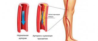
Vertebral artery syndrome is a collective term that unites a number of different diseases of a vascular, vestibular, traumatic, vegetative nature, accompanied by a narrowing of one or both vertebral arteries. This is accompanied by painful migraines, impaired hearing, vision, consciousness and can lead to stroke, disability, irreversible damage to the brain and disability.
Patients with this diagnosis fall out of the usual rhythm of life for a long time, lose their ability to work and require qualified treatment. But for it to be successful, it is important to accurately determine the cause of the development of vertebral artery syndrome and act on it. SL Clinic specialists will help you do this. With us you will receive a consultation and will be able to undergo all the necessary diagnostic procedures. As a result, we will determine exactly what caused the problem and develop the optimal treatment tactics.
What do the statistics say?
Statistical processing of data obtained from computed tomography showed that in almost 1/3 of patients (26% isolated and 3% in combination with other vessels) with ischemic stroke, the main focus is located in the vertebrobasilar zone of responsibility or basin. It is formed by the bilateral vertebral artery, which passes into the basilar (main) artery.
According to clinical findings, transient ischemic attacks in this zone occur 3–3.5 times more often than in other extracranial areas of the brain’s blood supply.
The cause of death from cerebral vascular insufficiency in 57% of cases is the atherosclerotic process in the vertebral arteries. The clinical picture of the lesion is associated with the characteristics of their location, shape, and participation in hemodynamics.
General information
The blood supply to the brain comes from two main basins: carotid and vertebral (carotid and vertebral arteries, respectively). The latter covers a quarter of the total need for oxygen and nutrients - it vascularizes the following structures:
- Brain stem.
- Cerebellum.
- Occipital lobes.
- A large part of the temporal lobes.
- Posterior part of the hypothalamus.
- Spinal cord (segments C1–Th3).
- Inner ear.
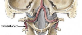
Damage to the vertebral artery in diseases of the cervical spine is determined by its anatomical and topographical features. The vessel, together with the nerve of the same name, passes through the canal, which is formed by openings in the transverse processes of the vertebrae. The latter is not static, since it changes according to movements in the neck. In the vertebral artery itself, according to its location, several segments are distinguished:
- 1 – from the subclavian artery to the entrance to the canal.
- 2 – in the canal at the level of C2–C6 vertebrae.
- 3 – from the exit from the canal to the entrance to the cranial cavity.
- 4 – in the cranial cavity (intracranial).
In the canal, the artery borders posteriorly with the uncovertebral joints, and laterally with the superior articular processes. After leaving it, the vessel bends twice: in the frontal and sagittal planes. It is in these places that disruption of blood flow through the vertebral artery often occurs.
The topographic and anatomical features of the vertebral artery make it vulnerable to the adverse effects of a number of external and internal factors that contribute to impaired blood flow through the vessel.
Anatomical features of the vertebral arteries
Normally, 30% of the required blood volume enters the brain through the vertebral arteries. Anatomy plays a significant role in creating the conditions for narrowing the diameter of blood vessels.
The vertebral artery branches from the subclavian towards the central part of the inner edge of the scalene muscle in the neck.
It is important that no more than 1–1.5 cm remains to the adjacent mouth of the thyrocervical trunk, which is also a branch of the subclavian artery. This creates an additional mechanism of “stealing” (redistribution of blood) in case of hypoplasia or stenosis of the vertebral artery.
Heading upward, the artery at the level of the sixth cervical vertebra (less often the fifth) enters a protected bone canal formed by the spinous processes of the vertebrae.
It is customary to distinguish sections or segments of the vertebral artery:
- I - the entire area from VI to II cervical vertebrae, where the vessel exits the foramen;
- II - outside the canal, at an angle of 450, it deflects posteriorly and goes to the transverse process of the first cervical vertebra (atlas);
- III - passing through the opening of the atlas on its back side, the artery forms loops, their role is to prevent disruption of blood flow when turning the head;
- IV - heading into the foramen magnum, the artery is located inside a dense ligament, with ossification of the receptacle or bone outgrowths on the occipital bone, conditions are created for traumatizing the walls of the vessel during movements in the cervical region;
- V - inside the foramen magnum (intracranial segment), the vertebral artery passes through the dura mater and lies on the surface of the medulla oblongata.
The fusion of the left and right arteries into a single trunk (basilar artery) ensures participation in the formation of the circle of Willis at the base of the brain
A special feature is the compensatory development of blood circulation due to the vertebral artery on one side if the other symmetrical branch is compressed. The asymmetry of blood flow through the vertebral arteries is leveled by the flow of blood through the basilar artery into the undamaged part.
Reasons for development
Many people mistakenly call vertebral artery syndrome a disease, whereas formally the syndrome is not a disease, but only a group of symptoms. In our case, the syndrome is a complex of symptoms that are caused by pressure (compressive effects) on the vertebral artery and the sympathetic nerve plexus located around it. Subsequently, blood flow is weakened. In this case, pathology can be observed in one or several vertebral arteries. Left-sided syndrome develops much more often. In any case, the pathology inevitably leads to disruption of the blood supply to the central nervous system (it is the arteries described that are responsible for approximately 30% of the blood flow to the brain).
What factors cause compression of the arteries? First of all, we should remember about degenerative-dystrophic lesions of the spine in the cervical region: osteophytes, intervertebral hernias and, more likely, osteochondrosis (another name for the pathology is posterior cervical sympathetic syndrome). In other words, osteochondrosis and this syndrome have an obvious connection.
Another possible cause of the problem lies in the atypical origin of the vertebral arteries from the subclavian arteries. Also, one should not exclude an abnormal structure of the atlanto-occipital joint. That is, the development of the disease can be facilitated by causes related to the spine and those not related to it. Predisposing factors for the development of pathology are sudden bending and turning of the head.
What anatomical pathology is most common?
20% of cases of pathology of vertebral arteries are due to developmental anomalies:
- originating directly from the aorta;
- entry into the bony spinal canal higher than usual (at the level of the third to fifth cervical vertebrae);
- displacement of the mouth outward.
More often, the lesions are combined and are divided into the following options:
- up to 34% is due to the combined effect of developmental anomalies and extravasal muscle compression;
- 39% are stenoses of atherosclerotic and thrombotic nature;
- the maximum part - 57% - represents compression by various displacements of the vertebrae in combination with atherosclerosis.
Diagnostics
For timely detection of pathologies in individual segments of the vertebral vessels, the doctor studies the picture of the course of the disease, and after analyzing the symptoms, prescribes a specific instrumental diagnostic technique.
To identify various pathologies in the arteries, the following types of examinations are prescribed:
- Dopplerography. This type of diagnostic examination makes it possible to measure the speed of blood flow in the vessels and its direction. Allows you to assess the degree of narrowing in each section of the spinal arteries
- Radiography. Using images, the doctor assesses damage to the vertebrae, tracks the places of external influence on the outer vascular walls, foci of inflammation and places of formation of sclerotic plaques and blood clots
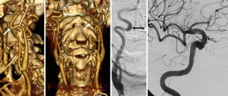
- Antiography. By introducing special solutions that tint the blood, the specialist visually obtains a more complete picture of pathologies in the arteries
- MRI. This method makes it possible to analyze the condition of not only the arteries of the spine, but also the entire vascular network of the cervical spine and bone tissue of the spine
The doctor may prescribe one type of examination, or recommend a comprehensive diagnosis. Having received comprehensive information about the pathologies of vascular pathology of the spine, the doctor chooses the most effective method of providing care to the patient.
Main causes and connection with damage location
All causes of pathology of the vertebral arteries are divided into 2 large groups:
- vertebrogenic,
- nonvertebrogenic.
Vertebrogenic are caused by the effects of changes in the spine. In childhood, the most common are:
- developmental anomalies;
- injuries in the cervical spine (including those received during childbirth);
- pathological muscle spasms with severe hypothermia, torticollis.
In adults there are more connections with vertebral diseases:
- osteochondrosis;
- Bekhterev's disease;
- tumors.
Injuries also matter.
Changed lateral processes of the vertebrae take part in the compression of the artery
Nonvertebrogenic diseases are represented by three groups of diseases:
- causing stenosis of the lumen of the arteries (inflammatory arteritis, thrombosis, atherosclerosis, embolism);
- contributing to disruption of the shape and direction of blood vessels (kinks, non-linear course from the sixth to the second vertebra, increased tortuosity);
- as a consequence of compression from the outside (spasmodic muscles, abnormal ribs, scar tissue in the postoperative period).
The level of narrowing of the vertebral artery correlates with the causes of the pathology.
If compression occurs to the point of entry into the bone canal, then this is due to spasm of the scalene muscle, enlarged stellate nerve ganglion. More often occurs with an abnormal location of the initial section of the artery. Here is the most vulnerable area for the deposition of atherosclerotic plaques (70% of cases).
Inside the bone canal of the transverse processes of the vertebrae, the following can be dangerous for the vessel:
- enlarged uncinate processes;
- subluxations in the vertebral joints, leading to pinching of one or both arteries;
- consequences of spondyloarthrosis, proliferation of articular surfaces;
- disc herniation (rare).
When leaving the canal, the artery is obstructed by:
- too deep groove above the upper edge of the atlas, which forms an additional bone canal (Kimerli anomaly);
- pressing against the vertebral bodies by the spasmodic inferior oblique muscle of the head;
- atherosclerotic plaques (it has been established that extracranial parts of the artery are more often affected by atherosclerosis than internal ones);
- increased tortuosity and additional kinks are more often formed at the level of the first and second cervical vertebrae, usually combined with similar changes in the subclavian and carotid arteries.
The main reason for increased tortuosity, which causes non-straightness of the course of the vertebral arteries, is the loss of elastic properties of the vessel wall due to age-related disorders in collagen metabolism, prolonged hypertension
Thrombotic changes in the vertebral arteries are found at autopsy in 9% of people who have suffered vascular diseases of the brain. As a rule, they are preceded by severe atherosclerosis. Without atherosclerotic changes, thrombosis is facilitated by the development of the “steal” syndrome with reverse vortex blood flows due to the subclavian artery and its other branches.
Main reasons
There are three main reasons for the development of vertebral artery stenosis:
- Congenital factor - predisposition at the genetic level leads to congenital changes in the structural structure of blood vessels. If the disease does not enter the acute phase, people with a similar diagnosis continue to live a full life.
- Acquired factor - can be attributed to the main reason causing the need to treat narrowing of the vertebral artery. The condition of blockage of blood vessels can cause diabetes, atherosclerosis and various metabolic disorders.
- Traumatic factor - the artery narrows due to force (fracture, bruise, hematoma). Surgery to eliminate the causes of the blockage is mandatory.
How does impaired patency of the vertebral arteries manifest itself?
Clinical signs of impaired blood flow in the vertebral arteries depend on the following factors:
- state of the circle of Willis;
- development of a network of collaterals and anastomoses with the subclavian artery;
- rate of increase in obstruction.
A combination of symptoms indicates damage to a specific part of the brain. The most common ischemia of the basin:
- posterior cerebral artery;
- brainstem or cerebellum zones (acute and chronic);
- nuclei and cranial nerves causing vestibular disorders.
The disease has a crisis course. Vertebral crises manifest themselves with a variety of symptoms. Most often stimulated by head movements. At the same time, damage to the brachial plexus and spinal cord is detected.
“Cervical” migraine syndrome accompanies cervical osteochondrosis and spondylosis. Characterized by:
- typical pain in the back of the head and neck, radiating to the supraorbital region;
- fainting;
- dizziness;
- tinnitus.
The duration of pain ranges from several minutes to hours
Vestibular crises are accompanied by:
- severe dizziness, a feeling of objects spinning;
- eye nystagmus;
- disturbed balance.
Atonic-adynamic syndrome appears with ischemia of the medulla oblongata:
- a sharp decrease in muscle tone;
- inability to stand independently.
Visual disturbances due to impaired microcirculation of the eyes:
- spots, dots, lines before the eyes;
- darkening;
- transient loss of visual fields;
- sensation of flashes in the eyes (photopsia), decrease in visible objects (micropsia);
- optical deception phenomena.
Less common:
- A syndrome of transient tonic cramps in the arms and legs without loss of consciousness, while the extensor muscles tense and the limbs stretch. The symptom of “intermittent claudication” in the arms is observed in 65% of patients.
- Transient speech disorders, spasm of masticatory muscles.
- A sudden contraction of the diaphragm, which is manifested by a paroxysmal cough, dilation of the pupil on the affected side, increased salivation, and tachycardia.
Outside of crises, the neurologist will notice in the patient some mild focal symptoms, paresis of some pairs of cranial nerves.
Vertebral artery syndrome (Barre-Lieu) - symptoms and treatment
For chronic neck pain, surgical and pharmacological treatment methods are used, as well as various methods of traction therapy —spinal traction with the help of special devices (blocks, belts, rings) [12].
The effectiveness of manual therapy methods - soft techniques, post-isometric relaxation, muscle stretching - is still being studied. To this end, two studies were conducted in 2020. A meta-analysis by Chinese scientists showed that manual techniques are less effective in eliminating pain than cervical spine traction [13]. However, a study by Canadian scientists found that manual therapy becomes more effective when combined with another method of active treatment for acute and chronic neck pain. Also, manual techniques cope better with chronic pain than massage, and more effectively combat acute and subacute neck pain than drug treatment. However, due to side effects from taking medications, manual therapy is preferable. In terms of effectiveness, manual techniques are similar to mobilization of the cervical spine, but mobilization as a separate intervention does not reduce pain [14].
As for the non-drug treatment method of acupuncture , a 2020 meta-analysis showed its effectiveness over sham acupuncture (acupuncture of non-acupuncture points) and inactive treatment [15].
An undoubted effective method of treating the syndrome is physical exercise . Their effectiveness in relieving acute and chronic pain is supported by a 2005 review. In this case, it is worth focusing on stretching the cervical region, shoulder girdle and chest. And the combination of exercises with mobilization and manual techniques on the cervical spine helps reduce pain in the short and long term [16].
Surgical methods include :
- puncture interbody fusion - combining and fixing several vertebrae to avoid their displacement;
- fenestration - partial removal of the arch of the intervertebral disc;
- autodermoplasty of intervertebral discs - replacement of the disc with one's own tissues.
During the operation, porous explants made of an alloy of titanium and nickel are implanted. Due to their porosity, bone tissue quickly grows in them. This makes the fixation strong and significantly reduces the patient’s period of incapacity for work and the neck being in a stationary position.
In addition to stabilizing operations of the cervical spine, other types of interventions are also performed:
- decompressive-stabilizing - removal of compression with subsequent fixation of the spine;
- decompressive-plastic (laminoplasty) - elimination of compression by enlarging the spinal canal while maintaining the integrity of the posterior elements of the vertebrae;
- decompressive operations - removal of the intervertebral disc or its arch, compressing the artery, etc. [9].
There is considerable experience in reconstruction of vertebral arteries . The following methods are used:
- transposition - displacement of the vertebral artery into the subclavian (common carotid);
- angioplasty - dilation of a vessel using an expanding balloon;
- Stenting is the expansion of a vessel by installing a stent.
If the first and second segments of the vertebral artery are simultaneously affected, bypass surgery is performed—creating a bypass at the level of the third segment [17].
Characteristics of the main symptoms
Headaches are present in 73% of patients. They have a shooting, contracting, pulsating character.
Intensifying:
- upon palpation of the cervical vertebrae;
- after sleeping in an uncomfortable position;
- as a result of local cooling.
Dizziness is more common in the morning after sleep, accompanied by impaired hearing, vision, and a feeling of noise in the head.
Such a symptom as tinnitus is felt on both sides in most patients.
With a unilateral murmur, it indicates the side of the lesion
Characteristically, the height of the audible noise increases at the onset of a vascular crisis and decreases in the interictal period. Patients note changes during the day with osteochondrosis (intensifies at night).
Numbness is observed on the skin of the neck, around the mouth, and on the hands.
Fainting conditions are provoked by hyperextension of the head back. They are usually preceded by other listed manifestations.
Nausea and vomiting are considered harbingers of a crisis.
The long course of the disease causes mental changes in patients and is accompanied by depression.
Symptoms and diagnosis
Signs of vasoconstriction are:
- Periodic headache. With vascular stenosis, basilar migraine often occurs. It is characterized by a severe, throbbing headache predominantly in the occipital region. Headache worsens after sleep, during shaking and when turning.
- Noise in ears.
- Dizziness.
- Vestibular ataxia (deterioration of coordination of movements, unsteadiness when walking, sleep disturbance, nausea, vomiting).
- Vegetative symptoms in the form of sweating, chills, chilliness of the extremities, lability of blood pressure, rapid heartbeat, redness of the face and pallor of the skin.
- Visual impairment (decreased visual acuity, nystagmus, fog, rainbow circles and stripes before the eyes, fatigue during visual work).
- Pain in the neck area. Most often it is felt from behind and radiates to the frontal and parietal lobes.
If transient ischemic attacks develop against the background of arterial stenosis, motor, speech and sensory disorders, visual disturbances in the form of hemianopsia (loss of visual fields), diplopia (double vision) and dysphagia appear.
When the left or right vertebral artery is narrowed, drop attacks (characterized by episodic weakness in the limbs and tilting of the head) and Unterharnscheidt syndrome (manifested by short-term loss of consciousness as a result of sudden turns of the head) often develop.
To make a diagnosis you will need:
- taking anamnesis;
- physical examination;
- assessment of neurological status;
- neurological examination;
- radiography of the spine in 2 projections;
- CT scan;
- Magnetic resonance imaging;
- rheoencephalography (assessment of blood filling of the arteries in the head and neck area);
- Doppler ultrasound;
- angiography;
- duplex scanning;
- ophthalmological examination;
- functional tests;
- general clinical tests;
- lipidogram.
Important information: How to strengthen weak walls of blood vessels (capillaries) in the brain with folk remedies and what to do to treat brittle (fragile) veins in the legs
The doctor’s task is to determine the causes of stenosis (narrowing) of the left or right vertebral artery.
What is the danger of violations?
Impaired patency of the vertebral arteries ultimately causes ischemia in various parts of the brain. Vascular crises are variants of transient ischemic attacks. Lack of attention to symptoms and improper treatment contribute to the rapid development of a “full-fledged” ischemic stroke with adverse consequences: paresis, paralysis, speech and vision impairment.
To miss important symptoms means dooming the patient to disability and his own helplessness. Recovering from a stroke is not easy for everyone and requires a lot of effort.
Why does narrowing of the vertebral artery develop?
Impaired blood supply to the brain depends on the degree of compression of the vertebral artery. The narrowing is caused by atherosclerosis, nerve spasm, external compression of the vertebrae, tumor, thromboembolism.
The cause of impaired blood supply to the vertebrobasilar area may be tortuosity of one or both vertebral arteries. To treat the pathology, stenting and balloon angioplasty are used.
The procedures are performed under local anesthesia. The stent is installed through a puncture in the femoral area. The introduction of an endoprosthesis is necessary to maintain the physiological lumen of the vessel. Until a few years ago, stenting was performed under X-ray control. A scopic cover was used to visualize the insertion of the balloon stent. The movement of the balloon from the femoral to the vertebral artery is clearly visible on the X-ray television screen. The procedure led to radiation exposure of the patient, so at the present stage the intervention is monitored under the guise of ultrasound.
How to identify pathology of the vertebral arteries?
Based on the presence of symptoms and the determination of its connection with neck movements, a suspicion of pathology of the vertebral arteries arises from a general practitioner or therapist. Timely referral to a neurologist and examination is a matter of experience.
Duplex scanning allows you to see the structure of the vessel, determine the nature of the stenosis, the degree of damage to the artery walls
Basic methods:
- Ultrasound Dopplerography - an assessment of all anatomical characteristics of the vertebral arteries on both sides, diameter along the length, speed of the blood flow wave is carried out; it is important as a way to determine cerebral circulatory reserve;
- Magnetic resonance imaging of the brain and neck vessels will indicate emerging lesions with impaired blood supply, the formation of cysts, aneurysms;
- An x-ray of the cervical spine can be used to judge the participation of pathological growths of bone tissue in pinching of the vertebral arteries;
- Angiography of the neck vessels is performed by injecting a contrast agent into the subclavian artery. The technique is informative, but is carried out only in specialized departments.
Causes of narrowing of the vertebral artery of the brain
Narrowing of the vertebral arteries of the brain can occur under the influence of many negative factors. They are divided into external and internal. First, let's deal with the internal ones, since they are less common in young and middle age. But the frequency of their occurrence increases sharply over the age of 55 years. Such negative factors include:
- diabetes mellitus and diabetic angiopathy;
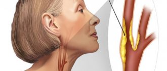
- high levels of cholesterol in the blood, which can provoke the development of cerebral atherosclerosis and deposition of cholesterol plaques in the lumen of the blood vessel;
- inflammation of the vascular wall (vasculitis), scar deformation and thickening;
- arterial thrombosis with increased blood clotting.
Other causes of narrowing of the vertebral artery are associated with the negative effects of external risk factors. These include the following diseases and conditions:
- inflammation of the muscles of the neck and collar area (when they are excessively tense and spasm, pressure is exerted on the vessels);
- excessive compensatory muscle tension due to pathologies in the spine;
- poor posture and curvature of the spine (provokes a change in the course of the artery and, accordingly, a sharp narrowing of its internal lumen);
- displacement of the vertebral bodies with compression of the blood vessel;
- development of osteochondrosis (degenerative dystrophic disease of the cartilage tissue of the intervertebral discs) with complications such as protrusion, extrusion and intervertebral hernia;
- deforming osteoarthritis of intervertebral joints;
- cicatricial deformation of the ligamentous, tendon and muscular apparatus;
- subluxation or dislocation of the first cervical vertebra;
- assimilation of the first cervical vertebra by the tissues of the occipital bone;
- traumatic impact on the neck area, leading to fracture, cracks, dislocation, displacement, stretching or rupture of tissue.
Much less often, narrowing of the vertebral artery is caused by the growth of tumors and infectious processes. These pathologies are excluded last.
Treatment options
One of the simple methods of treatment is to constantly wear a Shants collar. By the way, it is also used for diagnostics: if the patient feels improvement after using the collar, this confirms the connection with the pathology of the vertebral arteries.
The importance of exercise therapy and massage
We also recommend reading: Color duplex scanning of the brachiocephalic arteries
Rare vascular crises allow treatment to be done without potent medications. To do this, you need to master physical therapy exercises and massage techniques.
Movements should be done carefully, at a slow pace:
- turning the head to the sides, at first with a small amplitude, gradually increasing it;
- pressure on the ball with your forehead;
- head nods;
- shrug.
Massage is not performed in the acute period. Its main task is to relieve tension in the neck muscles and reduce pressure on the arteries. It is not recommended to trust the procedure to an inexperienced person.
Treatment with medications
Depending on the cause of the narrowing, the doctor chooses drugs:
- anti-inflammatory action (Nimesulide, Ketorol, Naisilate);
- to maintain vascular tone you will need Troxerutin and a group of venotonics;
- thrombus formation can be prevented with the help of Curantyl, Trental;
- for dizziness and vestibular disorders, Betaserc, Betahistine are indicated;
- To protect the brain from ischemia, neuroprotectors are needed (Mexidol, Piracetam, Gliatyllin).
Physiotherapeutic techniques have the same goals as massage and promote pain relief. Courses offered:
- magnetic therapy,
- diadynamic currents,
- phonophoresis with hydrocortisone.
Acupuncture and traction can only be used in specialized centers.
Exercise therapy is especially indicated for sedentary work
Complications
With thrombosis or dissection of the vertebral artery, a severe brainstem stroke can develop with a mortality rate of more than 80%. Other complications include the appearance of signs of dyscirculatory encephalopathy - decreased memory, performance, and sleep disturbances. With unstable plaques, symptoms of transient cerebrovascular accidents (micro-strokes) may be observed, associated with the tearing off of pieces of plaque and blockage of small arteries of the brain.
Depending on the causes of the symptoms of the disease, various complications may occur. Most often, this is a painful condition that causes depression in patients, but there can also be objective complications that are life-threatening.
- Thrombosis of the vertebral artery with the development of stroke
- Convulsive syndrome (epileptiform seizures)
- Drop attacks (falls without losing consciousness)
- Visual and hearing impairment
When is surgery necessary?
The first operation to reconstruct the vertebral artery was performed in 1956, and in 1959, a thrombus was first removed from the subclavian artery, involving the bed of the vertebral vessel.
The indications for surgery are determined by the results of conservative therapy. If the treatment is ineffective, as well as if the cause is established, related to compression of the artery by a tumor, a vertebral process, it is impossible to do without surgical intervention.
They operate on patients in neurosurgical departments. Bone formations, tumors, and sympathetic nodes are removed (to eliminate excessive spasm).
It is possible to eliminate abnormal tortuosity only if it is localized in segment I.
Narrowing of the left vertebral artery: symptoms
Narrowing of the left vertebral artery can be suspected if the following symptoms are present:
1. Headache syndrome is characterized by dull, burning pain localized in the parieto-occipital region. The symptom intensifies with severe physical activity. The probable location of pain is the superciliary, temporal, parietal areas; 2. Symptoms from the gastrointestinal tract. Nausea and vomiting occur in many patients with vertebrobasilar insufficiency. It is impossible to fight them with medications. The mechanism of dyspeptic disorders is compression of the vertebral artery with impaired blood supply to the intestines; 3. Disorders of the central nervous system - memory loss, changes in visual acuity, eye pain; 4. Vestibular disorders – disorientation, tinnitus; 5. Changes in the frequency of contractions of the cardiovascular system, instability of pressure, attacks of angina.
In addition to surgical and conservative treatment, patients with vertebrobasilar insufficiency need to undergo an additional set of treatment procedures - physiotherapy, massage, kinesiotherapy (treatment with physical movements).
Narrowing of both vertebral arteries is a dangerous pathology in which serious complications develop.
X-ray of the cervical spine in a lateral projection with a decrease in the height of the vertebral bodies at the lower level
Difficulties are caused by timely diagnosis of nosology. To identify the disease, not only x-rays are used, but also other radiation diagnostic methods.
Crisis prevention
Once the diagnosis is established, the patient is able to prevent vascular crises. To do this you need:
- do gymnastic exercises;
- wean yourself from sleeping on your stomach;
- undergo physiotherapy and massage courses at least twice a year;
- purchase an orthopedic pillow to ensure a level position of the cervical spine during sleep;
- wear a Shants collar;
- get rid of factors that narrow arteries (smoking, drinking alcohol).
The clinical picture of a stroke is not necessarily caused by intracerebral vessels. Extracranial disorders should always be kept in mind when making a diagnosis and prescribing treatment. This tactic helps prevent life-threatening complications.
Prevention ↑
Preventive measures are as follows:
- Perform exercises for the neck and shoulder girdle every hour: raise and lower your shoulders, gently move your head in different directions, perform exercises with counterpressure with your own palm. This is especially important for those who work in a sedentary position.
- Sleep on an orthopedic pillow in any position, just not on your stomach, and not in a position with your head thrown back.
- Take neck and collar area massage courses once a year – six months.
- Treatment in sanatoriums specializing in neurological diseases.
It is important to remember that vertebral artery syndrome and alcohol are incompatible things.
With this syndrome, the blood supply to a part of the brain is already impaired, and alcoholic drinks will further intensify the brain steal syndrome.
Treatment
In cases of uncomplicated development of pathology that has developed in various segments of the arteries, conservative treatment can be prescribed. It is based on taking medications and undergoing massage sessions, physical therapy and physiotherapy.
Massage and physiotherapy cannot be performed during a crisis, which can be treated with the following medications:
- anti-inflammatory drugs that eliminate foci of inflammation in the bone and soft tissues surrounding the arteries;
- drugs that stimulate blood flow to the brain;
- neuroprotectors;
- supporting vitamin formulations.
After the main symptoms are relieved, exercise therapy and physiotherapy will give a good result. Exposure of diseased areas to UHF currents and performing gymnastic exercises restores the level of cerebral blood supply. To eliminate the manifestations of osteochondrosis, you can use the skills of a qualified chiropractor.
In cases where conservative treatment methods do not give the desired result, as well as when it is necessary to correct congenital anomalies of the arteries, treatment is carried out in the surgical department.
The doctor performs the following types of surgical treatment:
- removal of formations affecting the outer walls of arterial segments;
- correction of congenital anomalies;
- replacement of a blocked section of a vessel with an artificial prosthesis.
To prevent exacerbation of the disease and avoid repeated crises, prevention of acute forms of the disease is very important.