Post published: 08/02/2016
In 25% of cases, patients with head or neck injuries experience damage to the vessels of the neck, while damage to the carotid arteries is observed in 5-10% of cases. Despite improvements in diagnostic and treatment methods, these injuries are still characterized by high mortality and complication rates.
The mortality rate for injuries of the carotid arteries is in the range of 10-31%, persistent neurological deficit occurs in 16-60% of cases.
Types of vascular injuries
Depending on whether there are external signs of vascular injury, they are divided into open and closed. The former may be accompanied by a bruise, tear or complete dissection of the vascular wall.
Closed injuries are not accompanied by external bleeding, but they lead to thrombosis, intense internal hemorrhage, tissue ischemia, and aneurysm of the wall of a vein or artery.
The danger to life increases if the main vessel is damaged; it is lower if secondary blood pathways are injured. Depending on the type of injured vessel, there are arterial, venous, capillary and mixed pathologies. Blood flow pathways in the arms, legs and neck, head and torso may be affected. Internal bleeding occurs when the organs of the chest or abdominal cavity are damaged . In the case of polytrauma, all these types are combined with each other.
Based on the nature of vessel ruptures, the following are distinguished:
- full,
- partial,
- through,
- tangentially,
- fenestrating (for puncture, wound by shrapnel).
We recommend reading an article about how post-traumatic thrombosis manifests itself and is treated. From it you will learn about the causes of post-traumatic thrombosis, the clinical picture of the development of the acute form, methods of diagnosis and treatment of the veins of the lower extremities, as well as recovery after treatment and the prevention of thrombosis after injury.
And here is more information about the prevention of blood clots and thrombophlebitis.
Causes and signs of venous bleeding
Venous bleeding can be characterized by the location of the blood leakage, since it determines the methods of first aid.
Venous bleeding can occur from:
- Deep veins;
- Superficial veins of the lower and upper extremities;
- Veins of the neck and head.
If there is a risk of venous bleeding, then only a doctor can diagnose and determine its location, since the type of bleeding cannot be determined only by external signs.
The following causes of bleeding can be distinguished:
- Superficial wounds and injuries (shrapnel, gunshot, knife, etc.);
- Phlebeurysm;
- Arterial hypertension;
- Pathologies of the hematopoietic system.
It is necessary to know by what signs different types of bleeding can be identified and how to separate venous from capillary or arterial.
Thus, venous bleeding has a number of signs, the most characteristic of which are the following:
Let's note a few points:
- If the superficial veins of the lower or upper extremities (feet and hands) have been damaged
, mild bleeding is observed, the duration of which does not take much time. However, in this case, first aid is still a necessary measure, since damage to the deeper veins, which are usually located on the inner surface of the limbs, may subsequently be revealed. - It should be taken into account that blood diseases, high blood pressure and alcohol intoxication
have a negative effect on the rate of blood clotting, which can cause increased bleeding.
Causes of injury to blood vessels
Factors that can lead to destruction of the vascular wall include:
- hit;
- wound - stabbing, cutting, laceration, gunshot;
- a fall;
- fracture, dislocation, bruise;
- fragility of blood vessels (age-related, atherosclerotic changes, inflammation);
- connective tissue diseases;
- arteriovenous malformations;
- aneurysms;
- tumor growth.
Damage to the main blood vessels during a fracture:
Types of neck injury
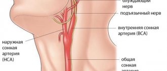
In the thickness of the soft tissues of the neck there are vital anatomical formations. These include: larynx, trachea, pharynx, esophagus, blood vessels supplying the brain and nerves providing vital functions of the body.
Neck injuries are classified depending on the mechanism of injury and which structures are damaged
.
Whiplash
This type of injury most often occurs to drivers and passengers during a car accident. Moreover, this injury is more typical for those who, at the time of a car collision,
with an obstacle,
was wearing a seat belt
.
In this case, a sharp flexion and hyperextension of the neck occurs. The tissues of the human body are not designed for such loads, so stretching of muscles, tendons, and damage to the vertebrae may occur.
Spinal
This is what they call a spinal cord injury.
regardless of its location. The spinal cord runs between the vertebral arches in the spinal canal. Any blow that lands on the back of the neck can cause damage to the spinal cord.
It is dangerous because with significant defects a complete lack of sensitivity
and movements in the upper limbs.
Sometimes sensitivity and motor skills of the lower extremities or the entire torso also suffer.
With a complete transection of the spinal cord at the level of the cervical spine, respiratory and circulatory arrest
.
Minor injuries can cause pain in the neck and paresthesia of the upper extremities. Photo 2. Spinal injury can lead to paralysis of the limbs. Source: Flickr (CDC Social Media)
Clinical signs of injury
The danger of vascular injuries depends on the intensity and type of injury received.
Open
Most often they manifest themselves in the form of external bleeding , but the vascular defect can be blocked by a thrombus or adjacent tissues, so in the presence of an open wound, sometimes there is no noticeable blood loss.
Injuries are also characterized by the passage of blood into soft tissues with the formation of a hematoma. Significant injuries lead to a drop in hemodynamics and the development of a state of shock. The most severe consequences are for arterial bleeding from large vessels.
Vascular injuries with open injuries come in three degrees of severity:
- Only the outer shell is damaged, the middle and inner layers are not affected.
- Through defect of the vascular wall.
- Complete rupture of an artery or vein.
Closed
With open injuries, the direction of injury goes from the outside to the inside, and with closed injuries it is the opposite, so the most severe cases are accompanied by complete destruction of the inner layer - the intima of the vessel. With minor injuries, cracks form in it. This is typical for impacts with blunt objects. There is no external bleeding, but an intravascular blood clot forms, leading to ischemia.
Formation of a blood clot due to a bruised vessel
The second degree of severity of closed vascular injuries occurs with a circular rupture of the intima and partially the middle layer. For example, in car accidents, a sharp impact leads to the formation of an aneurysmal sac in the area of the aortic isthmus. Severe injuries (third degree) are accompanied by extensive hemorrhages that compress the surrounding tissues. One option may be overextension with rupture due to a dislocated joint or a displaced fracture.
Signs of injury to the carotid artery
How to determine that the victim has a wound in the carotid artery? First, let's look at the differences between arterial bleeding and venous bleeding.
Arterial blood moves through channels away from the heart, so bleeding from the arteries is rapid and pulsating. The blood has a bright scarlet color and flows out of the damaged tissues like a fountain. The streams splash out gradually - simultaneously with each heartbeat. Those. synchronously with the pulse. This is why a person loses a huge amount of blood in a very short period of time. And the carotid artery, in addition to everything, has an impressive size, which further accelerates the fatal process.
Venous bleeding is characterized by other symptoms - the blood flows out calmly, not in fountains, and has a dark tint.
Thus, damage to the carotid artery can be diagnosed by abundant splashes of bright scarlet blood, the frequency of which corresponds to the pulse. Help for arterial injuries is fundamentally different from measures taken for venous injuries.
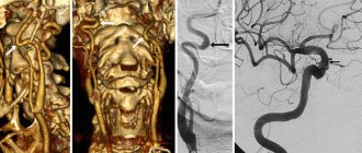
Diagnostics
Most often, the results of a doctor’s examination are sufficient to detect a vascular injury. Angiography, ultrasound with Doppler, CT and MRI are used to assess the consequences or choose surgical treatment tactics.
The diagnosis is made based on the following signs:
- visible bleeding;
- growing hematoma along the course of an artery or vein;
- swelling of tissues;
- wound or blunt trauma in the area of large blood vessels;
- symptoms of blood loss (increased thirst, tachycardia, darkening of the eyes, pale skin and mucous membranes, narrow pupils, partial or complete loss of consciousness);
- cold and pale (bluish) hands or feet, decrease or cessation of pulsation of the arteries below the affected area, ischemic pain, muscle spasm or contracture (wounds of the extremities);
- impaired blood supply to the brain with manifestations of stroke, pathological noises in the vessels of the neck (head and neck injuries).
Nose bleed
A strong flow of blood from the nose is stopped in this way:
- the victim must sit down so that blood can flow freely from the nose: tilting his head down slightly;
- to stop the bleeding, you need to clamp the damaged vessels by pressing on the wings of the nose on both sides for 5 minutes (if the cause is not a fracture);
- any cold object is applied to the bridge of the nose: a wet handkerchief, ice, snow;
- if the blood cannot be stopped within 15 minutes, turundas from a rolled-up bandage are inserted into both nostrils;
- It is strictly forbidden to throw back your head, suck in blood through your nose, or swallow it: vomiting may occur.
Even if first aid for venous bleeding is provided successfully, the victim still needs hospitalization.
First aid
The amount of assistance at the first stage depends on the degree and type of damage:
- bruise - apply ice, first placing a cloth on the site of injury;
- rupture of a capillary or small vein - a pressure bandage made of a bandage or any available fabric (belt, scarf, handkerchief, towel);
- arterial - pressing with a finger or fist, then apply a tourniquet to clothing or fabric in several layers, and under it a note with the time of application.
The use of a tourniquet is advisable only on the thigh or shoulder, since the vessels of the lower leg and forearm are located deep and cannot be compressed from the outside. The maximum time for which a limb can be clamped is 60 minutes for adults and up to 20 minutes for children.
Wounds to the neck are dangerous not only due to blood loss, but also due to the entry of air bubbles with subsequent embolism of cerebral vessels. Therefore, as soon as possible, you need to apply a rolled bandage or something similar to the bleeding site. To apply a bandage, the victim’s arm is raised up, and the turns of the bandage pass through it. This provides nutrition to the brain through the second, paired carotid artery.
First aid for venous bleeding from superficial veins
Includes the following measures:
| Location of damage | Measures |
| Distal segments (forearm, hand, foot) | 1) Pressing the bleeding vein under the wound site through the skin. If this measure is not effective enough, the vein above the wound is compressed in the same way; |
| 2) Giving the injured limb a temporary elevated position; | |
| 3) Rinsing the damaged area with hydrogen peroxide or any other water-based product, followed by covering it with a gauze bandage, which should cover the wound area under and above the wound. Before bandaging, you can place a gauze roll soaked in peroxide into the wound itself; | |
| 4) You can finally stop blood loss from the superficial veins either by simply suturing the wound, or by combining suturing with ligation of the ends of the damaged vessel. | |
| Proximal segments (hip, shoulder) | 1) Giving the injured limb a temporary elevated position; |
| 2) Pressing the bleeding vein under the wound site through the skin. If this measure does not have a sufficient effect, the vein above the wound is compressed in the same way; | |
| 3) Application of a tourniquet; | |
| 4) After removing the tourniquet, the damaged area is washed with hydrogen peroxide or any other water-based product, followed by covering it with a gauze bandage, which should cover the wounded area under and above the wound. Before bandaging, you can place a gauze roll soaked in peroxide into the wound itself; | |
| 5) To finally stop blood loss, you can simply suturing the wound or combining suturing with ligation of the ends of the damaged vessel. |
It is not advisable to apply a tourniquet in case of venous bleeding from the extremities, since such a procedure will only increase blood loss.
Surgery
After admission to the hospital, the patient first receives infusion therapy to restore circulating blood volume. For this purpose, droppers with isotonic solutions of sodium chloride, glucose, Albumin, Reopoliglucin, Voluven, Refortan are used. A blood transfusion of about 2 liters and 4 liters of solutions is indicated in case of damage to a large vessel.
Reconstructive surgery begins at a pressure of at least 100 mmHg. Art. and a pulse of about 100 beats per minute, but if the bleeding continues and threatens life, then the patient will be operated on immediately after hospitalization. Vascular operations are justified if there are signs of tissue viability - deep sensitivity is preserved, there is no muscle contracture. If these symptoms are present, the question of amputation is raised.
The integrity of the artery is restored in the following ways:
- side or circular seam;
- plastic surgery using your own vein or graft;
- connection of ends with a defect of no more than 2 cm.
If the vein is injured, a side suture is used, and if the damage is significant, then the femoral vein is isolated and used for plastic reconstruction.
Complications of injury to large vessels
Injuries to the cervical spine can provoke serious consequences with a fatal outcome, especially complications from injury to large vessels of the neck:
- swelling, inflammation in the spinal cord;
- a sharp drop in blood pressure;
- prolonged bleeding with provocation of hemorrhagic shock;
- cerebrovascular accident with increased spinal pressure;
- wounds of the larynx and trachea lead to disruption of the respiratory process;
- disruption of innervation, especially the vagus nerve, creates severe consequences with disturbances in heart rhythm until the heartbeat and breathing completely stop;
- fatal air embolism.
Injuries to the veins and arteries of the neck require emergency medical intervention; the risk of complications and threat to the patient’s life are very high. Even with minor injuries, it is worth applying a pressure bandage and seeking medical help at a medical facility.
The article has been verified by the editors
Prognosis for the patient
Factors that have an adverse effect on vascular injury:
- open damage;
- rupture of a large diameter artery;
- combined injuries (bones, soft tissues, nerve trunks are damaged, vital organs are affected);
- large blood loss;
- localization on the neck;
- More than 6 hours passed from the moment of injury to the operation.
We recommend reading the article about the reasons why blood vessels burst in the legs. From it you will learn about the symptoms, causes and conditions that cause hemorrhages under the skin, as well as dangerous diseases, treatment of pathologies and prevention of these hemorrhages.
And here is more information about the methods of checking blood vessels and veins.
Vascular injuries occur due to wounds, bruises, fractures, and pathological changes in the vascular walls. Their manifestations are determined by the open or closed nature of the damage, rupture of an artery or vein, the caliber of the vessel, and its location. Arterial bleeding from the main blood tracts is especially dangerous when the integrity of the vessels of the neck is violated.
At the time of first aid, it is important to stop the flow of blood by applying a bandage or tourniquet, after which the patient should be hospitalized immediately. Treatment is carried out only through surgery.
It’s easy to get a contusion, bruise, or hematoma, especially for children. There are effective remedies and ointments - Vishnevsky, Zinc, which will quickly solve the problem. In mild cases, you can use traditional methods, for example, iodine and banana. Medicines – tablets and injections – will help. What to do if the bruise does not go away, there is a lump? What to anoint a child with? How to quickly remove a bruise from your nose, under your eye, or on your leg?
Post-traumatic thrombosis occurs in the absence of adequate treatment. The acute form of damage to the deep vessels of the lower extremities is dangerous due to the detachment of a blood clot. The earlier a clot is detected, the higher the chances of success in treatment.
Deep vein thrombosis often poses a serious threat to life. Acute thrombosis requires immediate treatment. Symptoms in the lower extremities, especially the lower legs, may not be immediately diagnosed. Surgery is also not always required.
In case of injuries, as well as other problems, a vascular suture is required. Carrel overlay is often performed. There is also a circular, mechanical, and side seam, which is done using a special technique for children. The requirements for a doctor's qualifications are very high, and they use specific instruments.
The formation of a blood clot is not that uncommon. However, it can provoke thrombosis of cerebral vessels or embolism of cerebral arteries. What signs are there? How to detect thrombosis of cerebral vessels, cerebral embolism?
Complications of coronary angiography often occur, because the risks of reconstructing the heart vessels through the arm are quite high. Hematoma is the simplest among them.
If lameness and pain while walking suddenly appear, then these signs may indicate obliterating atherosclerosis of the vessels of the lower extremities. In an advanced state of the disease, which progresses to stage 4, amputation surgery may be required. What are the possible treatment options?
Only timely recognition of subarachnoid hemorrhage will save lives. The symptoms of traumatic and non-traumatic cerebral hemorrhage are clearly defined. Diagnosis involves CT scanning, and treatment involves surgery. With a stroke, the consequences are worse.
If atherosclerosis is suspected, the examination should be carried out in full. It includes a blood test, including a biochemical one, as well as many others. Which ones are still worth taking?
Bleeding in the veins of the neck
Why is damage to the veins in the neck dangerous?
- Without professional skills, it is impossible to apply a bandage without causing suffocation in the victim;
- vessels in the neck area are large in diameter, their injuries cause profuse and rapid blood loss, so first aid must be provided as quickly as possible;
- Air can be sucked into the lumen of a large vessel, resulting in the formation of an air plug (), which can cause death.
How to stop bleeding if your neck is injured:
- Lay the person down so as to provide easy access to the wound.
- If possible, apply a cotton or gauze cloth folded several times and soaked in antiseptic (hydrogen peroxide) to the wound.
- Press the area above and below the injury with three fingers (ring, middle and index) of both hands folded together.