CONTENT
- Notations and abbreviations
- Main part
- Ultrasound examination of the main arteries of the extremities
- Duplex scanning of the arteries of the lower extremities
- Sample protocol for ultrasound examination of the main arteries of the lower extremities
- Duplex scanning of the arteries of the upper extremities
- Sample protocol for ultrasound examination of the main arteries of the upper extremities
- List of sources used
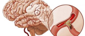
What pathology is the most common cause of vertebral artery syndrome?
About a fifth of all pathologies of the vertebral arteries consist of certain developmental anomalies.
Among them:
- High entry into the bone canal;
- When the mouth of the artery occurs to the outside.
Studies have also shown that vertebral disorders can be combined. And such options are much more common than others.
Examples include:
- One third of the lesions are anatomical abnormalities and muscle compression;
- About 40% are thrombotic and atherosclerotic in origin;
- Most are compression due to vertebral displacements and atherosclerosis.
Merger of the left and right arteries into a single trunk
ULTRASOUND STUDY OF THE MAIN ARTERIES OF THE LIMB
Ultrasound examination (duplex scanning, DS) of the vascular system is one of the leading diagnostic methods, allowing to detect both initial changes in the arterial wall at the preclinical stage and to identify more pronounced lesions, indicating their exact location and hemodynamic significance. The method also allows you to assess the condition of the distal vascular bed, collateral blood supply pathways and conduct a dynamic assessment of the results of drug and surgical treatment.
Some statistics
Data provided by brain CT studies have shown that one third of ischemic stroke patients had bilateral artery involvement at the point where it meets the basilar artery.
Clinical findings have shown that it is in this zone that ischemia occurs three times more often than in other areas.
One of the main causes of death due to cerebral ischemia is atherosclerotic pathologies of the vertebral arteries.
DUPLEX SCAN OF LOWER LIMB ARTERIES
Indications for the study
- clinical symptoms of acute or chronic arterial insufficiency;
- presence of risk factors for atherosclerosis;
- the presence of noise during auscultation of the common femoral arteries;
- the presence of asymmetry or absence of pulsation in the arteries of the lower extremities;
- genetic predisposition to the development of thromboangiitis obliterans;
- trophic changes in the lower extremities of unknown etiology;
- lower extremity injuries.
Research methodology
DS of the arteries of the lower extremities is carried out using B-mode, color Doppler mapping (CDC) mode and spectral Doppler, using multi-frequency linear and convex sensors with a scanning frequency from 3.5 to 12 MHz.
Inspection of the distal abdominal aorta and iliac arteries, due to their relatively deep location, is carried out with convex sensors with a frequency of 3.5-5.0 MHz. More superficially located vessels of the thigh are examined using linear sensors with a scanning frequency of 5-7.5 MHz, and the popliteal region and distal bed - 5-12 MHz. It should be taken into account that, depending on the characteristics of the patient’s constitution, the severity of the pathological process and the quality of the resulting image, the choice of sensor and scanning frequency in each individual case can be determined by the researcher independently. For example, in obese patients and with significantly difficult visualization, examination of even superficial arteries can be performed using a convex probe with an acceptable partial loss of information.
The standard locations for the main arteries of the lower extremities are: the distal aorta, the mouth of the common iliac artery (CI) and, if possible, the internal iliac artery (IIA), the external iliac artery (EIA), the common femoral artery (COA), the mouth of the deep femoral artery ( GFA), superficial femoral artery (SFA) in the upper, middle and lower third of the thigh, popliteal artery (PA), posterior and anterior tibial arteries (SPTA and PBTA) at the mouth and in the distal section, arteries of the dorsum of the foot and plantar arteries (the latter in are not studied in routine protocols) (Figure 1).
Each of the segments of the arteries of the lower extremities is examined sequentially in the gray scale, color flow and spectral Doppler modes.
Figure 1 - Standard locations of the main arteries of the lower extremities
In-mode evaluate:
- vessel diameter (unchanged, dilated, aneurysm); permissible asymmetry of the diameters of paired vessels is up to 20%. When measuring the diameter of a vessel, one should evaluate its lumen between the anterior and posterior internal walls, perpendicular to the axis of the vessel, without including the thickness of the intima-media complex and other layers in the measurement. The average normal values for the diameters of the main arteries of the lower extremities are given in Table 1;
- lumen of the vessel (homogeneity, echogenicity, presence or absence of additional inclusions);
- the condition of the vascular wall (the intima-media complex is smooth, clearly differentiated into layers, the thickness in the common femoral artery along the posterior wall is no more than 1.0 mm);
- condition of perivascular tissues (presence of edema, hematomas, etc.).
Table 1 - Normal diameter of the arteries of the lower extremities (Kulikov V.P., 2011)
| Author | Diameter, mm | |||||
| NPA | BOTH | PBA | PA | PBBA | ZBBA | |
| 7,9±1,3 | 8,2±1,4 | 6,0±1,2 | 5,2±1,1 | — | — | |
| 8,5±1,1 | 8,1±1,7 | 6,5±1,4 | 5,8±1,2 | — | — | |
| — | — | 7,8±1,3 | 5,7±0,9 | 1,6±0,2 | 2,2±0,3 | |
| — | 7,6±1,1 | 5,8±0,5 | 5,4±0,4 | 2,0±0,3 | 2,1±0,3 | |
In color Doppler mode, the following is determined:
- passable diameter of the vessel (its correspondence to the anatomical diameter);
- the presence of filling defects (indicate their location and measure the degree of stenosis of the vessel lumen);
- the nature of the color pattern distribution and the presence of turbulence zones.
When calculating the degree of stenosis, the image should be assessed in the longitudinal and transverse planes, since in the presence of an irregularly shaped plaque, a false impression may arise of a lesser degree of stenosis of the vessel in the longitudinal plane. Methods for measuring the degree of stenosis are shown in Figure 2.
Figure 2 - Methods for calculating the degree of stenosis by diameter (A) and area (B)
A
B
You also need to remember that the degree of stenosis calculated by area will be on average 15-20% greater than the degree of stenosis by diameter. Therefore, in the conclusion it is advisable to indicate the method for calculating the degree of stenosis. By default, the degree of stenosis calculated by diameter is indicated.
With spectral Dopplerography, the qualitative and quantitative characteristics of the Doppler curve are assessed.
Quality characteristics include:
- shape of the curve (main type of blood flow, main-modified, collateral);
- presence of a spectral window (laminarity of blood flow).
Normally, a mainline Dopplerogram has a three-phase waveform: an acute systolic peak, reverse flow in early diastole, a positive component in late diastole, and a clear spectral window (Figure 3).
Figure 3 - Dopplerogram of the main type of blood flow in the popliteal artery
The quantitative parameters of the Doppler curve include:
- peak (linear) systolic blood flow velocity (Vs, LSVR);
- end-diastolic blood flow velocity (Vd);
- time-averaged maximum blood flow velocity (Vtmax);
- time-averaged mean blood flow velocity (Vtav);
- resistivity index (RI);
- Pulsatility Index (PI);
- spectral broadening index (SB);
- systole-diastolic ratio (S/D).
However, in outpatient practice, only systolic and diastolic velocities are most often used; in some cases, pulsability and resistivity indices can be measured.
In the absence of pathology, Dopplerogram indicators are assessed only at standard locations of the main arteries of the lower extremities. In the presence of stenoses, quantitative measurements, in addition to standard points, are carried out at each point of maximum narrowing along the vessel.
Modern ultrasound scanners calculate all quantitative parameters automatically depending on the settings in the device menu. The units of speed measurement are also set at the discretion of the operator - cm/s or m/s.
Normal values for blood flow velocities in the great arteries are quite variable and depend on many factors, including systemic arterial pressure (BP), heart rate, peripheral vascular resistance and the age of the patient (Table 2).
Due to the significant improvement of modern ultrasound scanners and improved visualization quality, the degree of vessel stenosis will be more accurate when assessed in a gray scale image and using color doppler. If the use of these methods is limited or impossible, then it is permissible to calculate the degree of stenosis from changes in the Dopplerogram curve and systolic blood flow velocity (Table 3).
Table 2 - Normal parameters of blood flow in the arteries of the lower extremities (Kulikov V.P., 2011)
| Author | LSC, cm/s | |||||
| NPA | BOTH | PBA | PA | PBBA | ZBBA | |
| Jager KA,1985 | 119, ±21,7 | 114,1±24,9 | 90,8±13,6 | 68,8±13,5 | — | — |
| SchaeberleW, 2005 | 116,0±29,7 | 112,2±22,7 | 93,95±15,9 | 71,6±14,4 | — | — |
| Kuntsevich G.I., 1999 | — | — | 85,5±14,9 | 64,4±12,2 | 39,4±4,0 | |
| Lelyuk V.G., Lelyuk S.E., 2007 | — | 104±21 | 85±11 | 58±13 | 46±16 | 47±13 |
Table 3 - Hemodynamic criteria for the degree of stenosis of the arteries of the lower extremities (Jager K.A. et al. 1985)
| Stenosis degree, % | Changes in the spectrum of blood flow in the proximal segment | LSK at the site of stenosis | Spectral extension | Spectrum phase |
| Norm | Not changed | Not changed less than 150 cm/s | No | Three-phase |
| 1-19% | Not changed | Increased by 30% | No | Three-phase |
| 20-49% | Not changed | Increased by 100%, up to 150-200 cm/s | Occurs at peak systolic | Three-phase |
| 50-74% | Changed, biphasic, reduced BFV | Increased by 200-300%, up to 200-400 cm/s | Available | Two-phase |
| 75-99% | Changed, monophasic, decreased BFV | Increased more than 300% and more than 400 cm/s | Expressed | Monophasic |
| Occlusion | LSV is significantly reduced, disappears when approaching the site of occlusion | No blood flow | — | — |
Research technique
Before starting the study, you should ask the patient whether he has a history of reconstructive surgery on the vessels of the lower extremities, chronic diseases leading to damage to the vascular wall, or trauma. This will allow you to more fully analyze the results obtained and indicate in the conclusion the features that are fundamentally important for the attending physician.
Before the examination, the patient must be at rest for at least 15-20 minutes in a horizontal position. The examination should begin with measuring blood pressure in both arms and legs. This rule is often neglected due to the limitation of the time allotted for the study, however, it is the detected difference in blood pressure between the upper and lower limbs or its asymmetry between the right and left sides that allows one to suspect a stenotic lesion and suggest the level of its localization.
Visualization of the arteries of the lower extremities begins with the aorto-iliac segment, gradually moving distally along the main trunks.
The patient's position is lying on his back with his legs straight and slightly apart. The sensor is installed along the midline of the abdomen slightly below the navel, the location of the aortic bifurcation is determined and then, deviating slightly to the right or left of the bifurcation, the openings of the PCA, their bifurcations and the IPA, which are a continuation of the PCA, are visualized (Figure 4). The location of the VPA depends on the patient’s obesity and general visualization of the abdominal organs; in some cases it is not visible.
Figure 4 — Diagram of the sensor location when examining the aorto-iliac segment (A) and ultrasound image of the common, orifice of the internal and external iliac arteries (B)
A
B
The main points for studying the aorto-iliac segment:
- bifurcation of the abdominal aorta into the abdominal aorta – located at the level of the IV lumbar vertebra;
- The PPA at the level of the sacroiliac joint is divided into the IPA and the VPA; has only small muscle branches;
- The VPA gives off visceral branches to the organs and walls of the pelvis;
- The IFA below the inguinal ligament continues as the common femoral artery; has a number of muscular branches and anastomoses with the internal thoracic artery, lumbar arteries and the inferior epigastric artery.
After examining the iliac arteries, without changing the patient’s position, the femoral arteries are examined: the sensor is moved below the inguinal ligament on the BOTH, the thickness of the intima-media complex in it is measured, the mouth of the GIB is assessed, and then, gradually descending to the thigh, the SFA is examined to the level of its entry into Gunter channel (Figure 5).
Figure 5 — Sensor location diagram (A) and ultrasound image of the common, internal and superficial femoral artery (B)
A
B
Main research points on the hip:
- The OBA is a direct continuation of the IPA below the inguinal ligament and continues to the level of the origin of the GBA; has a number of branches that are sources of collateral circulation of the lower limb in case of stenosis and occlusion of the main trunk;
- GBA - the main vascular collateral at the level of the thigh, departs from the femoral artery 1-6 cm below the inguinal ligament; the branches anastomose between themselves and the adjacent branches of the popliteal artery; during the study, only a small proximal fragment of the GBA is most often visualized;
- The SFA is a continuation of the SFA on the thigh, it is directed downwards, entering the Gunter's canal in the lower third of the thigh; has only small branches involved, among other things, in the formation of the arterial network of the knee.
The VA, tibioperoneal trunk (TPT), and proximal tibial arteries are examined in the prone position, legs straight, foot flexed, and toes resting on the couch. The sensor is gradually moved down along the popliteal fossa, visualizing the initial sections of the PBBA and TJ (Figure 6).
Main research points in the popliteal region:
- PA – direct continuation of the SFA after exiting the femoropopliteal canal; located in the popliteal fossa, gives off a number of branches that form the arterial network of the knee
PA is conventionally divided into three segments:
- from the place of exit from the Gunther's canal to the upper edge of the patella;
- from the upper edge of the patella to the gap of the knee joint;
- below the gap of the knee joint to the level of the origin of the PBBA.
- TPS - a fragment of the SBAA between the origin of the SBAA from the VA to the level of the origin of the peroneal artery (MFA)
Figure 6 — Diagram of the sensor location when examining the popliteal artery (A) and ultrasound image of the popliteal artery, anterior tibial artery and tibioperoneal trunk (B)
A
B
The examination of the distal arteries of the leg is carried out with the patient lying on his back with his knees slightly bent and his legs slightly apart. To visualize the STBA, the sensor is placed along the lower third of the leg and slightly posterior to the medial malleolus. The PBBA is located along the lower third of the shin along the anterior surface in the projection of the continuation on the shin of an imaginary line drawn between the I and II toes of the dorsum of the foot (Figure 7).
Figure 7 — Diagram of the sensor location when studying the distal tibial arteries (A) and ultrasound image of the posterior tibial artery (B)
A
B
In everyday practice, MBA studies are not carried out. Its examination and measurement of blood flow parameters become relevant if the patient has chronic ischemia of the lower extremities or stenotic lesions of the tibial arteries. The MBA is visualized along the medial surface in the middle third of the leg.
The main research points when examining the arteries of the leg:
- PBBA - pierces the interosseous membrane in the upper third of the leg, goes down and in the lower third of the leg comes out to the front surface, continuing into the artery of the dorsum of the foot
- The STBA is a direct continuation of the VA on the lower leg, going downwards, giving off to the IBA in the upper third of the lower leg, and in the lower third the branches involved in the blood supply to the heel area; The continuation of the STBA on the foot is the common plantar artery
If ultrasound signs of lower extremity ischemia are detected in a patient, the ankle-brachial index (ABI) is measured.
ABI measurement is a reliable and effective method for quantifying blood supply to an extremity. Its positive predictive value is 90%, negative predictive value is 99%, and overall accuracy is 98%. The index is calculated using the formula:
ABI = tibial artery systolic blood pressure / brachial artery systolic blood pressure
Systolic pressure is determined using a tonometer with a pneumatic cuff and a linear ultrasonic sensor installed at the location point of the PLA and PA. If the latter is occluded, or it is impossible to clearly locate it due to damage, PBBA or MBA is used.
The measurement is carried out symmetrically on both arms and legs. The sensor is alternately installed at the location points of the indicated arteries and pressure is pumped into the cuff until the Doppler signal disappears in the vessel. At the moment of slow decompression of the cuff, the first beat of restored blood flow corresponds to the value of systolic blood pressure in the vessel.
Normally, a difference of up to 12-15 mm Hg is acceptable. Art. pressure on the hands. If pressure readings in the arms differ more significantly, then a stenotic lesion of the subclavian or axillary arteries on the side with lower blood pressure values should be suspected. In this case, to calculate the ABI, the blood pressure indicator from the arm where the obtained value is greater is used.
The normal pressure at the ankle is 10–15 mmHg. higher than on the shoulder, and the normal ABI value of systolic pressure is more than 1.0. A decrease in ABI of less than 0.9 is considered pathological. The index correlates with the stage of the lesion and the clinical picture of ischemia of the lower extremities:
- Stage I 0.8 ≤ ABI ≤ 0.9
- Stage II 0.5 ≤ ABI ≤ 0.7
- Stage III 0.3 ≤ ABI ≤ 0.5
- Stage IV ABI <0.3
It should be remembered that ABI values may be inaccurate in patients with distal angiopathy, severe calcification of the arterial wall and undetectable systolic blood pressure. In the case of incompressibility of the artery when pressure is injected into the cuff more than 200 mm Hg. Art., ABI is regarded as false positive.
There is also a technique for segmental measurement of pressure in the legs, which makes it possible to establish the approximate localization of a stenotic lesion based on pressure gradients in different parts. But in outpatient practice such measurements are usually not carried out.
When examining patients who have undergone reconstructive surgery on the arteries of the lower extremities, in addition to standard arterial location points, the patency of the shunt is assessed and the points of proximal and distal anastomoses are examined to identify stenoses or aneurysms. All measurements and registration of blood flow parameters are carried out similarly to other sections of the main arteries.
The study protocol should describe the diameters and velocities of blood flow for standard location points, the presence of stenotic lesions, their degree and location, and indicate the type of blood flow curve in each segment. In the case of ABI measurements, indicate the systolic blood pressure values for each limb, as well as the obtained ABI values for each side.
SAMPLE PROTOCOL FOR ULTRASOUND STUDY OF THE MAIN ARTERIES OF THE LOWER LIMB
FULL NAME.
Age
research Date
Right left
OPA diameter, mm; type of blood flow (main/main altered/collateral); blood flow speed, cm/s; stenosis, %
BOTH diameter, mm; type of blood flow (main/main altered/collateral); blood flow speed, cm/s; stenosis, %
GBA diameter, mm; type of blood flow (main/main altered/collateral); blood flow speed, cm/s; stenosis, %
PBA diameter, mm; type of blood flow (main/main altered/collateral); blood flow speed, cm/s; stenosis, %
PA diameter, mm; diameter, mm; type of blood flow (main/main altered/collateral); blood flow speed, cm/s; stenosis, %
ZBBA diameter, mm; type of blood flow (main/main altered/collateral); blood flow speed, cm/s; stenosis, %
PBBA diameter, mm; type of blood flow (main/main altered/collateral); blood flow speed, cm/s; stenosis, %
BP system on the shoulder, mm Hg. Art. on the shoulder, mmHg Art.
on the lower leg, mm Hg. Art. on the lower leg, mm Hg. Art.
ABI
Conclusion________________________________________________________
What is antegrade blood flow?
Have you been struggling with HYPERTENSION for many years without success?
Head of the Institute: “You will be amazed at how easy it is to cure hypertension by taking it every day...
When cerebral symptoms appear in young patients, neurologists must take into account the possibility that this is a change (hypoplasia) of arteries such as the left vertebral, the right, or both. The disease is considered a birth defect. Without treatment, it contributes to impaired blood circulation in the vessels of the brain, often in the posterior sections.
Since the nuclear structures of the brain determine the rhythm and normal functioning of the entire body, the patient may experience signs of cardiovascular disorders and vestibular changes.
OUR READERS RECOMMEND!
Our readers successfully use ReCardio to treat hypertension. Seeing how popular this product is, we decided to bring it to your attention.
What is hypoplasia?
First of all, let us remember that the task of the vertebral arteries is to bring up to 30% of the required blood volume into the brain’s bloodstream. It is these vessels that form the circle of Willis at the base of the brain, from which branches extend along a lobar principle deep into the medulla.
In a state of underdevelopment of an organ or tissue, they speak of hypoplasia. The most common hypoplasia is in the right vertebral artery. According to statistics, it occurs in 10% of the population. A left-sided lesion or two at the same time is rare.
The causes of impaired vascular development in the fetus may be:
- uncontrolled treatment of a pregnant woman with drugs that have a toxic effect on the child (antibiotics, anti-influenza drugs, diuretics);
- work of the expectant mother in harmful conditions (with pesticides, dyes, household chemicals);
- drug use, alcohol use, smoking during pregnancy;
- previous injuries;
- acute infectious diseases and exacerbation of chronic ones;
- finding a pregnant woman in a radioactively contaminated area;
- hereditary predisposition to pathology of the heart and blood vessels.
Such changes entail:
- decrease in total blood flow (usually through the right, less often through the left arteries) to the brain tissues;
- favorable conditions for thrombus formation (local decrease in the speed of platelet movement);
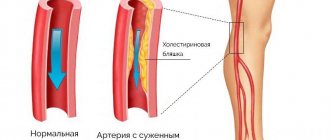
- with subsequent disruption of fat metabolism with age, they create a convenient place for the deposition of cholesterol plaques and the development of the atherosclerotic process.
DUPLEX SCAN OF ARTERIES OF THE UPPER LIMB
Indications for the study
- the difference in blood pressure in the arms is more than 12 mm Hg. Art.;
- clinical symptoms of acute or chronic ischemia of the upper extremities;
- presence of risk factors for atherosclerosis;
- presence of noise during auscultation of the subclavian arteries;
- vasculitis, neurovascular syndrome;
- clinical signs of angiodysplastic processes;
- trophic changes in the upper limb of unknown etiology;
- upper limb injuries.
Research methodology
DS of the arteries of the upper extremities is carried out using gray scale, power Doppler mapping (EDC), color doppler and spectral Doppler modes, using multi-frequency linear and convex sensors with a scanning frequency from 3.5 to 14-16 MHz.
Scanning of the mouth of the left subclavian artery (LSA) is carried out with convex or sector sensors with a frequency of 3.5-5.0 MHz. Segments of the main arteries of the upper extremities that are more accessible for visualization, such as the mouth of the right PlA, the first and second portions of both PlA, axillary, brachial (PlA) and the initial sections of the ulnar (LoA) and radial arteries (LA) are examined using linear sensors with a scanning frequency of 7 -12 MHz.
When studying more superficially located vessels, it is advisable to increase the scanning frequency to maximum values. Thus, the distal sections of the arteries of the forearm, the superficial and deep palmar arches and arteries of the fingers are examined with linear sensors with a frequency of 14-16 MHz. But, as with any other research, the operator, depending on the conditions and quality of visualization, can use the most optimal sensors and scanning frequency for each case.
The screening technique is based on assessing the qualitative and quantitative parameters of blood flow at standard research points, where the artery is as close as possible to the surface of the skin and its location is associated with constant anatomical landmarks.
The standard points for studying the main arteries of the upper extremities are: the mouth, the first and second portions of the PCA, the axillary artery, the PCA along and at the site of bifurcation into the LoA and PA, the mouths and distal sections of the LoA and PA. If necessary and depending on the purpose of the study, the palmar superficial and deep arterial arches and digital arteries are examined (Figure 8).
Each segment of the arteries of the upper extremities is examined sequentially in the longitudinal and transverse planes using gray scale, color flow and spectral Doppler modes, and small arteries of the palmar arches and digital arteries can be visualized using EDC.
Figure 8 - Standard locations of the main arteries of the upper limb
In B-mode they evaluate:
- vessel diameter (unchanged, dilated, aneurysm); permissible asymmetry of the diameters of paired vessels up to 20%; average normal values of the diameters of the main arteries of the upper extremities (Table 4);
- lumen of the vessel (homogeneity, echogenicity, presence or absence of additional inclusions);
- the state of the vascular wall (the intima-media complex is smooth, clearly differentiated into layers, the thickness in the PLA at the level of bifurcation along the posterior wall is no more than 0.6 mm);
- condition of perivascular tissues (presence of edema, hematomas, etc.).
Table 4 - Normal diameter of the arteries of the upper extremities (Lelyuk S.E., Lelyuk V.G., 1999)
| Artery | Diameter, mm (M±m) | Diameter, mm min-max |
| Subclavian | 5,7±0,8 | 4,2-8,2 |
| Shoulder | 3,3±0,8 | 2,3-5,5 |
| Elbow | 1,8±0,3 | 1,5-2,4 |
| Radial | 1,8±0,4 | 1,0-2,5 |
In the color flow mode the following is determined:
- passable diameter of the vessel (its correspondence to the anatomical diameter);
- the presence of filling defects (indicate their location and measure the degree of stenosis of the vessel lumen);
- the nature of the color pattern distribution and the presence of turbulence zones.
With spectral Dopplerography, the qualitative and quantitative characteristics of the Doppler curve are assessed.
Quality characteristics include:
- shape of the curve (main type of blood flow, main-modified, collateral);
- presence of a spectral window (laminarity of blood flow).
Normally, the Dopplerogram of the arteries of the upper extremities of the main type has a three-phase curve: an acute systolic peak, reverse blood flow during early diastole, a positive component of late diastole and a clear spectral window (Figure 9). In some cases, LA and LoA in healthy individuals can record a low-amplitude positive diastolic component, while the peak systolic blood flow velocity corresponds to the age norm. To exclude a pathological decrease in peripheral resistance, a test of clenching the hand into a fist is performed; Normally, diastolic blood flow becomes retrograde.
Figure 9 - Normal three-phase Dopplerogram in the subclavian artery
The quantitative parameters of the Doppler curve include:
- peak (linear) systolic blood flow velocity (Vps, LSVR);
- end-diastolic blood flow velocity (Ved);
- time-averaged maximum blood flow velocity (Vtmax);
- time-averaged mean blood flow velocity (Vtav);
- resistivity index (RI);
- Pulsatility Index (PI);
- spectral broadening index (SB);
- systole-diastolic ratio (S/D).
In routine practice, only systolic and diastolic velocities are usually measured; in some cases, pulsability and resistivity indices can be measured.
Modern ultrasound scanners calculate all quantitative parameters automatically depending on the settings in the device menu.
If a violation of the hemodynamic parameters of blood flow is detected at any of the standard points, a study of the arterial bed is carried out along its entire length to identify the causes of changes.
Normal values of blood flow velocities in the main arteries of the upper extremities depend on many factors, including systemic arterial pressure (BP), heart rate, peripheral vascular resistance and the age of the patient (Table 5).
Table 5 - Normal parameters of blood flow in the arteries of the upper extremities (Lelyuk S.E., Lelyuk V.G., 1999)
| Artery | Blood flow speed | P.I. (М±m, min-max) | |
| Vps, cm/s (М±m, min-max) | Vedcm/s (М±m, min-max) | ||
| Subclavian | 112±20,5; 90-163 | 15.7±5.2; 9-30s | 5,3±0,57; 4,89-7,24 |
| Shoulder | 65±10,8; 45-82 | 12,3±3,0; 6-16 | 3,7±0,98; 2,64-4,9 |
| Elbow | 50,5±5,4; 43-61 | 9,6±3,4; 2-13 | 3,06±1,4; 1,6-9,3 |
| Radial | 53±9,4; 35-63 | 13,1±4,6; 7-20 | 4,14±3,3; 1,3-14,8 |
Research technique
Before examining the main arteries of the upper extremities, especially the distal arteries, the patient should be in a room with a comfortable temperature for at least 20 minutes.
The patient's examination should begin with measuring blood pressure in both arms. The detected difference is more than 12 mm Hg. Art. allows one to suspect a stenotic lesion on the side with lower scores.
Vascular examination begins with the proximal parts of the upper limb. The PCL on the right is a branch of the brachiocephalic trunk, and on the left it arises directly from the aortic arch distal to the common carotid artery. At the first rib, the PclA penetrates into the interscalene space, where it lies in the groove of the rib, and then, going around it, lies under the collarbone and, going down to the axillary fossa, continues in the form of the axillary artery.
Examination of the PCL is carried out with the patient lying on his back with his arms straightened along the body, the head slightly tilted in the opposite direction. PclA location is carried out from the supraclavicular and subclavian approaches, using convex, sector or linear sensors, depending on the constitutional build of the patient and the quality of visualization. The orifice and the first segment are better visualized from the supraclavicular approach. To do this, the sensor is placed in the supraclavicular fossa slightly outward from the jugular notch. When examining the second segment of the PclA, better visualization can be achieved from the subclavian region, where the sensor is placed parallel to the course of the artery (Figure 10).
Figure 10 — Sensor location diagram (A) and ultrasound image of the subclavian artery (B)
A
B
Main research points for PclA:
- the first segment – from the mouth to the interscalene space; the main branches in this segment are: the vertebral artery, the internal thoracic artery and the thyrocervical trunk;
- the second segment is the interscalene space, where the PclA gives off the costocervical table;
- the third segment is from the interscalene space to the axillary artery.
It should be remembered that segments I and II of the PclA artery are the main sites of localization of initial atherosclerotic changes in the arteries of the upper extremities. In case of stenosis of more than 70% or occlusion of the PCA in the first segment, main altered or collateral types of blood flow will be recorded in its distal parts. In addition, with stenosis of 50% or more in the vertebral artery on the affected side, hidden, transitional or complete vertebral-subclavian steal syndrome (steel syndrome) occurs. Stenotic lesions of the II and III segments of the PCA are not accompanied by the development of vertebral artery steal syndrome, but an altered type of blood flow will be recorded below the site of stenosis.
It should also be remembered that segment II of the PclA, located in the interscalene space, can be subject to compression, clinically manifesting itself in the form of superior thoracic outlet syndrome (syndrome of compression of the neurovascular bundle at the exit from the chest). To diagnose this syndrome, there are many tests with different positions of the arms and head, aimed at identifying the source of compression (Adson's test, a test with moving the hands behind the back and squeezing them “into a lock”, a test with throwing the hand on the back of the head, etc.). However, the most effective test is for the patient to assume a position in which he has complaints and at this very moment to register blood flow or record its absence in the distal part of one of the arteries of the forearm.
The axillary artery is a direct continuation of the PclA after its transition to the axillary fossa; below the edge of the pectoralis major muscle it passes into the PclA. To examine it, the patient’s arm is retracted outward and rotated slightly (Figure 11).
Figure 11 — Sensor location diagram (A) and ultrasound image of the axillary artery (B)
A
B
The PLA is located from the lower edge of the pectoralis major muscle to the level of the ulnar fossa. On the shoulder, it lies along the medial groove of the biceps muscle, where it gives off a number of branches, the largest of which is the deep brachial artery. The PLA is examined with the arm abducted and slightly rotated, moving the sensor distally along its course, up to the level of the bifurcation, which is usually located 1-3 cm below the cubital fossa (Figure 12).
Figure 12 — Sensor location diagram (A) and ultrasound image of the brachial artery and vein (B)
A
B
From the level of the bifurcation of the PLA along the anterolateral and anteromedial surfaces of the forearm, the LoA and PA are located, which, going down, are traced to the level of the distal parts of the forearm, and when reaching the hand, they participate in the formation of the superficial and deep arterial arches of the hand. When examining the arteries of the forearm, the patient's hand is turned palmar upward. The sensor is placed in the lower third of the forearm along the radial and ulnar surfaces (Figure 13).
Figure 13 — Diagram of the sensor location when studying the arteries of the forearm (A) and ultrasound image of the radial artery (B)
A
B
When the sensor is positioned along the medial and lateral surfaces of the phalanges of the fingers, the common and proper digital arteries are located. Their study in outpatient practice is carried out extremely rarely. When assessing the distal arterial bed (palmar arches and digital arteries), the B-mode is not informative; Small arteries are more often visualized using CD or EDC. In addition, the determination of quantitative parameters in the digital arteries requires certain operator skills in conducting this study.
At the end of the examination, the protocol should indicate the diameters and velocities of blood flow for standard location points, the presence of stenotic lesions, their degree, location and hemodynamic significance, and indicate the type of blood flow curve in each segment.
SAMPLE PROTOCOL FOR ULTRASOUND STUDY OF THE MAIN ARTERIES OF THE UPPER LIMB
FULL NAME.
Age
research Date
Right left
PklA diameter, mm; type of blood flow (main/main altered/collateral); blood flow speed, cm/s; stenosis, %
Axillary artery diameter, mm; type of blood flow (main/main altered/collateral); blood flow speed, cm/s; stenosis, %
PLA diameter, mm; type of blood flow (main/main altered/collateral); blood flow speed, cm/s; stenosis, %
LoA diameter, mm; type of blood flow (main/main altered/collateral); blood flow speed, cm/s; stenosis, %
LA diameter, mm; type of blood flow (main/main altered/collateral); blood flow speed, cm/s; stenosis, %
Conclusion________________________________________________________
When is surgery necessary?
The first operation to reconstruct the vertebral artery was performed in 1956, and in 1959, a thrombus was first removed from the subclavian artery, involving the bed of the vertebral vessel.
The indications for surgery are determined by the results of conservative therapy. If the treatment is ineffective, as well as if the cause is established, related to compression of the artery by a tumor, a vertebral process, it is impossible to do without surgical intervention.
They operate on patients in neurosurgical departments. Bone formations, tumors, and sympathetic nodes are removed (to eliminate excessive spasm).
It is possible to eliminate abnormal tortuosity only if it is localized in segment I.
Forecast
Without treatment, atherosclerotic occlusion of the subclavian artery leads to a gradual decrease in working capacity, and the likelihood of ischemic stroke and gangrene of the hand increases.
After restoring blood circulation by any method, the problem is completely eliminated. Normal blood flow promotes normal hand function and eliminates brain steal syndrome.
Relapses after stenting of the subclavian artery occur in approximately 10% of cases, due to the development of new plaques inside the stent (restenosis). If a carotid-subclavian bypass operation was performed, then the probability of relapse is no more than 2% of cases.