Characteristic
Ultrasound examination of cerebral vessels is one of the most common diagnostic methods today. It is based on the reflection of an ultrasonic wave from the area under study. It is used to detect anatomical pathologies of the vertebral, basilar, carotid arteries, anterior and internal jugular veins, subclavian artery and vein, and facial vein. Ultrasound of cerebral vessels shows the diameter of the lumen, internal formations, and the condition of surrounding tissues.
The procedure, supplemented by Doppler ultrasound, allows you to detect areas of blood vessels with impaired blood flow due to their narrowing, blockage, and neoplasms. With its help, the functioning of the circuitous blood flow pathways is checked, the treatment being carried out and the results of the surgical intervention are monitored.
Today, when giving a referral for ultrasound diagnostics, a doctor separately indicates the type of examination: ultrasound, Doppler, duplex, triplex or transcranial. In most cases, a simple ultrasound is not performed, but is combined with Doppler ultrasound in order to obtain a complete anatomical and functional picture.
The advantages of ultrasound diagnostics include safety, non-invasiveness, painlessness, good quality of information obtained, breadth of use, and low price. It is also important that the study does not require administering a contrast agent or irradiating the patient. In addition, ultrasound imaging provides a picture in real time.
The study also has disadvantages: with its help it is easy to find out about the condition of large vessels, but small branches can be hidden behind the bones of the skull. This prevents us from getting the full picture. The deposition of calcium salts during atherosclerosis also interferes with obtaining accurate information. Difficulties also arise when carrying out the procedure in obese people. When conducting transcranial triplex scanning of cerebral vessels, the quality of the information received may deteriorate due to the specifics of the equipment.
How are scans done in clinics?
Often the most dangerous disorders and diseases are completely asymptomatic and appear at the most unexpected moment. On the other hand, circulatory disorders make themselves felt one way or another, however, many patients simply do not go to clinics. If you experience frequent and worsening headaches, ringing in the ears, impaired hearing, vision, or coordination of movements, it is important to consult a doctor as soon as possible to check your health and identify a dangerous disorder in time.
As already mentioned, scanning the neck vessels is safe for any patient. In addition, it does not require expensive equipment from clinics, so the cost of diagnostics is quite affordable. Among the positive qualities of research is simplicity. Being a non-invasive procedure, ultrasound scanning of the head and neck does not require any preparation from the patient and does not bring him any inconvenience. This ultrasound examination in clinics is done like this: the patient is asked to undress to the waist and special ultrasound sensors are attached to certain points in the upper body. The doctor then displays the data on the screen and takes medical readings.
And yet, despite the fact that intensive preparation of the patient for conducting the study on the territory of the hospital center is not necessary, it is better not to eat spicy food the day before, and on the day of the study not to drink drinks that can have a significant effect on blood supply. It is very important that the medical indications during the procedure are correct.
Research in the centers can also be done in color. This procedure is often called triplex scanning in clinics. In this case, the veins and arteries have different colors, which helps to most accurately identify their condition and detect pathologies. A clear color image is a more modern way to identify abnormalities even in small vessels.
Triplex scanning of the vessels of the neck and brain is an addition to the duplex procedure. The only difference is that color is used to depict blood flow. In this case, the red color characterizes the blood that moves towards the sensor, and the blue color characterizes the blood moving away from the sensor. When a color triplex ultrasound is done, all turbulent movements are colored green. It is the triplex test that best detects reverse blood sampling. Also, during the procedure, blood clots and plaques are better visible.
Indications
Ultrasound is scheduled and performed on a regular basis for people over 40 years of age, patients leading a sedentary lifestyle, prone to severe emotional stress, depression and often experiencing stress. Regular examination is also important for those who have undergone surgery and who are suspected or have already been diagnosed with the following diseases:
- problems with blood circulation in the brain;
- traumatic brain injuries;
- cervical osteochondrosis;
- arterial hypertension;
- diabetes;
- neoplasms in the head or cervical spine;
- past diseases associated with inflammatory processes;
- atherosclerosis.
Indications for ultrasound diagnostics are the appearance of disorders such as constant headaches, dizziness, darkening of the eyes, tinnitus, weakness, tingling, numbness in the arms and legs. Other important reasons include: loss of consciousness, which happened even once, disturbances in speech, vision and hearing, attention, performance, and memory. An ultrasound is required before surgery on the brain or heart.
Dopplerography
This study performs only one function - determining the speed of blood flow and its direction. A graph with the results of the study appears on the monitor. There is no visualization of blood vessels.
Direct Dopplerography of the brain allows you to obtain the following information about the vessels:
- elasticity of walls;
- characteristics of the internal cavity;
- violation of the integrity of the walls;
- formations inside the lumen;
- change of course;
- branch of a branch in the wrong place.
What is the essence of the method?
Ultrasound scanning is based on the Doppler method - the property of ultrasound to change its frequency, reflecting from the surface of tissues of different densities. Discovered by the Austrian physicist Christian Doppler in the 40s of the 19th century. Observations of waves on the surface of the water gave the scientist the idea to derive a mathematical formula that is universal for the characteristics of waves of any nature. Several years later, experiments were carried out that confirmed the scientist’s theoretical conclusions. In this case, the change in frequency (wavelength) turned out to be completely valid for acoustic and ultrasonic waves.
The discovery was called the “Doppler effect”, and its diagnostic application in the field of medicine began to be called Dopplerography. In a broad sense, this is the study of body tissues using reflected radiation: electromagnetic, magnetic, ultrasound, etc.
Dopplerography of blood vessels reveals mainly the nature of blood flow inside the veins and arteries. It can be used to judge the elasticity of the vascular walls, the width of the lumen, the integrity of the vessels, etc., but there is no visualization of the data. With duplex scanning, diagnostic technology is based on the reflection of an ultrasonic wave from the area under study with visualization of the results on the monitor. The name “duplex” indicates that two tissue characteristics are detected simultaneously. On the computer screen you can see the structure of vascular tissues and monitor the characteristics of blood flow (direction, speed, presence of obstacles in the movement of blood through the vessels).
Duplex scanning
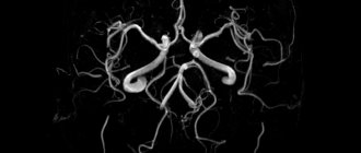
Duplex scanning of cerebral vessels is an ultrasound examination that combines a two-dimensional picture - the anatomical structure of the vessels, the tissues around them and the speed of blood flow. Using this method, they find atherosclerotic plaques, blood clots in arteries and veins, and check the condition and integrity of the vascular wall.
There are extracranial studies, aimed at checking the main highways, and scanning, studying the intracranial vessels located in the skull. During the procedure, the common carotid arteries are examined along their entire length, the internal carotid arteries up to the entrance to the skull, and partially the external carotid and vertebral arteries.
Duplex scanning of the vessels of the head and neck makes it possible to diagnose the disease at an early stage of development.
In what cases is a study prescribed?
Duplex ultrasound scanning is used for various diseases and pathological conditions of cerebral vessels:
- congenital vascular malformations;
- deformations of head vessels;
- atherosclerosis;
- injuries;
- when blood clots form in the vessels of the brain (thrombosis);
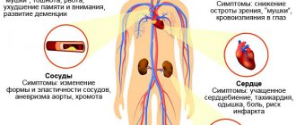
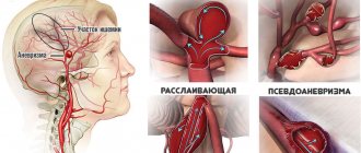
- if you suspect the presence of an aneurysm (expansion of part of the vascular wall);
- inflammation of blood vessels (vasculitis);
- damage to blood vessels (angiopathy);
- with discirculatory encephalopathy (brain damage that occurs due to circulatory failure);
- in the presence of diseases in nearby adjacent tissues and structures.
Diagnostic measures are also prescribed for patients who have the following symptoms:
- migraine;
- constant feeling of heaviness in the head, accompanied by tinnitus and
- frequent headaches;
- dizziness;
- motor coordination disorders, unsteadiness when walking;
- fainting;
- feeling of weakness and numbness in the legs and arms;
- speaking disorders.
Duplex scanning is prescribed to patients who have previously suffered a stroke or surgery on the vessels of the brain, as well as people suffering from chronic cerebrovascular accidents. In this way, the condition of blood vessels and blood flow is monitored. Duplex scanning most informatively reveals secondary pathological changes in the vessels of the head in the presence of various diseases (for example, with a persistent increase in blood pressure or in the presence of diabetes mellitus).
The study is prescribed in the presence of large tumors inside the brain (cystic, cancerous formations and hematomas). In this case, the goal of diagnosis is to evaluate the tumor and its blood supply. Ultrasound scanning is also required before surgical intervention on cerebral vessels or to determine methods of conservative treatment.
Duplex scanning is of high importance in neurological practice and makes it possible to diagnose cerebral circulatory disorders in the early stages of development. This allows you to take timely measures to prevent an acute process. In this case, the possibility of a detailed study of cholesterol plaques and their density is of great importance.
Triplex scanning
The results of intracranial and extracranial triplex scanning of arteries and veins of the brain reflect their anatomical structure. The blood flow is presented in color depending on the speed in a particular area. Depending on the subject of study - veins or arteries, the image is colored blue or red.
This is not a separate research method, but an extended duplex scanning of cerebral vessels with an additional function. The vessels are viewed in two longitudinal and one transverse planes.
How the procedure works
This diagnostic method is absolutely painless and does not require general or local anesthesia. The patient is examined standing, lying or sitting, depending on which organ needs to be examined.
The surface of the skin is lubricated with a special gel, which facilitates signal transmission. The ultrasound technician then directs the probe to the desired area.
During duplex scanning of the brain, the sensor is placed in places called “ultrasonic windows” - parts of the skull where the bones are thinner or where there are physiological openings in them.
Through such zones, the ultrasound beam easily enters the cranial cavity.
During the procedure, the patient may be required to adopt a more comfortable position for the examination. The doctor may also ask you to hold your breath or do other similar actions. The manipulation lasts approximately half an hour and causes absolutely no discomfort.
Transcranial
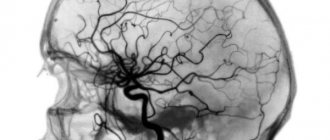
Transcranial Dopplerography of cerebral vessels is a type of duplex study. Its main purpose is to study the speed and direction of blood flow in intracranial vessels. The goal is to identify hematomas, large lesions and control previously detected disorders. It is impossible to examine the walls of the vessels located in the skull. Information about the structure and lumen of the artery is available only in color mode, the change of which depends on the speed of blood flow.
With transcranial duplex scanning, brain vessels can be seen in two planes.
TCD of cerebral vessels is indicated in the following cases:
- Indirect signs of damage to the arteries in the skull were found.
- Symptoms of cerebral ischemia, the causes of which are unknown, have been identified.
- A duplex scan of cerebral vessels showed signs of stenosis and blockage.
- Constant headaches.
- The patient has a complex vascular disease leading to cerebrovascular accident.
- With brain pathology, which leads to vascular deformation and impaired blood flow.
How is the procedure performed?
Duplex scanning can be done by referral from your attending physician in the neurological departments of large city hospitals or you can go to a clinic according to your area of residence. The manipulation occurs according to the general rule. The patient is placed on the couch, a hard pillow or cushion is placed under the head, and the head is moved to the side opposite to the sensor.
Before starting the procedure, the doctor applies a little special gel to the area under study, with which you can easily “move” the transducer over the surface of the skin, analyzing the arterial and venous beds. The blood vessels of the brain are checked through the bones of the skull. The skin is first treated with a water-soluble gel, then the doctor places sensors on the following areas:
- temples;
- above the eye sockets;
- alignment of the occipital bone with the spine;
- occipital bone.
Neurosonography
A separate type of ultrasound examination aimed at checking pathologies of the newborn’s brain is neurosonography. Recently, in many maternity hospitals, this examination is carried out even before the baby is discharged, and a pediatrician or neurologist prescribes it when the baby reaches 1 month or according to indications.
It is necessary to carry out it if the child was born prematurely, received an Apgar score of less than 7/7 points at birth, or there are suspicions of hydrocephalus, cerebral palsy, malformations or developmental delays, intrauterine infections, genetic pathologies or diseases of the nervous system.
Another group of indications for neurosonography are prolonged or, conversely, rapid labor, birth injuries, Rh conflict and monitoring the dynamics of the baby’s treatment.
Currently there are 4 types of research:
- Transfontanel NSG is performed through the large fontanel. This technique provides a complete examination of the brain cavity, and therefore is the most common. However, it is carried out only up to a year - by this time the fontanel usually closes. The most informative examination is immediately at birth or during the first few months.
- When performing transcranial USG, data is obtained through the temporal and sometimes parietal bones.
- The combined method includes examination through the fontanelle and the bones of the skull.
- USG is also performed through bone defects.
There is no need to prepare your child for the examination. The procedure is performed without anesthesia or sedatives.
NSG allows you to identify signs of increased intracranial pressure. An increase in the size of the ventricles of the brain indicates the accumulation of cerebrospinal fluid in them and the development of hydrocephalus. The detected focus of ischemia indicates probable oxygen starvation. Detection of hemorrhage is an indication for urgent hospitalization.
During the examination, various cysts may be found. Subependymal cysts look like fluid-filled cavities and are located near the ventricles of the brain. Such formations require treatment. Appear due to lack of oxygen or hemorrhage.
Vascular cysts look like small bubbles with fluid, located at the site of cerebrospinal fluid secretion. Formed during childbirth or in the prenatal period. Usually do not require treatment.
Arachnoid cysts occur as a result of infections, hemorrhage, trauma, and can be located in any part of the head. Their rapid growth leads to compression of nearby tissues. Treatment is necessary.
Fetal ultrasound
Signs of some diseases diagnosed in infancy can be detected in the perinatal period. During pregnancy, 3 ultrasound examinations are performed, each of which reveals signs of brain pathology.
First trimester screening is performed at 12-14 weeks. It allows you to detect acrania, anencephaly, exencephaly, cranial hernia, as well as signs of certain chromosomal pathologies, such as Down syndrome.
In acrania, there are no bones of the skull. Anencephaly is characterized by the absence of not only the bones of the skull, but also the brain. In exencephaly, there is no bone tissue, but brain tissue is partially present. A cranial hernia is diagnosed when fragments of the meninges protrude through defects in the bone tissue.
During screening in the second trimester, features of the formation of the brain and face are checked. By this time, all anatomical structures and organs have been formed. Much attention is paid to the head circumference and its shape, calculated as the ratio of the biparietal and fronto-occipital dimensions. A lemon-shaped, strawberry-shaped shape is determined. Look at the size of the head - small or disproportionately large. The lateral ventricles are measured. Their increase indicates hydrocephalus.
The study of the cerebellum is of particular importance - the size of the hemispheres and the degree of development of the cerebellar vermis are determined. Its underdevelopment leads to an inability to maintain balance, muscle inconsistency, abruptness of movements, and trembling of the limbs. The visual thalamus, corpus callosum, horns of the lateral ventricles and many other areas of the brain are studied.
Attention is also paid to the facial skeleton. Often the shape of the nose and cleft lip are a symptom of chromosomal diseases.
The purpose of the third screening is to confirm or exclude defects detected in the first two studies. At the same time, CTG is performed - registration and analysis of the fetal heart rate. This study shows signs of oxygen deprivation, which may have a negative impact on brain development.
How to prepare for a brain ultrasound
Ultrasound of cerebral vessels is usually prescribed by a neurologist or therapist. When receiving a referral, it is necessary to discuss the use of vasoconstrictor or vasodilator drugs with a specialist. Your doctor will probably ask you to stop taking them temporarily.
The day before the procedure, it is recommended to exclude from the diet foods that can affect the tone of the walls: alcohol, pickles, caffeine-containing drinks and foods, including coffee, tea, chocolate, energy drinks. Drinks with ginger and ginseng are also contraindicated.
You should have your last meal 4-5 hours before the examination. It is not recommended to take a hot bath two hours before the ultrasound. There is also no need to smoke - smoking a cigarette leads to a narrowing of the arteries and veins.
Immediately before the procedure, you need to remove all jewelry from your head and neck and tie your hair in a ponytail. To examine the cervical region, it will need to be freed from clothing.
Preparation for the procedure and contraindications
This technique is especially popular precisely because it is absolutely harmless and has no contraindications. This procedure can be performed on patients regardless of age: both children and the elderly. You can repeat the manipulation as many times as necessary until the diagnosis is definitely established.
In addition, the diagnostic examination does not require any preliminary preparation. But there are some recommendations that will make this procedure much more effective.
On the day of the examination, stop nicotine and any other substances that to one degree or another affect vascular tone. This::
- coffee;
- energy;
- strong tea.
If you use any medications, such as adaptogens (schisandra chinensis, ginseng, etc.), magnesium, piracetam, vinpocetine, you should first notify the neurologist who prescribed this procedure. Before scanning, you need to get rid of jewelry from your neck and head: earrings, chains, hairpins and others. After the examination, you must wash your hair thoroughly.
Features of the procedure in children
Children under one year old undergo neurosonography. Indications for ultrasound examination in children, including Doppler sonography of the brain, are speech delay, restlessness, behavioral problems, increased fatigue, decreased attention, and memory impairment.
In general, the preparation and sequence of actions for the procedure itself are the same as for adults. However, if adults are asked not to eat 4-5 hours before the procedure in preparation for an ultrasound, then it is better to feed infants and preschool children an hour before the examination. There is nothing wrong if babies sleep during the procedure.
Another difference is that it is difficult to keep children, especially younger ones, in place, so you must be prepared to calm them down during the examination. Perhaps it will be enough to follow the picture that appears on the monitor. In some cases, it is better to take a toy or book with you.
Decoding
Usually, a few minutes after completion of the examination, the diagnostician gives the patient a conclusion and an image. In the transcript, the obtained indicators are compared with normal ones. Brain ultrasound results describe:
- Structure and size of the ventricles of the brain.
- The distance between pools filled with liquor.
- The condition of the walls of blood vessels, their pathological changes, features of blood flow in different phases of heart contraction, index of resistance and pulsation.
Dopplerography of veins provides descriptive information about their shape, structure, patency, diameter, formations in the lumen, and the speed of blood outflow. Numerical values are practically not used. The results of ultrasound scanning of arterial vessels, on the contrary, are presented in the form of digital analysis and comparison with normal values.
Indicators that are used to guide the interpretation of ultrasound results: no signs of deformation, compression of blood vessels, or turbulence at the branch site of small arteries. Arteries and veins must have sufficient clearance for blood movement; vertebral arteries have the same structure. The usual wall thickness is 0.9-1 mm.
Based on the characteristics obtained during the procedure, suspicions of pathologies such as:
- Stenosis is a narrowing of the vascular walls that restricts the passage of blood through them.
- Atherosclerosis - cholesterol plaques appear on the walls, thickening of the vascular tissue is characteristic, echogenicity is changed.
- An aneurysm is a protrusion of the vessel wall due to thinning.
- Turbulent flow is the formation of turbulence along the blood flow.
- Occlusion – obstruction of blood vessels.
- Vasculitis - the vascular wall is thickened, the differentiation of its layers is noticeably impaired, and echogenicity is changed.
- Arteriovenous malformation is a vascular wall between the bed of an artery and a vein.
Decoding the results
In addition to duplex scanning, there is also triplex scanning. Its difference is that during the procedure, the areas under study on the device screen are highlighted in bright color for better viewing. The entire picture remains black and white, but the blood flow on the monitor is highlighted. It can be observed in dynamics as a stream of red, blue or another color. The results of classical Doppler ultrasound look like rows of graphic information that can be printed. Based on the study of these lines and graphs, the specialist draws conclusions about the possible presence of blood clots, plaques, aneurysms, etc. violations. Duplex scanning is a more advanced and accurate method. Its only disadvantage is that the resulting dynamic image cannot be transferred to a static sheet of paper. The specialist assesses the condition of the vascular system based on visual information obtained during the procedure. The same applies to triplex, which differs from duplex only in color.
Only a specialist can correctly interpret research data. This information contains characteristics of the condition of the vessels in many respects: the patency of the vessels, the size of their lumen, hemodynamics, elasticity of the walls, blood pressure values. The difference between its upper (systolic) and lower (diastolic) indicators is also informative. These values reflect the degree of elasticity of blood vessels, dehydration of the body, the initial stage of many diseases, and the level of stress.
Contraindications
There are no contraindications for ultrasound examination, it is safe and can be performed several times in a row. Limitation – damage, wounds, exacerbation of dermatitis, psoriasis, eczema. They make it impossible to apply the gel to the scalp and attach the sensor.
Difficulties may arise if the vessel being examined is covered by bone tissue or is located under a thick layer of subcutaneous fat. The study is difficult in patients with cardiac pathologies, in particular arrhythmia and slow blood flow.
Ultrasound diagnostics of the brain is an accurate, safe way to obtain information about the condition of this organ, its parts and vessels. It is prescribed for suspicion of various pathologies associated with changes in the vascular pattern, the appearance of plaques, neoplasms, impaired blood flow, before preparing for operations and in the postoperative period. Usually, based on the results of this study, the doctor can prescribe drug therapy and speed up recovery.