What is ultrasound of cerebral vessels
Ultrasound of the vessels of the brain and neck is a diagnostic technique that evaluates the efficiency of blood movement in the arteries and veins of the head. During the procedure, the patient does not feel discomfort; the result of the study can be copied to a portable device or the image can be printed.
Now there are other diagnostic options, such as MRI, but ultrasound is an affordable and universal method.
A timely ultrasound of the vessels of the head makes it possible to identify:
- disturbances in the blood flow to the brain;
- oxygen is supplied in insufficient quantities;
- signs of stroke;
- vascular disorders;
- effectiveness of prescribed drugs.
Vascular examination allows us to determine pathologies:
- atherosclerotic disorders in the arteries - the size of plaques is determined, the level of vasoconstriction is determined - if the indicator is high, the connective tissues of the neck are examined;
- blood flow disturbance;
- deformation of arterial walls;
- destruction of blood vessels and arteries;
- possible vasoconstriction;
- degree of tissue compression;
- abnormalities received at birth;
- pathologies of blood supply in the brain.
Preparing for an ultrasound of the brain
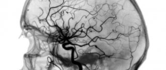
Ultrasound is reflected at the interface between solid and liquid media, and it is these signals that are recorded.
The substance of the brain itself and its structures and vessels are not visualized through such a study.
Neurosonography is performed as an emergency study that does not require any preparation in cases where there is a suspicion of gross organic pathology in the cranial cavity.
Who needs echo-EG
Indications for this study are:
- brain tumor
- hemorrhage in the hemisphere
- brain abscess
- if the child had a head injury, but an x-ray of the skull did not answer the question of whether there was brain damage
- Ultrasound examination of the brain can be carried out as a control over the treatment of the above diseases, so as not to carry out such expensive procedures as MRI or computed tomography each time
- if an ischemic stroke is suspected, when it is not possible to conduct a more accurate and informative diagnosis.
Indications for diagnosis
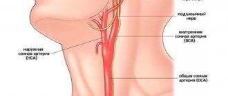
Diagnosis of the vascular system of the brain is performed after an ultrasound of the arteries and veins of the neck, because the causes of the disease are sought in this area. The fact is that the vascular system of the head is based on the main arteries, which include the carotid and vertebral veins.
There are clear indications for ultrasound:
- osteochondrosis in the cervical spine;
- arrhythmia;
- VSD;
- high cholesterol;
- pressure;
- atherosclerosis;
- fatigue;
- excess weight;
- planned heart surgery;
- aneurysm.
An ultrasound scan of the blood vessels of the brain and neck should be performed if symptoms occur:
- recurrent headache;
- tinnitus;
- loss of consciousness;
- sudden loss of attention and hearing;
- impaired coordination of movements;
- insomnia.
Some people are advised to undergo testing regularly, regardless of the appearance of certain symptoms:
- age exceeding 45 years;
- diseases of the musculoskeletal system;
- presence of bad habits, including smoking;
- stroke;
- brain injuries and concussions;
- high blood pressure;
- diabetes.
Who is the procedure indicated for?
An ultrasound of the head and neck is performed at the discretion of a neurologist or other doctor based on examination and questioning. Even if there are symptoms indicating problems, you should not select research methods on your own. You shouldn't be afraid of manipulation either. It is considered one of the safest in the entire impressive list of diagnostic approaches.
Ultrasound of the brain is a common procedure in newborns.
The main indications for ultrasound are the following:
- osteochondrosis and other lesions of the cervical spine;
You can learn more about osteochondrosis and related problems of blood circulation in the brain here.
- hypertonic disease;
- a history of stroke, heart attack, traumatic brain injury, or diabetes;
You can learn about the relationship between hypertension, stroke and diabetes here.
- excess of normal blood cholesterol levels;
- obesity;
- heart and vascular diseases (ischemia);
- presence of signs of infectious damage to the membranes or substance of the brain;
- in childhood - prematurity, trauma during childbirth, convulsions, suspicion of pathologies of brain development or hydrocephalus, symptoms of cerebral palsy and detection of an intrauterine infection in the mother.
From time to time, it is useful for heavy smokers to assess the condition of cerebral vessels.
Sometimes the study is indicated for upcoming surgery on the heart or other internal organs. Especially if we are talking about an elderly person or a representative of one of the risk groups. Men over 60 years of age are recommended to undergo examination every year.
Often this type of examination is prescribed if children or adults have at least one of the following symptoms:
- cephalgia of varying localization and intensity of unknown origin;
- impaired coordination of movements and decreased functionality of the musculoskeletal system;
- noise in the head or ears;
You will learn about the causes and treatment of noise in the head with the help of medications here.
- frequent dizziness, confusion, fainting;
- muscle weakness, numbness in fingers or limbs;
- chronic weakness, increased fatigue, unexplained drowsiness;
- heaviness in the head, decreased quality of memory or attention;
- nausea for no reason - especially if it ends with vomiting and does not go away even after it;
- unusual persistent decrease in blood pressure;
- sleep disturbance, problems with orientation in space;
- decreased clarity of vision, spots before the eyes, hallucinations.
Echoencephalography is indicated for adults, schoolchildren and preschool children.
There are several methods for performing ultrasound of the brain. Echoencephalography is indicated for adults, schoolchildren and preschool children. Children under one year old or a little older undergo neurosonography, for which the parietal fontanel is used. Separately, Doppler ultrasound and duplex/triplex scanning are distinguished. These are modern referral methods that show the maximum degree of information content.
You can learn more about echoencephalography of the brain here.
Are there any contraindications?
Any method of examining blood vessels does not pose a health hazard, so it can be repeated. It is not possible to make a diagnosis if the area being examined is covered by fat or bone tissue.
Difficulties may also arise in the following cases:
- pathologies in the work of the heart;
- circulatory disorders;
- abrasions, cuts on the skin;
- the patient’s inability to assume a horizontal position due to illness;
- serious condition of the patient.
Advantages and disadvantages
Ultrasound of the vessels of the brain and neck is characterized by safety, accessibility, and the absence of contraindications. Ultrasound is indicated even for infants.
The advantages of this research method are:
- no pain during the procedure;
- three-dimensional image of the study area;
- repeated examination if necessary without harm to health;
- accurate result;
- affordable price;
- possibility of soft tissue analysis.
Ultrasound of the vessels of the brain and neck is so safe that it can be performed even on children.
The disadvantage of ultrasound examination is the inability to visualize some areas due to projection overlap.
Among the disadvantages are:
- low resolution compared to MRI;
- the patient's excess weight may make diagnosis difficult;
- difficulty in visualizing bone tissue.
Kinds
Ultrasound examination of the brain, which is done to examine the circulatory system, can be of the following types:
- Doppler study. In this type of diagnosis, the sensor is installed on the arterial or venous area, without their preliminary visualization.
- Duplex examination. Dopplerography of this type is combined with conventional ultrasound. During the study, the specialist examines the thickness of the vessels using an ultrasound sensor and Doppler sonography.
- Triplex view is combined with duplex scanning and color differentiation.
- TKDG. During research using this method, the sensor is placed on areas of the skull where there is insignificant bone thickness. This method allows you to assess the condition of the circulatory system, which is located in the brain.
Any study of cerebral vessels is based on ultrasound. However, if an ultrasound examination of blood vessels is prescribed, they usually mean Doppler ultrasound combined with Doppler.
Ultrasound shows even a minor pathological focus
How is ultrasound better than MRI?
There are main differences between the two research methods:
- Different sensitivity of the equipment - ultrasound can be used to monitor the course of the disease.
- An ultrasound detects a large tumor, while a tomograph allows one to detect tumors at an early stage, as it is highly accurate.
- Ultrasound is a more affordable method of examination.
Comparative analysis of ultrasound and MRI:
- Both research methods are completed in 20 minutes, are painless, have no side effects, and are safe.
- Ultrasound can be done during pregnancy, while MRI is excluded in the first trimester.
- MRI determines the cause of the disease, ultrasound allows you to identify the disease without identifying the cause.
- Excellent visualization in two methods.
Ultrasound of the blood vessels of the brain and neck is used when it is necessary to quickly diagnose a disease. The effectiveness of vascular patency and the nature of blood movement are revealed. The result of the analysis is presented in the form of an image of 3 waves: initial, middle and final - from which the doctor determines the disease.
In the absence of pathologies, the graph will be symmetrical with equal distances.
If a detailed study is required, then MRI is used - diagnosis of brain diseases by obtaining a 3D image in real time, provides an accurate determination of abnormalities, identifies multiple sclerosis and hidden disorders. Using MRI, you can determine the likelihood of developing the disease.
Ultrasound standards:
- the thickness of the walls of the artery of the head and neck should not exceed 1.1 mm;
- free flow of blood through the vessels;
- absence of vortex blood flows;
- artery diameter – 2 mm;
- no decrease in the speed of blood flow in the vessels;
- no vasoconstriction.
After decoding, the neurologist makes a diagnosis and prescribes treatment.
Decoding of ultrasound parameters
After performing an ultrasound analysis of the cerebral vessels, the sonologist can compare the patient’s indications and complaints. Assess the degree of damage to the organ, the impact of defects on other processes in the body. There is an understanding of how to carry out further treatment, or how effective the treatment that is currently being carried out is.
Standards for performing ultrasound of cerebral vessels:
| Study area | Indicators are normal |
| Artery wall thickness | 0.8-1.2 mm |
| Vascular lumen | The value must be specified as "norm" |
| Turbulence | Should not be detected in the bloodstream |
| Presence of arteriovenous malformations | Should not be found in the circulatory system |
| Vertebral artery diameter | No more than 2 mm, the diameter must be the same for all arteries |
| Presence of compression | Must not be detected |
Types of ultrasound examination of blood vessels of the head and neck
Ultrasound is performed on a special machine, consisting of a console with an electronic display, a screen and a signal converter necessary for scanning. The sensor is presented in the form of a device connected to the scanner with a cord. Based on the type of study and the depth of passage of the vessels, different sensors are used.
Linear transducers are usually used; they are effective when analyzing closely located vessels. The resulting picture of the vessel being examined is correlated with the sound signal that comes from the pulsating artery in different phases of heart contractions. The patient and doctor will hear this sound.
Modern equipment is equipped with a recording function for recording more important stages of research and printing images. All methods of analysis are performed on the principle of ultrasonic waves; with their help, static movements in the body are detected.
There are three types of procedure:
- Carrying out a study of a vessel in a two-dimensional system shows the structure of the vessels. It is performed in two ways: ultrasound of large arteries and vessels of the neck. To carry out the procedure, the transducer is placed in the area where large arteries are located. If the veins are in non-standard places, the procedure is not performed. The disadvantage of this method is the inability to determine the speed of blood flow. Advantages – detection of pathology at an early stage.
- Duplex scanning – used when the diagnosis is already known, reveals the causes of pathologies, allows you to see the picture of blood supply in the arteries.
- Triplex scanning – allows you to obtain compressed data on the speed of blood flow. The advantage of the method is the accurate reflection of vascular patency. Disadvantage: it does not allow determining blood flow parameters; additional research is required.
Preparing for an ultrasound
No special preparation is provided. Before prescribing the procedure, the patient should obtain recommendations from a therapist to determine whether it is necessary to stop using heart medications. Ultrasound of the vessels of the brain and neck suggests excluding strong tea, energy cocktails, coffee, and drinks containing ginger from the diet on the day of treatment.
Approximately 4 hours before the appointed time you need to stop eating. The fact is that in a well-fed body, blood flow increases, which can negatively affect the diagnosis. If an ultrasound is scheduled for a child, then he needs to be fed within an hour.
One hour before the diagnosis, you should stop smoking. Before the procedure, you must free your head from the chains to secure the sensors; long hair must be pulled back into a ponytail.
What does a normal echographic picture look like?
Brain in normal condition on ultrasound (top view/lateral view)
Traditionally, the following indicators are the norm:
- The structures of the brain are symmetrical.
- The ventricles of the brain have a homogeneous structure, clear contours, and are anechoic.
- The subcortical nuclei have moderate echogenicity.
- No M-echo offset.
- There should be no foreign liquid in the interhemispheric space.
- There are no inclusions in the brain structures.
- There are no neoplasms.
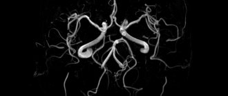
- Absence of aneurysms of large cerebral vessels.
- Vascular walls have smooth contours and structure.
- Vascular patency is within the age norm.
How is vascular ultrasound performed?
The patient needs to lie down and turn his head towards the ultrasound machine. The carotid arteries are examined first. The physician turns the patient's head away to provide access to the neck. Using sensors, the lower region of the carotid artery is diagnosed.
A subsequent study is carried out along the neck in order to identify the work of the vessel, its patency and determine the area where it splits into many arteries. By turning on a special mode, the doctor diagnoses the artery and outgoing veins, which makes it possible to determine vascular pathology.
If defects are detected, another study is prescribed, with its help the level of vascular damage and the further course of the disease are determined.
Next, the vertebral arteries are examined, directing the transducer along the neck. During the procedure, the sensor passes through the scalp, so a gel is used for easy glide. The speed of blood flow in the veins is analyzed, which makes it possible to identify emerging pathologies.
Applying a sensor to the temporal zone allows you to analyze the condition of the vessels, determine their thickness and patency. Analysis of the occipital zone is used to determine the pathology of the veins and arteries of the spine.
When performing the procedures, the doctor will give recommendations:
- stand up;
- not breathe;
- blink quickly;
- breathe deeply.
Another doctor’s recommendation is to squeeze the vessel with a finger - it determines the nature of the blood flow. Even children can easily tolerate ultrasound; the procedure is not accompanied by pain.
In rare cases, thirst or increased heart rate may occur. During the study, it is possible to hear pulsating sounds that change in pitch and appear in time with the heartbeat.
Classification of ultrasound examinations
With the development of medical technology, specialists have moved far ahead, offering several types of ultrasound today. They all differ in the quality of the information provided, but use the same operating principle. The manipulation algorithm is identical for both checking the cerebral ventricles and bone structure after mechanical trauma to the skull.
First, the patient will be asked to remove metal jewelry and then lie down on a medical couch. There is no need to worry, since the non-invasive examination does not cause any physical discomfort or pain.
The doctor directs the sensor to the area being studied, to where the blood vessels that need monitoring are located. For ease of manipulation, a person should turn his head to the side and tilt it back a little.
Then the diagnostician checks the referral brought by the patient. It will tell you in which mode the assessment should be carried out. There are 2 options:
- two-dimensional, which is also called B-mode;
- duplex color.
The first aims to monitor only the vascular network that extends outside the skull. This is about:
- internal artery;
- common artery;
- external carotid artery;
- jugular vein;
- vertebral arteries;
- small surrounding branches.
For the conclusion, information regarding arterial and venous patency is used. It is also worth noting the location of large vessels in relation to generally accepted standards. The general tone of the vessels, their diameter, the elasticity of the walls, possible intraluminal formations, and the health of the surrounding tissues are taken into account.
Color scanning using the duplex principle has a larger number of readings. The difference concerns the ability of the duplex to cover the state of the vessels in the cranial cavity and beyond. The first option in medical terminology is called transcranial, and the second is also called extracranial.
This scanning format using ultrasound provides the most complete picture of:
- arterial blood flow velocity;
- uniformity of filling of vessels;
- blood flow turbulence;
- gaps;
- venous valves;
- geometry of blood vessels;
- cross-country ability;
- elasticity of venous walls;
- condition of the venous sinus.
If the doctor additionally indicates the need for a transcranial examination, the sensor is applied only to certain areas of the head. Among them:
- temporal bone, including behind, in front of the ear, and also above it;
- connection of the head with the neck;
- eyelid of closed eyes.
But here it is necessary to take into account the peculiarities of the bone structure of each individual patient. For example, with severe osteochondrosis of the cervical spine, it is problematic to examine the structure. In this case, alternative methods are used.
It should also be taken into account that when switching the ultrasound machine to the Doppler effect mode, it is not always possible to conduct a high-quality assessment. The obstacle is insufficient blood flow.
To clarify the data to obtain more extensive information, auxiliary methods can be used. These are functional tests involving two methods:
- hyperventilation;
- finger pressure.
With the help of such measures, it is possible to better diagnose the mechanism of blood flow regulation.
It is very rare that specialists have to use the so-called extended Doppler ultrasound. It helps convert conventional ultrasound signals into sound signals. After listening to the areas being studied, the expert will be able to form a more detailed picture of what is happening in the bloodstream. The information obtained will become the basis for making a diagnosis regarding blockage or vascular narrowing. Additionally, a rare technique determines the degree of disruption of blood transportation.
On average, one manipulation lasts about half an hour. But the time can be extended if it is necessary to study health in several modes using functional tests. If you resort to a portable Doppler mode, you will be able to save almost a third of the time allocated for the appointment.
What does an ultrasound scan of the vessels of the head and neck show?
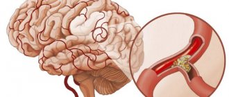
Ultrasound reveals deformed veins, congenital abnormalities, blood flow rate and indicators that assess tissue nutrition. Ultrasound of the vessels of the head determines the structure of the veins, branching, and the speed of blood circulation in the brain. The analysis provides information about existing obstacles: plaques, blood clots - allows you to systematize the data and find pathology, inflammation, aneurysm.
Using data obtained during spasm of neck vessels, their functionality and hidden potential for normal blood supply are revealed.
Based on the research obtained, the neurologist determines the pathology and its progression depending on the patient’s symptoms. The information received is systematized and a recommendation is made about the course of the disease, treatment methods and possible consequences.
The following data is used for decryption:
- blood flow rate;
- heart rate;
- thickness of blood vessels.
The results contain hidden data on the functionality of blood vessels and the presence of abnormalities. If instability of the veins is detected, then the wall thickens - stenosis; if the indicator is less than 15%, the presence of atherosclerosis is stated. Ultrasound helps identify plaques and determine whether they are causing a lack of oxygen to tissues - this information will help prevent the development of a stroke.
Increased wall thickness can also indicate inflammation of the veins. Identification of an unusual vascular network indicates an anomaly of the veins.
Main signs of deviations:
- Affected by atherosclerosis - plaques are recorded in the area of the carotid artery, which will subsequently lead to narrowing and obstruction of the veins. The initial stage of blockage of blood vessels is characterized by thickening of the vessel up to 1.5 mm, above this indicator - the presence of a plaque.
- Destruction of the arteries - atherosclerotic changes, occur from a sharp drop in pressure.
- Vein disease is a lesion of the arteries of a circular nature, affecting the entire wall of the vessel, which does not allow identifying individual components during the study.
- Structural changes - appear in patients with diabetes, characterized by impaired metabolism.
- Disintegration of arterial walls - occurs after injury, characterized by detachment of the upper part of the wall and blood entering there, after which blood clots form.
- Lack of circulation of venous blood in the brain - appears due to a reduction in the diameter of the vein, high speed of blood movement.
- Arterial thrombosis is a high level of vascular obstruction; arterial pathology develops as the disease progresses.
An ultrasound of the head and neck identifies dangerous abnormalities and prevents the development of the disease.
Precautionary measures
Many people are interested in whether manipulation is done only after certain preparatory measures. In fact, nothing significant is required from a person. Doctors ask you to stop drinking alcohol and put cigarettes aside a day before your appointment. You should also refrain from drinking tea or coffee. All of the above can somewhat distort the final image.
Before the examination, you should definitely consult with your specialist whether you can continue taking previously prescribed medications. Some medications affect the condition of blood vessels, so sometimes you should stop taking pills a couple of days before the ultrasound.
But this is allowed only after prior agreement with the doctor. Unauthorized refusal to follow the established course of therapy can significantly worsen the current well-being of the victim. At the preliminary consultation, it is necessary to announce all the medications that the person is taking, regardless of which organ they are intended for.
The listed standards are aimed at avoiding the occurrence of increased vascular tone. To obtain the most detailed image, you need to remove jewelry and other metal items from the upper body.
The collected information will help to identify possible pathology at its early stage of development. Most often, following a doctor’s recommendation, which includes going for an ultrasound, allows you to identify:
- vascular aneurysms;
- arterial stenosis;
- atherosclerotic signs.
The examination records even minor deviations in blood flow. Incoming information in real time is the basis for establishing the presence of an abnormal blood supply. Complaints, data from other examinations, information about hereditary predisposition, coupled with ultrasound results, help make a diagnosis and prescribe treatment, and reduce the risks of developing serious complications.
Ultrasound results can be used in surgery, neurology, therapy, and pediatrics.
Decoding the results
The sonographer or radiologist outlines the information obtained during the procedure. Next, the doctor enters the data into the card and makes a conclusion. In especially severe cases, additional research is required to confirm the pathology, taking into account a certain period of time. This research method is required after surgery or medical therapy.
The standard ultrasound result determines:
- the diameter of the vessels and its freedom from plaques;
- condition of the vein walls;
- presence of destruction;
- the amount of constriction of the veins;
- speed of blood flow;
- artery diameter;
- type of blood flow;
- general condition of the veins.
Healthy vessels have good permeability, are located straight, layers can be distinguished in the walls, by which pathology and developing atherosclerosis are easily determined. The size of the paired arteries is analyzed - there are no significant differences. The diameter of the spinal arteries directly affects the blood supply to the brain.
The normal value is 3-4 mm. If a value of less than 2 mm or more than 5 mm is detected, pathology is determined. The ultrasound protocol contains an analysis of the anatomical characteristics of the arteries of the neck, the movement of blood flow, the presence of deformities, and vascular obstruction.
The jugular veins should have an oval shape and should be easily compressed when applying pressure, otherwise a blood clot may be detected.
The course of the veins is straight, the size is equal, not exceeding three times the size of the carotid artery, the veins of the spine should not be more than 2.5 mm. The movement of blood in the neck should correspond to the rhythm of the sigh, the indicator should be within 30 cm/s.
Addresses of Moscow clinics and cost of the study
Ultrasound is recommended in any clinic or medical center equipped with equipment. In the clinic, vascular pathology is determined using ultrasound before identifying symptoms. The best way is to undergo research in special centers at the highest level, obtaining a competent transcript and prescribing the correct treatment.
The cost of diagnostics depends on the area of the procedure. When choosing a medical center, you should focus on the optimal price and quality. Treatment of identified vascular diseases by surgical means is carried out by large clinics; it is preferable to choose such institutions.
You can sign up for an ultrasound scan at any clinic in Moscow:
| Clinic name and address | Price |
| Miracle Doctor – Shkolnaya street, 49 | 1500 rub. |
| Orange Clinic Medical Center – Novoyasenevsky Prospekt, 13, building 2 | 2100 rub. |
| Euromedklinik – Sirenevy Boulevard, 32a | 2420 rub. |
| Clinic Cecil Plus – 1st Tverskoy-Yamskoy Lane, 13/5 | 2500 rub. |
| MEDFamily - Krasnodarskaya street, 57a | 1400 rub. |
| Clinic ViTerra Belyaevo – Profsoyuznaya street, 104 | 3300 rub. |
| Health Clinic – Klimentovsky Lane, 6 | 2500 rub. |
| MEDEO Medical Center – 1st Mashinostroeniya Street, 2/7, Building 1 | 2000 rub. |
| AvroMed Children's Center - Tolbukhina Street, 13, building 1 | 2800 rub. |
Addresses of clinics in St. Petersburg and cost of the study
In St. Petersburg, ultrasound is performed in the following medical centers:
| Clinic name and address | Price |
| Altermed - Bolshevikov Avenue, 7, building 2 | 2200 rub. |
| Treatment and diagnostic center – Lenskaya street 19, building 1 | 2800 rub. |
| LabStory - Basseynaya Street, 45 | 2500 rub. |
| Valmed Clinic - Moskovsky Prospekt, 73, building 4, room 27-N | 1850 rub. |
| Treatment and diagnostic medical center CMRT - Tipanova street, 12A | 3750 rub. |
| Altermed Clinic - Oleko-Dundicha Street, 17, building 1, letter A | 2200 rub. |
| Medical clinic Family Doctor - Petrogradsky district, Akademika Pavlova street, 5E | 2200 rub. |
| First Nevskaya Clinic - Yesenina street, 1k1, 1st floor | 1400 rub. |
| International clinic MEDSI - Marata street, 6 | 3996 rub. |
Vascular ultrasound determines abnormalities in the functioning of blood flow in the brain and neck, allowing the doctor to develop a treatment method and prevent the development of complications. The study is harmless, does not require prior preparation from the patient and is an effective diagnostic method.
Article design: Oleg Lozinsky