Rheoencephalography (REG) of cerebral vessels is a research method that allows you to determine vascular tone in the area of interest and assess blood supply. A special device takes into account the difference in electrical conductivity of tissues caused by vibrations of the vascular wall during heartbeats and the corresponding change in blood supply.
Rheographic study of the vascular system of the brain
Decoding the head REG often causes difficulty for patients. But this is not necessary, because a certified specialist is responsible for deciphering the result and drawing up a conclusion.
Questions that can be answered using REG:
- How fast does a pulse wave travel?
- What is the intensity of blood flow?
- How viscous is blood?
- Are blood vessels elastic?
- Is there sufficient outflow through the veins? And etc.
Also during the study, regional vascular reactions are determined.
A specialist can give advice on how to decipher the REG of cerebral vessels in each specific case, even online, if you download the result and send it to the doctor.
Characteristic
The doctor writes a mysterious abbreviation in the direction and, without going into too much detail, sends home the patient who forgot to ask what it is - REG.
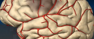
REG, rheoencephalography, is a non-invasive type of study of cerebral vessels, which is based on measuring the electrical resistance of tissues at the moment a weak high-frequency impulse passes through them. Blood is a conductor of electric current and, filling the vessels, reduces their resistance, as can be seen on the rheoencephalogram.
Clinical rheography is used to show cerebral blood supply. Another type of study, integral, is aimed directly at assessing vascular resistance.
The information obtained from REG allows you to see:
- tone of the walls of blood vessels;
- time of change in their resistance;
- elasticity;
- condition of arteries and veins;
- blood volume;
- viscosity;
- speed of pulse wave propagation.
The rheoencephalogram shows signs of impaired blood flow, but this is not enough to reliably say about the presence of a particular pathology and, moreover, to differentiate between diseases. In this regard, recently there has been a lot of debate about the advisability of conducting REG and replacing it with more informative research methods. So, if REG records a weakening of blood flow and an increase in resistance, then magnetic resonance imaging allows you to see the exact location of a blood clot or a damaged area of a vessel.
However, it is too early to write off rheoencephalography. It is indispensable in cases where MRI and CT are contraindicated: during pregnancy, lactation, the presence of pacemakers or other implants, obesity, claustrophobia, contrast agent intolerance, and the need to avoid radiation. Sometimes, to clarify the diagnosis, this procedure is complemented by echoencephalography.
REG is often confused with EEG. To carry out these studies, electrodes are placed on the head. The main difference is in the purpose: electroencephalography examines brain activity, and REG examines the state of blood vessels.
Advantages of REG
The rheoencephalographic examination method is absolutely safe and can be performed on patients of all ages, even small children. It is painless, does not require surgical intervention, and does not pose a risk to the patient’s health. The procedure takes a few minutes, and the result can be obtained almost immediately after its completion. REG provides accurate information about the condition of the arteries and veins of the brain.
An important factor is the availability of REG. The survey not only has a low price. Equipment for its implementation is usually available in all clinics and paramedic centers.
What the study shows
The value of the survey is that:
- Based on rheoencephalography of the vessels of the head, specialists receive significant information about the condition of the object of examination. Among them is the possibility of studying vascular tone, their elasticity, blood circulation speed and blood inflow/outflow.
- The use of rheoencephalography makes it possible not only to identify abnormalities in the vessels of the brain, but also to monitor blood flow after complex operations or severe injuries.
- With the help of REG, various pathologies are detected, and the severity of the pathological process is established.
In this case, the high speed of obtaining results is of no small importance.
What problems are being identified?
During the examination, the following are diagnosed:
- presence of traumatic brain injuries;
- localization of hematomas formed as a result of head trauma;
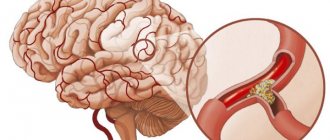
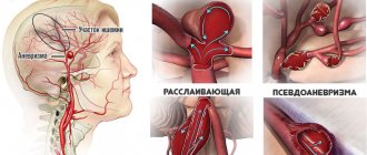
- pre-stroke condition;
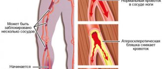
- damage to blood vessels by atherosclerotic plaques (atherosclerosis);
- thrombus formation in the vessels of the brain;
- predisposition to increased blood pressure;
- diseases associated with circulatory disorders.
The procedure facilitates the task of making an accurate diagnosis, on the basis of which the doctor prescribes an adequate course of treatment. With the help of it, he subsequently monitors the effectiveness of therapy.
Due to the complete safety of such an examination for the patient’s health, it can be performed repeatedly.
One of the most significant advantages of encephalography is the ability to distinguish between pre-stroke indicators, which have certain differences for men and women.
Other features of the method
Specialists obtain even more information by conducting functional tests.
The simplest and most accessible of them is with nitroglycerin. This substance helps reduce vascular tone. This test is used to differentiate organic and functional disorders.
Indications
Normally, human blood vessels are elastic, smooth, without any formations or plaques. They allow blood to pass through well, do not interfere with its movement, and provide adequate nutrition to the brain. However, sometimes their shape changes, the walls lose elasticity, and obstacles arise in the path of blood flow. Because of these changes, frequent headaches, dizziness, loss of consciousness, injuries, causeless disturbances in vision, hearing, speech, movement coordination occur, and health worsens when the weather changes. You may experience tinnitus, and “spots” flying before your eyes.
If these symptoms are present, the doctor gives a referral for rheoencephalography. This study is also prescribed as a complementary method in diagnosing the following diseases:
- arterial hypertension;
- vegetative-vascular dystonia;
- stroke;
- cervical osteochondrosis;
- diabetes;
- atherosclerosis;
- cerebral ischemia;
- Parkinson's disease.
REG shows the dynamics of changes in the postoperative or post-stroke period.
What is rheoencephalography of cerebral vessels
Rheoencephalography is a term derived from three ancient Greek words: (rheo) - flow, (encephalo) - brain, (graphos) - write. Putting this together, we get a description of the flow of processes in the brain.
The literal translation of the term cannot be called convenient, but it is so apt that the essence immediately becomes clear. To summarize: REG displays objective information about what is happening in the center of the nervous system.
Based on this, one can imagine how productive this brain study can be from a diagnostic point of view.
What does REG of the brain show?
How to prepare
Excitement and emotional stress affect the examination results, increasing vascular tone. This is a reflex process and is difficult to regulate. Therefore, the main rule is to try not to be nervous and calm down. There is nothing wrong with the planned procedure. Heavy physical activity should be avoided. Go out for research early, don’t run or rush. Plan your arrival time so that you have 15 minutes to recover. It is better to rest before the examination in a ventilated area where there are few people and noise.
Carrying out REG
Rheoencephalography of cerebral vessels is carried out in a special room, protected from noise and bright light. For the study, a rheograph device is used, consisting of an electric current generator, electrodes (sensors), a detector for signal recognition and a writing device. The device has from 2 to 6 channels according to the number of areas being tested. Polygraphs are sometimes used. Modern devices are connected to a computer.
The patient is placed on a couch or seated in a chair. It is important that he is comfortable. Before starting, blood pressure is measured. An elastic band securing the paired electrodes is placed on the head so that it passes at the back of the head, above the ears and eyebrows. Instead, a cap with installed electrodes is sometimes used. The sensors conduct current and are usually made of steel, nickel or aluminum. A special gel or paste is applied under the electrodes to facilitate contact with the scalp. During the procedure, it is recommended to sit or lie with your eyes closed and do not move.
The nurse or doctor turns on the rheograph. One of the electrodes emits a signal, which, after passing through the brain tissue, is recorded by the opposite sensor. After this, the data is processed by the rheograph and transmitted to a monitor or paper tape in the form of a curve. The resulting graph, a rheoencephalogram, displays the characteristics of tissue electrical conductivity.
Electrodes are placed on the head depending on the area being examined. They are attached to the bridge of the nose and in the area of the mastoid processes to diagnose the internal carotid arteries. At the temples - to study the external carotid artery. On the back of the head and mastoid processes - to examine the vertebral vessels, on the forehead - to study the blood flow of the middle cerebral artery.
REG with functional tests
In some cases, when recording a rheoencephalogram, functional tests are performed:
- Nitroglycerin test. The drug dilates the blood vessels, so the amplitude on the rheogram increases and is soon restored. If this happens, they speak of functional damage. Minor changes or their absence indicate an organic abnormality, for example, sclerotic vascular lesions.
- Carbon dioxide test. The patient inhales a 5% mixture of oxygen and carbon dioxide for 5 minutes. It reduces tone, dilates blood vessels, improves blood supply, and reduces peripheral vascular resistance.
- Study with clamping of any main vessel. The rapid restoration of the amplitude of the curve on the graph is a sign of the intact capabilities of the roundabout (collateral) blood flow.
- Hyperventilation test. The patient is asked to breathe deeply for 3-5 minutes. If at this time there are pronounced changes in the amplitude of the curve on the graph, they speak of high reactivity. Minor changes indicate decreased reactivity.
- Movement test. The patient changes position, perhaps standing up or sitting down from a lying position. The change in amplitude on the rheogram indicates a functional disorder.
- Head turn test. The subject is asked to turn his head to one side for 5 minutes, then to the other. Asymmetry of the curves is a sign of stenosis or other irreversible changes in the vertebral artery.
Time: 10-15 minutes. With additional samples it may increase.
Features of REG in children
Rheoencephalography has no restrictions on the age of the patient. The difficulty is that during this procedure you need to sit or lie quietly for several minutes. Not every child of primary school age is capable of this, not to mention preschoolers and infants. For this reason, it is better to conduct the examination in the presence of a parent. It is important that there are no distractions in the room. It is permissible to hang a picture that would attract the child’s attention for a while. Cartoons should not be shown.
Putting a hat or elastic band on your baby's head can also be difficult. Children under 3 years old will take it off. It’s better to practice putting on a pool cap or tying a ribbon around your head at home in advance. This way the child will get used to the discomfort and perhaps stop being afraid.
Before the examination, you need to pay attention to the fact that some children feel discomfort from the sticky cool gel applied to the skin. Electrodes can cause irritation or interest - it is better to remove them away from children's hands.
The best way to distract your child's attention is to tell him a fairy tale or read a book.
About contraindications
Due to the absolute absence of harm to the body, rheoencephalography has virtually no contraindications or side effects.
The main prohibition against performing the procedure is damage to the scalp.
Such examination is contraindicated for newborn children . This is explained by the small amplitude of the reflected waves, the large size of the anacrote and the complete absence of incisura. Such readings do not provide an accurate picture of the condition of the vessels of the head.
Rheoencephalography is an effective and affordable method for examining cerebral vessels. Its widespread use is due to the presence of the device in every hospital and, of course, the absence of side effects and contraindications for use.
Decoding
When analyzing the rheogram, the data obtained as a result of the examination, as well as age, health status and chronic diseases are taken into account. The graph that appears on the screen or paper tape when examining a teenager will be different from the graph of an adult. In the first one, it looks like a series of uneven waves with clearly defined periodic peaks. In an adult, it looks like a straight line with evenly spaced vertices. The graph shows anacrotes, catacrotes and incisuras.
Anacrota is a wave that shoots sharply upward with a slightly rounded top. Catacrota is a wave that goes down smoothly. An incisura is a notch between a downward wave and a tooth that anticipates the next wave.
Decryption is carried out only by a specialist. During the analysis, the regularity and type of waves, features of the rounding of the top, the dicrotic tooth located after the descent of the wave, and the position of the notch are assessed. Based on the results of the analysis, the doctor determines the signs of pathologies:
- With the hypotonic type, the waves have sharp, high peaks, the tooth or additional waves are displaced, the incisura and amplitude are increased.
- The hypertensive type is characterized by prolonged anacrosis with displaced waves, additional waves are possible. The amplitude is reduced and uneven. The teeth have different shapes.
- Cerebral atherosclerosis is indicated by flat tops, soft vibrations, and the absence of additional waves on the catacrota. In severe pathology, the waves have a dome shape.
- Spasm of the walls is characterized by round peaks.
- With stagnation of venous blood and difficulties in its outflow, increased catacrota and a large number of small waves between the main ones are observed.
- Angiodystonia is manifested by floating teeth and waves on the catacrota.
Advantages of CT in the clinic of JSC "Medicine" (clinic of academician Roitberg)
JSC "Medicine" (academician Roitberg's clinic) in Moscow is equipped with modern high-precision electrocardiographs that allow diagnostics. Data processing is carried out using computer programs, and therefore decryption requires a minimum of time. Rheoencephalography allows you to obtain high-precision results that reflect a comprehensive picture of the dynamics of the development of diseases affecting the brain. The procedure takes place in comfortable conditions. For patients with limited mobility, rheoencephalography can be performed at home. Optimal price, professionalism of doctors and individual approach are the main advantages of JSC Meditsina.
Go through rheography at JSC "Medicine" (clinic of Academician Roitberg) with comfort!