Vascular formations, especially in such an important location as cerebral structures, are relatively common. According to statistics, this kind of disorder is present in almost 3% of the world's population.
There are even more cases when the pathological condition is just developing, taking shape, but the prerequisites already exist. As a rule, diagnosis is greatly delayed, since the disease hardly makes itself felt until the critical moment.
It turns out that the patient understands what is happening so late that the treatment itself becomes dangerous.
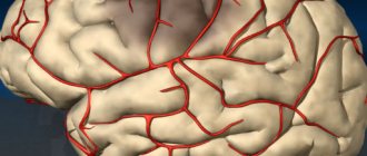
A cerebral aneurysm is a wall protrusion of large arteries of cerebral structures, which is accompanied by a decrease in the elasticity of the internal walls of the vessel, a change in the density and strength of tissue at the local level.
The condition is dangerous and has its own code I67.1 according to ICD-10. If nothing is done, death from aneurysm rupture and massive hemorrhage cannot be avoided. The only question is how soon this will happen.
Unfortunately, the pathological process either does not produce any symptoms, or the signs are so scant that it is impossible to discern such a dangerous condition right away.
The prospects for recovery directly depend on the stage of the disorder and the moment of initiation of therapy. There is absolutely no time to hesitate. It's a matter of life and death.
Development mechanism
The formation of the pathological process is based on a group of common pathogenetic factors. Which ones:
Congenital vascular anomalies
They occur more often than you might think. Another thing is that such conditions and characteristics of the body do not always lead to something as dangerous as an aneurysm.
We are talking about a pathological process that is formed in the prenatal period.
Whether this is due to a mutation or something else does not have much clinical significance. Since this does not affect the consequences in any way, and there is no point in clarifying the issue yet: prevention of genetic abnormalities is not yet possible.
Previous brain injuries
Cerebral structures are not only nerve tissue, but also an abundance of blood vessels. When the fibers are damaged, the arteries themselves undergo changes.
This can have a very negative impact, although it is impossible to guarantee the development of an aneurysm. These are just theoretical risks.
It takes some time: a year or two to monitor the patient’s condition.
Tissue irradiation
Radiation causes cell destruction and changes at the molecular level. The result is decay, gradual or rapid. Depends on the dose.
The result is softening of the vascular wall, tissue destruction at the local level.
Attention:
Especially often this consequence occurs against the background of radiation therapy.
Patients who deal with radiation as part of their professional duties are also at risk. Submariners, nuclear power plant employees and others.
Past infectious pathologies
Mostly viral. For example, some forms of herpes, as has already been proven by theorists of medical science. There are also risks from bacteria, for example, after meningitis or encephalitis.
Atherosclerosis
Chronic pathology. It is typically characterized by the deposition of cholesterol on the walls of blood vessels, the inner lining of the arteries of the brain.
Gradually, fats turn into full-fledged plaques, clogging the blood flow.
Pressure at the local level is growing significantly. The vessels are not designed for such loads. A slow or rapid degenerative process begins.
If nothing is done, an aneurysm will form within a few years.
Toxic destruction of vascular tissue
For example, with systematic smoking, consumption of alcohol or drugs. It makes sense to give up the bad habit as quickly as possible.
How quickly the change will develop is difficult to say. How to predict whether it will happen at all. But there are such risks and they are more than real.
Genetic predispositions
If there were patients with aneurysms in the family, the likelihood of a pathological process becomes several times higher.
You need to look at relatives of the direct, ascending line. That is, father, mother, grandparents are involved in the probabilistic analysis.
These are the basic mechanisms. As a rule, they do not intersect. Although certain combinations are possible. For example, long-term smoking causes injury.
The risk of developing an aneurysm in this case will be prohibitively greater, since the pathogenetic factors are numerous.
Digestive problems
If you have an aneurysm, you may experience nausea or vomiting. These symptoms are usually accompanied by a headache caused by a cerebral hemorrhage. The pain begins first, and then digestive disorders begin, sometimes even diarrhea. You may also experience dizziness, bleeding, sensitivity to light, and a stiff neck. All of them are directly related to bleeding in the brain tissue, in some cases they are caused by the pressure created by a large aneurysm on the brain. If sudden indigestion is combined with such sensations, do not ignore your condition and do not attribute it to banal poisoning - consult a doctor as soon as possible. It is always better to take action than to regret inaction later.
What Alsou's rich husband looks like: he's handsome (photo)
"Let the kids out" and other tips from small business owners in Asia
I’m making this for the second time: recipe for ice cream cake with strawberries and blueberries
Classification
The pathological process should be divided according to three bases: size, location and shape.
By size
Based on the criterion of the volume of altered tissue, the following types of pathological process can be distinguished:
- Miliary or microaneurysms. They represent the initial stage of the disorder or a full-fledged variety of it. They do not reach three millimeters in size. But they still pose a danger.
Therefore, it makes sense to check the patient every six months. If there is no dynamics as such, medications are prescribed, special living conditions and a diet are prescribed.
However, a microvariety may be a transitional variant. That is, as was said, the initial stage of the disorder. Then you cannot do without surgical correction.
- Small ones. Size up to 10 mm. They occur much more often. They pose immediate risks to the life and health of the patient. It is from this variety that the pathological process begins.
Treatment is strictly necessary. It is carried out under the supervision of a vascular neurosurgeon.
- Average. Up to 1.5 cm in diameter. These are serious entities. Enough physical stress is enough for the structure to rupture and lead to bleeding. Therapy is prescribed on an emergency basis. It is carried out strictly in stationary conditions.
- Large ones. 16-25 mm. Such forms are relatively rare. The size of the vascular formation leads to enormous risks of complications. The structure can rupture due to minor mechanical activity.
- Macroaneurysms. Over 2.5 cm in diameter. The danger lies not only in the possibility of rupture. Also, such a vascular formation compresses the surrounding tissues, leading to neurological deficits. Treatment is urgent, surgery is needed.
By localization
Regarding the second criterion, location:
- Anterior aneurysms. Located in the area of the parietal lobes. They are especially common. They do not pose great difficulties in terms of access, therefore the chances of recovery and a good course of the postoperative period are high.
- Medium varieties. Localized in the area of the parietal and temporal lobes. Access needs to be worked out as the issue is somewhat more complex.
- Localized in the area of the vertebrobasilar region. That is, closer to the occipital lobes.
- Located in the area of the internal carotid artery. It is difficult to reach such an aneurysm straight away. The correction method needs to be worked out. The task is not the easiest; it requires the participation of an experienced and qualified neurosurgeon.
The location must be clarified in order to carry out a competent and safe operation for the patient.
This classification is rather arbitrary, since an aneurysm can mature in almost any part of the brain.
By shape
The third basis for classification is the actual form of education. The following types of pathological structures are distinguished:
- Saccular. They occur most often. And account for more than 80% of the total number of situations. Externally they look like lateral wall protrusions of the artery, strictly in one direction. Such swelling became the basis for calling the aneurysm saccular.
- A fusiform change develops much less frequently. As the name suggests, the artery bulges in both directions. That is, there are no healthy, unaffected areas left.
A pathological process of this kind is much more dangerous. Because there is no restraining or compensating factor. Both sides of the vessel are involved in the breach and are at risk of rupture at any moment.
Doctors use all three classifications to describe the pathological process and plan treatment.
Symptoms
The clinical picture depends on the location of the aneurysm in the brain and its size. As a rule, a pronounced complex of manifestations occurs with formations larger than 10-15 mm. Before this, the patient does not suspect that he is sick with something so serious. Often such “dumbness” of altered tissues costs a person his life.
Symptoms of cerebral aneurysm can be divided into prodromal, subacute and acute, depending on the degree of development of the arterial formation itself.
This classification of signs applies to all types of pathological structures without exception, regardless of location.
Prodromal or precursors
They develop against the background of a stable course of the disease. When the risk of rupture is minimal. They accompany the patient constantly, almost never leaving him for a second.
- Pain in the eyes. Manifestations occur with large aneurysms. The discomfort is pressing, bursting. It intensifies in the morning and after physical exertion or any mechanical activity.
- Headache. Average in strength. Periods are much more intense. They become unbearable after stress, attempts to play sports, or other types of activity. The patient does not connect the symptom with the aneurysm.
- Decreased vision. It does not always occur, but in about half of the cases.
- Hearing loss. The same. A focal clinic that is not observed in all clinical situations.
- Anisocoria. A condition in which the pupils have different sizes are typical symptoms of a brain aneurysm. This occurs when cranial nerves are damaged. This is the result of direct ischemia, insufficient nutrition of brain tissue, or as a result of compression. Options can be different, including combined.
- Paresis and paralysis of the facial muscles. When you can't control your facial expressions.
- Nausea.
- Vomit. Several times a day or during an acute attack of headache. When ischemic processes increase.
- Weakness, drowsiness. Accordingly, a decrease in performance.
- Dizziness. A sign of a cerebral aneurysm is the inability to coordinate movements and move normally. This is the result of damage to the cerebellum, frontal lobe, and parietal areas.
Subacute
As the pathological process develops, the risk of aneurysm rupture increases almost every day. Harbingers or subacute symptoms of this condition are:
- Double vision. Visual symptoms of a cerebral aneurysm are characterized by a decrease in picture clarity, the disappearance of part of the image on the left or right in both eyes, flickering spots, and photopsia. The entire analyzer is affected, since the problem is central and localized in the brain.
- Noise in ears. Squeak, rustle, hum. Options may vary. As a rule, its intensity changes before our eyes. Because the blood flow itself becomes stronger and weaker. The faster it is, the higher the frequency of the sound and vice versa.
- Dizziness. Sudden and very strong. Aneurysmal protrusion of cerebral vessels with a risk of rupture is accompanied by acute loss of coordination of movements, problems with orientation in space, and sudden falls are possible.
- Speech disorders. It becomes difficult to speak, the tongue is literally twisted. The perception of information suffers: it is difficult to understand what others are saying.
- Muscle weakness. A sharp decrease in skeletal muscle tone. May cause falls and injury.
Symptoms of this stage of the pathological process sometimes last up to several days. On average, the count goes by minutes or hours.
The episode almost inevitably ends with acute symptoms, an emergency condition.
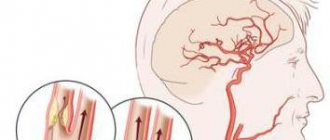
Signs of aneurysm rupture (acute)
Hemorrhage occurs in the meninges. As with a hemorrhagic stroke. The processes differ little in essence, except perhaps in origin.
The clinic will be the same:
- Unbearable headache due to irritation of vascular tissue. Characterized by extremely high intensity. The episode lasts up to several tens of minutes. May end in fainting.
- Intolerance to light and sound. Any irritants cause a lot of discomfort to the patient. This is the result of hyperactivity of all analyzers.
- Loss of control over the fulfillment of natural needs. Urination, defecation.
- Paralysis, paresis. Typically, half the body is affected. Left or right, opposite to the location of the rupture and hemorrhage.
- Acute mental disorders. Increased motor activity, agitation, behavior change, anxiety, panic attack.
The victim rushes about and cannot find a place for himself. Inadequate and does not respond to words addressed to him. The opposite phenomenon is also possible: complete loss of activity. Depression, lethargy.
- Loss of the ability to coordinate one's own movements.
- Often - coma or stupor. The depth of the disturbance of consciousness depends on the volume of hemorrhage and the degree of development of the pathological process. The more severe the condition, the worse the matter.
The patient needs to be urgently hospitalized in neurosurgery. It is important to pump out the blood, restore normal brain function, and nourish the affected area.
Attention:
There is very little time: first the count goes by minutes, then by hours. With proper promptness there are chances.
Sudden sharp headache
Headaches often signal a serious problem, and this is a symptom that definitely should not be ignored. With an aneurysm, the discomfort is especially severe. Patients often note that this is the most acute headache of their life. What happens at such a moment? Hemorrhage irritates the lining of the brain, causing a feeling of severe discomfort. The difference from a regular headache or migraine is that the sensations appear suddenly and are very pronounced. Unbearable discomfort can be associated with other serious diseases, so diagnosis in this case will definitely be useful, even if you do not have an aneurysm.
Causes
As for specific provocateurs of the pathological process:
- Congenital anomalies, mutations. They occur relatively often, but the degree of change is so small that it is very difficult to suspect deviations. Not always, but it happens.
- Complicated anamnesis. If there was at least one elder in the family with a similar problem, the risks automatically increase by 50-70%. It is worth taking a closer look at the people of the side branch: brothers, sisters. The probability increases by a quarter or a little less. It makes sense to undergo regular examinations with a neurologist or therapist; this is an important factor in prevention.
- Encephalitis. A type of inflammation of brain structures.
- Meningitis. The same thing, but of a different origin. Both pathological processes lead not only to an aneurysm, but also to immediate destruction of nerve tissue, deficiency, and focal symptoms. Be it dementia or speech, hearing, or vision disorders.
- Brain injuries. TBI. From concussions to open injuries, penetrating wounds and more. The probability is calculated based on a specific pathological process.
- Smoking. Nicotine and harmful substances affect blood vessels, their strength, and the elasticity of the internal lining.
- Alcohol consumption.
- Drug use.
- Arterial hypertension. Stable increase in pressure in the bloodstream. At any stage, it increases the likelihood of a pathological process.
- Heart diseases. IHD, angina pectoris.
- Tumors. They can compress blood vessels, thereby reducing their regenerative capabilities and adaptive potential.
- Atherosclerosis of cerebral vessels.
- Vasculitis. Autoimmune inflammation of the internal lining of blood vessels. Potentially lethal disorders. They manifest themselves in dozens of ways. Extremely dangerous in themselves. They require treatment under the supervision of a rheumatologist and as quickly as possible.
The causes of aneurysm are vascular, autoimmune, traumatic, cardiogenic pathologies.
If there is at least one of these disorders, and the aneurysm has not yet formed, the patient is classified as a high-risk group and is closely monitored.
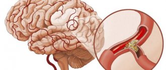
What is a cerebral aneurysm?
An aneurysm is a noticeable enlargement of a blood vessel in the brain. A swelling appears that resembles a berry hanging on a stem. In most cases, the aneurysm does not burst and may not even cause severe symptoms for a while, but sometimes bleeding does occur and this can be extremely dangerous to your health. The thing is that hemorrhage can lead to a stroke, which sometimes leads to death - only an experienced doctor can prevent such an outcome. If you are experiencing symptoms of an aneurysm, do not hesitate - call an ambulance or ask someone close to you to take you to the hospital as quickly as possible. Do not drive under any circumstances - you may lose consciousness along the way and get into a serious accident, this will be dangerous for both you and the people around you.
Expensive mistake: Police officer returns money to man that fell out of ATM
4 lipstick colors that will rejuvenate the face of women over 40 (red is among them)
The cat with white eyebrows became famous on Instagram
Diagnostics
The examination is not particularly difficult. Neurosurgeons work with patients of this profile.
The list of events will be as follows:
- Questioning a person.
- Anamnesis collection.
- MRI.
- CT.
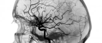
- Classic angiography, contrast-enhanced x-ray.
- EEG. To assess the degree of functional activity of the brain, to identify disturbances in the activity of cerebral structures.
Basically, this is enough to make a diagnosis. Additional measures are determined by a medical specialist.
Vision problems or drooping eyelids
If you suddenly start to see double or notice that your eyelid is drooping, the cause may be a rapidly enlarging aneurysm. It puts pressure on the nerve that controls your eye movements and causes problems with its functioning. When you notice this symptom, you should consult a doctor immediately. This is not a situation that you can simply wait out - this is a very serious symptom that indicates that the body’s condition is becoming critical.
Just think about the facts: how to get rid of anxiety in 5 minutes
I make a “braid” out of eggplants, and put minced meat, peppers and pasta inside: recipe
Sergei Zhukov and his wife celebrated 14 years of marriage: his wife is beautiful
Treatment
If the pathology does not pose a real danger, a wait-and-see approach is chosen. Drugs are used to strengthen blood vessels and normalize blood pressure. Also vitamin and mineral complexes.
But this is an exceptional measure. As a rule, therapy is surgical. It is necessary to eliminate the risks and remove the aneurysm. The issue is resolved in different ways.
There are several methods:
- Clipping. Conducted open access. After opening the skull, a special clamp is applied to the base of the aneurysm. The formation dies and the area is replaced by scar tissue. This is a complex, but at the same time effective solution.
- Microinvasive intervention. A special catheter is inserted along the vascular bed. A stent is inserted into the aneurysm, and then a coil is inserted, which closes the volume of the bulging wall of the vessel, or a substance is infused, which leads to sclerosis of the walls of the structure. Education is dying.
Both options are good when it comes to unilateral, saccular lesions. But what to do when an entire area of the artery is changed?
Fusiform forms of the pathological process are eliminated using open access. The affected segment of the vessel is removed. The healthy ends are connected. The junction or so-called anastomosis is sutured. Everything returns to normal, blood flow is restored.
Treatment of an aneurysm is not limited to surgery; you need to change your lifestyle and overcome the etiological factor, the one that led to the development of abnormal protrusion of the artery.
Patients are strongly advised to henceforth give up strenuous types of physical activity, minimize the amount of fat in the diet, and fortify the menu.
Correcting the root cause of the disorder is of great importance. After all, an aneurysm does not develop out of the blue. They look for the culprit: hypertension, atherosclerosis and correct it. This is a guarantee that education will not appear again, but in a different place.