How does pulmonary edema occur in heart failure?
With the development of heart failure, various disorders appear in the body. The heart has a large number of compensatory mechanisms. They allow, even under working conditions, to forcefully eject enough blood in one contraction to provide oxygen and nutrients to the organs in the systemic circulation.
But these capabilities cannot be maintained constantly. When compensatory mechanisms exhaust themselves, acute heart failure develops or chronic exacerbation occurs. Typically, pulmonary edema develops as a result of myocardial infarction.
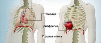
Since the left ventricle, despite vascular resistance, ejects blood into the aorta and works at the limit of its capabilities, it is the first to suffer during ischemic disorders due to insufficiency.
The contractility of the heart decreases, and the left ventricle cannot eject all the blood that comes to it from the left atrium. As a result of insufficiency, it can push out a small amount of blood, because of this the volume of blood in the ventricle gradually increases, which contributes to an increase in pressure in the pulmonary circulation and the development of congestion in the lungs. Hydrostatic pressure increases and the hydrostatic mechanism by which the vascular bed retains water is disrupted.
Vascular permeability increases and fluid enters the interstitial pulmonary space. After some time, it is absorbed by the lymphatic vessels. But if treatment is not carried out, the amount of fluid increases and it enters the alveoli. They fill up and gas exchange cannot occur in them.
When a lot of venous blood accumulates in the vessels of the lungs, another compensatory possibility begins to operate. Shunts open between the arteries and veins of the lungs. Blood saturated with carbon dioxide passes past the alveoli and into the vessels that carry it out of the lungs. This contributes to a slight unloading of the alveoli, but blood saturated with carbon dioxide enters the heart and then to other organs, exacerbating oxygen starvation.
Due to the high permeability of blood vessels, blood proteins can also leak out of them. A decrease in colloid pressure is observed.
Reasons for development
Pulmonary edema is a form of acute failure that most often occurs due to circulatory problems. The main anatomical part of the lungs are the alveoli. They contain a large number of small vessels, which lead to its contraction and normal gas exchange.
Heart rhythm disturbances cause some alveoli to stop functioning. This leads to a significant deterioration in the gas exchange process and all organs and tissues suffer from a lack of oxygen. Such deviations contribute to swelling of the alveoli and cause stagnation of the liquid part of the blood in the lungs. Gradually its quantity increases, which leads to the death of the patient.
The development of disturbances in the functioning of vitally important things can be provoked by:
- acute heart attack;
- cardiosclerosis;
- systolic dysfunction;
- heart disease;
- weak blood circulation in the left ventricle;
- chronic failure of the body.
A complication such as pulmonary edema in people with cardiac pathology can be caused by:
- impaired contractility of the left ventricle due to coronary insufficiency;
- blood volume overload as a result of mitral or aortic valve insufficiency;
- an increase in vascular resistance in the systemic circulation due to a hypertensive crisis;
- blocking blood flow at the level of the pulmonary veins.
The pathology is very dangerous to health and requires urgent treatment.
Course of treatment in hospital
While in the hospital, the patient is under the full control of doctors, which allows him not to do unnecessary contraindicated actions that contribute to pulmonary edema. In addition to this control, the patient is also subjected to additional pharmacological therapy, which completely eliminates the pathology and prevents its further manifestation.
Typically, the above-mentioned therapy consists of the use of drugs in four main categories:
- Glycosides - affect the functioning of the myocardium, stabilizing and restoring its natural rhythm.
- Nitrate medications - reduce reverse venous circulation and redistribute blood flow in the affected area. This category of drugs is administered intravenously and significantly dilates blood vessels.
- Adrenergic blockers - reduce the rate of work of the heart to reduce the speed of blood circulation in the body, which in turn allows the respiratory organs to normalize their natural functioning,
- Diuretics are drugs aimed at immediately removing excess fluid from the body. This allows you to reduce the current swelling, and after it completely disappears, reduce the chance of recurrence of the pathology.
This course of treatment can be used as a preventive measure, but only after consulting a doctor.
Stages
Depending on the course of the process, swelling can be:
- Hydrostatic. Fluid in the lungs during heart failure accumulates due to the fact that the pressure in the vessels of the pulmonary circulation increases and the walls of the capillaries burst. This leads to the flow of blood into the alveoli, their constant filling and a decrease in lung volume.
- Membrane. Its development is associated with a violation of the integrity of the capillaries that make up the structure of the alveoli. The lungs fill with fluid in a short time. Half an hour is enough for this.
Therefore, at the first manifestations, it is necessary to visit a doctor and undergo treatment.
The pathological process can occur in different ways:
- With fulminant edema, a pronounced clinical picture is observed, which develops very quickly.
- The development of acute pathology is observed within an hour.
- In case of prolonged illness, the process progresses within two days. This is associated with a gradual exacerbation of chronic heart failure.
- With subacute edema, symptoms either increase or decrease in severity.
In different cases, manifestations differ in severity.
Symptoms
Fluid accumulation in the lungs occurs in heart failure:
- Shortness of breath. Its development occurs with little physical activity or at rest. This symptom is characteristic of heart failure, but the swelling is accompanied by an increase in the frequency of respiratory movements per minute. The state of health worsens when the patient takes a supine position. Therefore, patients always try to sit down and fix the shoulder girdle. If left untreated, the severity of shortness of breath increases.
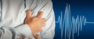
- Painful sensations in the chest, which is associated with insufficient oxygen supply and ischemia of the heart muscle.
- Wheezing in the lungs. During listening, moist rales are detected above the surface of the lungs. As the condition worsens, moist wheezing in the lungs becomes clearly audible even at a distance.
- Increased heart rate. Due to the lack of oxygen, the compensatory capabilities of the heart try to provide the body with the necessary volume of blood. Since the organ cannot expel the normal volume during a contraction, contractions become more frequent. In this case, there is weakness in the filling and tension of the pulse.
- Severe cough. During it, foamy pink sputum is released, whose color is due to blood entering the alveoli.
- Total cyanosis. Blue discoloration of the skin surface is due to lack of oxygen.
- Discharge of cold, sticky sweat.
- Swelling of large vessels of the neck. This is due to stagnation of blood in the pulmonary circulation.
Also read: How does pericarditis manifest in adults
? As oxygen deficiency develops, the appearance of motor or emotional agitation is first noted. But it is replaced by lethargy and confusion. In the absence of help, a coma develops.
Symptoms and clinical manifestations
Symptoms of pulmonary edema are specific and allow a diagnosis to be made already at the stage of physical examination of the patient:
- frequent coughing with foamy pinkish sputum (as the condition worsens, sputum is released from the nose);
- increasing bubbling hoarseness, suffocation;
- blue tint of skin and mucous membranes;
- pain in the chest area;
- dyspnea;
- vertigo;
- weakness;
- encephalopathy;
- faintness;
- hyperhidrosis;
- fear of death;
- tachycardia up to 200 beats/minute, which can suddenly turn into life-threatening bradycardia;
- blood pressure surges.
Pulmonary edema does not occur on its own; it is a symptom of many diseases.
Diagnosis
Congestion in the lungs in acute heart failure can be easily determined by a doctor during an external examination.
Research to confirm the diagnosis should be carried out as quickly as possible. The pathological process is determined using:
- X-ray of the chest organs.
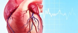
- Electrocardiography. The study reveals pulmonary infarction, cardiac arrhythmias, enlargement of the left ventricle and borders of the heart, and other pathologies that can provoke heart failure.
- Percussion. By tapping the surface of the chest, the presence of a dull percussion sound above the surface of the lungs is noted. But this is not an accurate method, since the sound appears as a result of any process that causes compaction of the lung tissue.
- Auscultation. While listening, you can hear that breathing has become harsh and moist rales have appeared.
- Blood pressure measurements. With the onset of pathology development, there is an increase, and gradually a decrease in indicators.
- Biochemical blood test. The presence of enzymes in the blood is determined, which will confirm infarction of the heart muscle.
- Pulse oximetry. It will show that the oxygen concentration has dropped below 90%.
- Determination of central venous pressure. Indicators are detected using central vein catheterization.
The most effective diagnostic method is x-ray. This is the simplest and most common procedure. Using an x-ray, the doctor determines whether water has begun to accumulate in the lungs due to heart failure. This can be seen from the following signs:
- vagueness of the vascular network, which indicates fluid on its surface;
- slight increase in cardiac tissue;
- long and short lines in the center of the pulmonary zone;
- the presence of a large number of infiltrates in the peribronchial areas;
- darkening of certain areas of the organ, making the image look like a bat.
By assessing the data obtained, the doctor can determine the presence of pulmonary edema in the initial stages of development. To obtain more information about the patient’s health status, other methods are prescribed to confirm the pathology.
Is there prevention?
Prevention of pulmonary edema due to heart failure, of course, exists. It consists in treating this very deficiency. A person diagnosed with CHF should be regularly monitored by a specialist - a cardiologist, and undergo a course of inpatient treatment and examination 2-3 times a year. If your condition worsens at the slightest level or if you experience difficulty breathing, you should immediately consult a doctor.
Taking medications prescribed by a doctor must be regular, since violations in taking medications can worsen the condition. Often, elderly relatives suffer from forgetfulness, so family members should monitor the process of drug treatment.
Sufficient exposure to air is also an excellent prevention of hypoxia; elderly people are advised to take evening walks before bedtime. The diet should contain all the necessary nutritional components, but be quite light and easily digestible.
Timely and correct treatment of any disease is the key to the absence of complications, therefore even a mild cold with CHF requires examination by a doctor. Take care of yourself and your loved ones and take care of your health.
Did you like the article? Save it!
Still have questions? Ask them in the comments! Cardiologist Mariam Harutyunyan will answer them.
Ivan Grekhov
Graduated from the Ural State Medical University with a degree in General Medicine. General practitioner
Treatment approaches
Treatment of pulmonary edema in heart failure should be carried out in the intensive care unit.
Intensive therapy consists of the following measures:
- If the patient is able, he is helped into a semi-sitting position.
- An oxygen mask is placed on the face or a nasal catheter is placed so that the patient breathes air with a high oxygen content. If indicated, transfer to artificial ventilation.
- With blood pressure above 100 mm. rt. Art. A Nitroglycerin tablet is placed under the tongue or infused intravenously. Thanks to the drug, the ischemic zone in the heart muscle decreases, the preload on the heart decreases and the blood pressure inside the vessels of the lungs decreases.
- A catheter is installed in the area of the central vein. If difficulties arise with its installation, then catheterization of the peripheral veins is performed.
- To relieve pain and reduce excitation of the respiratory center, the patient is given Morphine.
- Diuretics are used. Under their influence, the amount of fluid in the body decreases, as a result of which its volume in the lungs decreases.
- If the swelling continues to increase, shortness of breath increases, the skin turns blue, the patient is unconscious and cardiac performance decreases, there is a need for artificial ventilation.
They also establish infusion administration of medications that will correct and support cardiac activity. During treatment, cardiac activity and respiratory function are constantly monitored.
Actions of doctors during first aid
Having arrived, doctors begin to carry out the first urgent actions, namely:
- Two catheters are placed to be able to monitor the patient’s arterial and pulmonary pressure.
- The drug morphine is administered intravenously to dilate the arteries and blood vessels.
- Using a special device, doctors artificially fill the patient's lungs with oxygen to avoid attacks of hypoxemia.
- Furosemide solution is injected into a vein to reduce pulmonary edema and reduce venous reverse circulation.
- To relieve the heart, doctors administer the drug Aminophylline, also intravenously.
After the above procedures, the patient is taken to the hospital - to the nearest clinic for further treatment.