Features of the child's cardiovascular system
The heart muscle of a child has its own anatomical features depending on age. For example, at birth the weight of the heart is only 0.8% of the baby’s total weight (22 g), in adults it is 0.4%. The ventricles (right and left) are almost identical in mass, the wall thickness is only 5 mm. By the age of one year, the mass of the heart doubles, by the age of 3 – three times, by the age of six – by 11 times. With age, the load on the left ventricle increases, the thickness of its walls reaches 10 mm by the age of 14, and the thickness of the right ventricle is only 6 mm.
As we grow older, tissues also mature, connective tissue thickens, and arteries that provide blood supply grow. Until the age of 12, a child’s aorta is narrower than the pulmonary artery, then the lumens become identical, and the boundaries of the heart in children are formed until the age of 15. Blood flow volume also increases with age.
There are also differences in the anatomical location of the heart in children and adults:
- In babies, the heart is located higher; as they grow older, it lowers. This is primarily due to the structure of the diaphragm.
- At birth, the baby's heart resembles a ball. During the first years of life, it moves inside the cell and gradually descends.
- There is also a difference in heart rate; children have a more frequent pulse than adults, since the heart muscle contracts faster and metabolism occurs intensively.
What is the normal size of a person’s heart, valves and chambers?
You can study the structure of the heart using ultrasound. Its size is determined by age and physique. With developmental defects, cardiomyopathy, hypertension, disturbances occur - deviations up or down. Data obtained from instrumental research are needed to make a diagnosis and choose treatment tactics for the patient.
In the fetus
You can hear the heartbeat at 8 weeks, but detecting atria and ventricles in it is possible only closer to the twentieth week. It is usually recommended to have an ultrasound to measure the fetal heart by 24 weeks of pregnancy. The organ itself is easily detected in the chest cavity, but studying its structure and volume can be difficult, especially with high fetal mobility or a decrease in the amount of amniotic fluid.
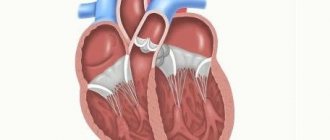
With normal development of the heart, its characteristics are:
- location in the left half of the chest;
- hollow ball shape;
- detection of four chambers, valves between them, septum, aortic arch.
Ultrasound of the fetal heart (color Doppler)
Since the fetal heart resembles a ball, the main measurements are taken in any direction. The diameter at week 24 is 2.5 cm, and by birth it increases to 4.5. All these indicators are averages and depend on the child’s weight. The valves are visualized, and their range of motion can be assessed.
The thickness of the heart muscle is also considered an important parameter - during contraction it is 4 mm, and in the relaxation phase it is 2.9 mm. An essential feature of the fetal heart is the equal size of the ventricles.
We recommend reading the article about the structure of the human heart. From it you will learn about the internal anatomy of the heart and the structural features of the valves, atria, ventricles, as well as the diagram of the blood circulation. And here is more about the heart of an athlete.
In children and adolescents
As the child grows, the heart gradually takes on the characteristics of an adult. This process is finally completed only by the age of 11–14 years. In one-year-old children, the weight of the organ increases by 2 times compared to the parameters after childbirth, and by three years it is 3 times higher. By the age of 5-6 years, growth slows down slightly, and in adolescents it accelerates again. In a 17-year-old boy or girl, the size of the myocardium is 10 times larger than in newborns.
First, the left ventricle enlarges, since it bears the main burden of pumping blood. Its size in a four-month-old baby is 2 times larger than that of the right one. Myocardial thickness from 5 mm reaches 12 (left) and 6 mm (right). The relative volume of the heart (compared to the chest) is larger in children than in adults.
Boys' hearts up to 10 years of age are larger than those of girls, then increased development by the age of 16 allows girls to get ahead of boys, then growth slows down again.
At one year, the heart has the following average parameters (diameter in cm):
- ventricular diameter – left 3.2 and right 1.4 (at maximum filling);
- atrium – left 2.4 and right 1.1;
- partition – 0.5.
For adolescents 15 years old, the left half of the heart normally has the following dimensions:
- end diastolic diameter of the ventricle is 4.3 cm, systolic – 3.5 cm;
- left atrium – 3 cm;
- aortic diameter – from 2 to 3 cm.
The right ventricle reaches only 1.8 cm.
In an adult
The length of the heart varies from 9 to 16 cm. Most often it is close to 12.5 cm. The base of the heart is about 10.5 cm wide, and the size from the front to the back wall is 6 - 7.5. Heart chamber parameters (cm):
- diameters - left ventricle about 4.6, right 1.95, left atrium 2.9 - 3.1, right 1.9 - 2.5;
- valve circumferences - aorta 0.8 - 0.85 and pulmonary artery 0.57 - 0.98, and atrioventricular ones average about 1 cm;
- the thickness of the wall of the ventricles is up to 0.5 for the right, and up to 1.5 for the left.
Expert opinion
Alena Ariko
Expert in Cardiology
At the same time, well-trained athletes may have a larger heart than normal, while short, thin women may have a smaller heart size. If there are no dysfunctions, then such deviations can be neglected. Diagnosis cannot be made based solely on echocardiography or radiography measurements. Clinical symptoms, laboratory data, and ECG are taken into account.
How to study heart function in children
Before determining the boundaries of the heart in children, it is necessary to examine the main organ for performance. The first indicator is heart rate. It should be symmetrical on both hands. The characteristics of the pulse also include such properties as rhythm, tension, filling, size and shape.
It is taken into account that heart rate indicators can be individual. Depending on age, there are certain norms, deviations from which may be within 20%. If the pulse is more frequent, there is tachycardia; a slow heart rate indicates bradycardia. The heart rate is calculated per minute during breaks between physical activities. Rhythmicity is when it is measured at certain intervals.
Deviations from normal readings can be caused by various diseases, even those not related to the cardiovascular system. But this is a reason to see a doctor. You can determine the work of the heart using a one-time cardiogram or measuring indicators throughout the day to notice how and when the condition of the heart muscle changes.
Features of the location of the heart in children by age
There are indicators of the functioning of the heart muscle that determine the normal development of the child. First of all, this relates to the size and location of the heart, which is determined by the anatomical structure of both children and adults.
Determining the boundaries of the heart in children allows one to assess the functionality of the organ, as well as identify possible pathologies in its development. The boundaries of relative and absolute dullness of the heart are distinguished by age:
- up to two years, the organ is located 2 cm from the left nipple line, at the top at the level of the second rib;
- by the age of seven – 1 cm from the left nipple line, and above in the area of the 2nd intercostal space;
- by 12 years - at the level of the left nipple line and third rib.
The boundaries of absolute dullness of the main organ, regardless of age, look like this: the right side is located at the level of the left sternal line, at the top up to two years at the level of the 3rd rib, up to seven years - at the 3rd intercostal space, by 12 years - at the level of the 4th. th ribs.
The acquired knowledge about the location of the heart, depending on age, allows you to listen to the organ with a stethoscope and evaluate the functioning of its components. This physical diagnostic method is called cardiac ausculation and is performed at 5 different points:
- The mitral valve can be heard in the area of the apex beat.
- The aortic valve is in the 2nd intercostal space on the right side.
- Pulmonary valve - in the 2nd intercostal space on the left side,
- The tricuspid valve is at the base of the xiphoid process of the sternum.
- The aortic valve is at the Botkin-Erb point (4th intercostal space to the left of the sternum).
When assessing ausculation, you can evaluate the work of the heart using the following indicators:
- volume, clarity and purity of tones;
- rhythm;
- tone ratio;
- presence or absence of noise.
In healthy children, loud, clear and rhythmic tones are heard, and there are no noises. If noise is present, this indicates pathology, which is examined for location, intensity and duration.
Limits of absolute cardiac dullness in healthy children.
| Age | Right border | Upper limit | Left border |
| Reference point | Left edge of the sternum | Rib or intercostal space | Left midclavicular line |
| Up to 2 years | At the level of the left edge of the sternum | 2nd intercostal space | 1 cm outward |
| 2-6 years | 3 rib | On line | |
| 7-12 years | 3rd intercostal space | 1 cm inwards | |
| Over 12 years old | 4 rib | Inset 1.5-2 cm from the line |
Percussion data of relative cardiac dullness can be used to quantitatively measure cardiac diameter in children. The diameter is measured by the sum of two terms. The first term
is the distance from the middle of the sternum to the right border of the heart - in children 1.5 years old, most often along the third intercostal space, in children older than this age along the fourth intercostal space.
The second term is
the distance from the middle of the sternum to the most distant point of the left border of the heart - in young children - along the fourth intercostal space, after 1.5 years - along the fifth intercostal space. The boundaries of the heart shift as the baby's position changes. Therefore, percussion should be performed in a lying position. Repeatedly, to monitor changes in heart size, when the child’s condition allows, you can percussion in a standing or sitting position. The boundaries of the heart in healthy children change with age in accordance with changes in the position of the heart in the chest, the size of the heart, chest and organs bordering the heart.
Auscultation. Listening to the heart is carried out in a child who is as calm as possible in various positions: lying on his back, lying on his left side, standing. It is advisable to carry out auscultation at the height of inspiration while holding the breath and during full exhalation. The heart of children over 9 years of age is also listened to after slight physical activity. The procedure for listening to the heart in children can be the same as in adults, however, after the main points of auscultation, it is necessary to listen to the entire area of the heart. When listening, it is advisable to focus only on the features of the tones at a given point, and then on the characteristics of the noise.
Listening is carried out with special pediatric flexible stethoscopes with a small bell diameter - no more than 20 mm.
With a relatively small thickness of the bone and soft tissue insulating layer that absorbs heart sounds in children, a much brighter sound picture of heart activity reaches the stethoscope and, accordingly, the doctor’s ear than is the case when listening to adults. Because of this, auscultation in children seems simpler. For the same reasons, children often hear the third tone, accentuation and splitting of the second sound on the pulmonary artery, and a richer range of “functional”
or
“accidental”
heart murmurs. In addition, a feature is the lability of the sound picture of cardiac activity due to the greater sensitivity of the heart to changes in gas exchange, vascular tone, electrolyte balance, etc. In children, changes in heart sounds and the appearance or disappearance of murmurs most often reflect changes in the contractile properties of the heart muscle or the tone of the notochordal muscles that occur in other diseases of the respiratory tract, gastrointestinal tract, urinary system, etc.
Auscultation of the heart in young children is carried out only in a horizontal position, in children after 1.5-2 years - in vertical and horizontal positions, in the presence of auscultatory changes in school-age children - additionally on the left side, at the height of inspiration, at the height of exhalation, after moderate physical activity.
Auscultation points : 5 standard points, the entire heart area, left axillary, interscapular and subscapular areas.
1 point of auscultation – the area of the apex beat – the place where the mitral valve is heard.
2nd point of auscultation - at the xiphoid process or slightly to the right - the place of listening to the tricuspid valve.
3rd point of auscultation - 2nd intercostal space on the right at the sternum - the place where the aortic valves are heard.
4th point of auscultation - 2nd intercostal space on the left at the sternum - the place where the pulmonary valves are heard.
5 additional auscultation point (Botkin-Erb) - along the 3-4 intercostal space (depending on age) at the left edge of the sternum - the place of additional listening to the aortic valves and the place of the most frequent listening to functional noises.
Components of the formation of the first tone:
| 1) | muscular – contraction of the ventricles; |
| 2) | valvular – closure of the mitral and tricuspid valves; |
| 3) | vascular - vibration of the walls of the aorta and pulmonary artery. |
Components of the formation of the second tone:
| 1) | valvular – closure of the semilunar valves of the aorta and pulmonary atreria; |
When auscultating the heart, the following are assessed :
| 1) | the ratio of the volume of I and II tones at the apex (1 point of auscultation); |
| 2) | the ratio of the volume of I and II sounds at the base of the heart (2 and 3 points of auscultation); |
| 3) | the ratio of the volume of the second tone on the aorta and on the pulmonary artery (2 and 3 points of auscultation). |
In children of different ages, heart sounds have their own characteristics.
In children of the first year of life, heart sounds are weakened due to insufficient structural differentiation of the myocardium.
At the apex, in the first 2-3 days of life, tone II is louder than tone I. Then they level out in sonority (loudness) and from 2-3 months at the apex tone I is louder.
| 1. | At the base of the heart in the first month of life (newborn period), the second tone is louder. Then the volume of tones I and II levels out and from 1 to 1½ years the volume of tone II again predominates. |
| 2. | The second tone at the base from 2-3 to 11-12 years is louder at the pulmonary artery. Often it is split. By the age of 12, the volume of the second tone in the aorta and pulmonary artery levels off. After 12 years, the second tone is louder in the aorta. |
In order to determine which tone is I and which is II, it is necessary to correlate heart sounds with the apical impulse or pulsation of blood vessels. Coincides with the pulse and apical impulse of the 1st sound.
The third tone is determined above the area of the apex of the heart and in the zone of absolute dullness after a deep breath and after slight physical activity, but can also be heard when the child is lying down.
This tone is usually short, dull in timbre and soft.
The melodic pattern of heart sounds in the neonatal period and in the first 2 months of life may be close to embryocardia .
With
embryocardia
, listen to a rhythm resembling the beats of a metronome, i.e. equality of the volume of tones (I and II) along with equality of intervals between I–II and II–I tones. Embryocardia in a premature and newborn child reflects insufficient structural differentiation of the myocardium, but in other age periods it always reveals a pathological condition. Embryocardia makes it difficult to differentiate between the first and second heart sounds. Their identification in such cases should be carried out by the connection of tones with the apical impulse or pulse impulse on the carotid artery.
Other changes in the melody of tones in children are associated with the emergence of three-part rhythms with the appearance of opening tones of the mitral valve, the pericardium - a tone or gallop rhythm (presystolic or protodiastolic). The greatest diagnostic significance is the weakening of the first sound at the apex of the heart or its selective strengthening.
The first occurs with weakening of the heart muscle and mitral insufficiency, the second with mitral stenosis.
A significant increase in the third tone
often indicates a decrease in the contractility of the left ventricle. In these cases, equalizing the volume of the I and III tones will create a peculiar type of heart melody, called a systolic gallop. Atrial, or diastolic, gallop, caused by a pathological increase in the fourth atrial heart sound, is extremely rare in children.
Heart murmurs in children
like tones, they are heard more loudly and clearly. Noises are distinguished by intensity (loudness).
In addition, there are very significant differences in timbre, duration, point or zone of maximum audibility, connection with systole or diastole, and area of preferential conduction. Based on a set of these characteristics and other data from direct and instrumental research, a conclusion is made about the mechanisms of noise occurrence, its organic or functional nature. There are two fundamentally different categories of heart murmurs. First
of these includes “
organic ” noises - with a fairly definite and constant connection of the sound phenomenon with the anatomical substrate in the form of changes in the walls, openings or valves of the heart.
These include noises caused by acquired and congenital heart defects, inflammation of the endocardium, and sound phenomena such as clicks, extratones, and sounds of prolapsed valve leaflets. Along with noises that occur when valves and leaflets are damaged (valvulitis, congenital defects), children with high frequency and regularity (up to 100%) hear various noises that are classified as “inorganic”, or “atypical”, or “functional ” in due to the absence of a simple anatomical cause for their occurrence (second group).
The auscultatory characteristics of functional noises are very diverse, but most of them are characterized by low intensity, systolic character and its changes when the child’s position changes, physical activity, and sometimes simply lability over time - disappearance or intensification when listening at short intervals. According to the characteristics of this large group of noises and their interpretation, they, in turn, can be divided into 3 subgroups:
1. The most common and, of course, “ functional ” or, in the terminology of foreign colleagues, “innocent” murmurs include three types of heart murmurs:
a) venous “buzz” or “murmur”
sounds like a long, somewhat blowing noise at the base of the heart, often just below the collarbones. Very inconsistent in timbre and duration. It changes depending on the position of the head and the phases of breathing. When lying down it completely disappears. The noise is associated with the movement of blood in the main veins leading to the heart. An inexperienced physician may mistake the noise for a symptom of a patent ductus arteriosus;
b) noise of transpulmonary acceleration of blood flow
occurs in the area of the pulmonary valve as a soft systolic ejection murmur or sluice in the second intercostal space to the left of the sternum. The noise intensifies in any situation that leads to an acceleration of blood flow - tachycardia after exercise, in a feverish state, anemia, cardiac arrhythmia;
c) heart vibration noise:
short, low-timbre systolic murmur - “humming” along the left edge of the sternum or directly at the apex of the heart. The noise is not constant and varies in timbre and duration depending on the position of the body.
2. Noises caused by changes in muscle tone.
When the tone of the papillary muscles or the entire myocardium decreases, noise may occur.
They usually appear due to incomplete closure of the valve flaps and regurgitation of blood. As a rule, this is observed in the area of the left atrioventricular orifice. The murmur is heard above the apex of the heart and in the third to fourth intercostal space, near the left edge of the sternum. The reasons for the decrease in the tone of the cardiac or papillary muscles are acute and chronic dystrophic changes in the myocardium, often with metabolic disorders of the heart muscle. Another group of causes of changes in muscle tone are neurovegetative dysfunctions. Disorders of the autonomic nervous system lead to heart murmurs due to changes in muscle tone, especially the papillary muscles. At the same time, changes in vascular tone are important in their occurrence. In addition, with vegetative disorders, an increase in the tone of the papillary muscles can be observed, which causes their shortening, resulting in the creation of conditions for incomplete closure of the valve leaflets - “hypertensive functional murmurs”
according to A.L. Myasnikov. Often such conditions occur in adolescents who, along with autonomic dysfunction, have increased activity of the thyroid gland.
3. Heart formation murmurs.
These murmurs arise due to the fact that the different parts of the heart grow unevenly, which causes relative discrepancies in the sizes of the chambers and openings of the heart and blood vessels. This causes turbulence in the blood flow and noise. In addition, there is uneven growth of individual valve leaflets and chords, which leads not only to a temporary failure of the valves’ closing function, but also to a change in their resonant properties (vibration).
4. Murmurs of “small” anomalies of the heart and blood vessels can be classified as “borderline” noises adjacent to organic ones.
By “minor” anomalies we mean such disorders of the development of the heart that cannot be interpreted as heart defects, since they are not accompanied by changes in systemic hemodynamics, heart size, or its contractility. Most often, these are additional chords, an abnormal arrangement of the chords, a violation of the architectonics of the trabecular surface of the myocardium or the original location of the chords, the structure of the papillary muscles that create blood turbulence, as a result of which murmurs occur.
The attitude towards atypical or functional murmurs should be careful, since this group also includes murmurs associated with diseases of the heart muscle and possible cardiac anomalies, which can subsequently lead to hemodynamic disturbances. In many cases, it is necessary to conduct a primary cardiac examination of the child, including echocardiography, electrocardiography and phonocardiography.
Conventionally, children with atypical noises can be divided into three categories:
1) healthy children with definitely functional murmur;
2) children with noises of muscle origin requiring immediate or planned in-depth examination;
3) children with noises requiring dynamic monitoring.
Murmurs with acquired and congenital heart defects. For mitral insufficiency
in children, the systolic murmur is heard with the greatest volume at the apex of the heart immediately after the weakened first sound. It can be heard throughout systole. In terms of timbre, it is most often blowing, carried out to the left axillary region. In addition, it can sometimes be heard on the back, at the angle of the left shoulder blade.
For stenosis of the left atrioventricular orifice
an amplified (popping) first sound is heard at the apex and a diastolic or presystolic uneven rumbling noise of varying volumes is heard. This noise is best heard with the child lying on the left side.
Aortic valve insufficiency
accompanied by the appearance of protodiastolic noise with a maximum at the V point.
This noise is quiet and gentle. The timbre of this noise is pouring. You can imagine it very roughly if you exhale briefly through your mouth in complete silence. Stenosis of the aortic mouth
is accompanied by the appearance of a rough systolic murmur, clearly audible over the entire region of the heart and in the region of the jugular fossa, as well as on the back.
For tricuspid valve insufficiency
the murmur is heard in the lower third of the sternum. It can be carried out to the right and upward. This systolic murmur is often loud and prolonged, but with a low degree of blood regurgitation or severe circulatory decompensation, it is weak and slightly radiating.
Stenosis of the right atrioventricular orifice
is accompanied by a flapping first sound over the lower third of the sternum and a soft, short presystolic murmur, which is better heard when the child is lying on the right side and intensifies with inspiration.
Murmurs associated with changes in the surface of the heart valves during their inflammatory edema or erosion (murmurs of valvulitis) are heard in the projection areas of the affected valves and, according to their auscultatory characteristics, are characterized by low volume and inconstancy.
With mitral and aortic valvulitis - sounds of blowing and pouring water. Their timbre is similar to the timbre of noise when these valves are insufficient. In most cases, the murmurs of valvulitis are subsequently transformed into louder and more constant murmurs of the corresponding heart defects, but their disappearance can also be observed when recovery occurs without the formation of a heart defect.
Murmurs and sounds with pericarditis
heard in both phases (systole and diastole). They are best heard at the sternum. They are usually of a scraping nature, but are sometimes tender and fickle.
is very peculiar .
This is either a single
“click”,
determined after the first sound at the apex of the heart, or a whole series of clicks, creating a picture of crackling or drumming. Along with clicks at the apex, a late systolic murmur of either blowing or harsh-musical character is heard. The noise intensifies when standing. After physical activity, the click may merge with the first sound.
Murmurs associated with congenital heart defects are much more variable in character and intensity. All defects characterized by the discharge of blood (septal defects, patent ductus arteriosus) are characterized by systolic murmurs of varying loudness, localized in the area of the discharge site (PDA) or spreading throughout the heart region (VSD). The defects that determine the presence of a “gateway”, i.e. obstructions to blood flow in large vessels also produce systolic murmurs, but of less intensity. Dilatation of the heart chambers can cause relative valve insufficiency, and less commonly, relative stenosis of the orifices of the heart. Then noises appear, simulating valve damage.
Auscultation of blood vessels. Carry out at points of visible pulsation or palpation of the arterial pulse. Aorta
can be auscultated with a stethoscope applied to the jugular fossa or to the right of the manubrium of the sternum.
A systolic murmur may be heard over the aorta as it dilates in cases of coarctation or aneurysm. The carotid artery
is listened to at the inner edge of the sternocleidomastoid muscle or at the level of the upper edge of the thyroid cartilage of the larynx, the subclavian artery
is listened
under the clavicle in the deltoid pectoral triangle (Mohrenheim's fossa);
femoral -
under the Pupart ligament with the patient lying on his back with the thigh turned outward.
Tones above the vessels occur in the event of a sharp drop in their tone or with an increase in pulse pressure, which is observed with insufficiency of the aortic valves. Noises in the arteries begin to be detected when they narrow or widen, as well as when there is a sharp increase in the speed of blood flow. With insufficiency of the aortic valves,
sounds and noises may become double.
Traube's double murmur and Durosier's double murmur are well heard over the femoral artery. Blood pressure is measured using a standard 13 cm wide cuff or special cuffs for children of different ages.
If blood pressure is measured with a standard cuff in children with an upper arm circumference from 15 to 26 cm, it is necessary to correct the obtained data: a certain number is added to systolic blood pressure, and subtracted from diastolic blood pressure. In children in the first year of life
systolic blood pressure can be calculated using the formula
76+2n , where n is the number of months.
Diastolic pressure ranges from ½ to 2/3 of systolic pressure. For boys over 1 year old
Average blood pressure values can be calculated using the formula:
| · | systolic 90 + 2 n; |
| · | diastolic 60 + n, where n is the number of years |
To determine the upper border blood pressure we add 15 , to determine the lower border blood pressure we subtract 15 .
At 5 years, the average blood pressure is 100/65 mm. rt. Art., at 10 years – 110/70 mm. rt. Art., at 15 years old – 120/75 mm. rt. Art.
For girls, 5 must be subtracted from the obtained systolic pressure values.
In all age periods, the sum of the pulse rate and systolic blood pressure is about 200.
Normally, blood pressure on the right arm is 5-10 mm higher than on the left. rt. Art. In the legs, blood pressure is 10-20 mm higher than in the arms. rt. Art.
Methods for determining the boundaries of the heart in children
The percussion method can determine the size, position and configuration of the heart in both children and adults. It can be carried out in a vertical and horizontal position. There is also direct percussion, which determines the size and configuration of the organ in children, and indirect percussion, which is used in adolescents.
Percussion of the borders of the heart in children, basic rules:
- The absolute boundaries of the heart are determined by the quietest percussion, the relative boundaries by quiet percussion.
- The research method is carried out along the intercostal spaces, moving from the lungs to the heart (the pulmonary sound is clearer, then dull), while the finger moves parallel to the border of the heart, which is determined in advance.
- The absolute boundary is determined by the inner edge of the finger, the relative – by the outer edge.
- In the ortho-sagittal plane, percussion determines the left border of relative cardiac dullness.
- The procedure is carried out in a certain sequence.
Determination of the boundaries of the heart in children by percussion is carried out according to the following sequence:
- the height of the diaphragm is determined;
- the right border is determined one rib above (4th intercostal space), then the left border;
- the apex beat is found by palpation and along this intercostal space (4 or 5) the left border of the organ is determined.
Borders of the heart and vascular bundle – cardiac percussion – BSMU
(46 2,22 of 5) Loading...
Percussion is the main clinical method for determining the boundaries of the heart and vascular bundle, their size and position. When percussing over the heart area, a dull sound occurs, since the heart is a muscular organ.
But the heart is surrounded on both sides by the lungs and partially covered by them, therefore, when percussing over this part, a dull sound appears, that is, relative dullness of the heart, the definition of which corresponds to the true size of the heart.
Dullness, which is determined by percussion over the area of the anterior surface of the heart not covered by the lungs, is called absolute dullness of the heart and is formed by the right ventricle.
The procedure for performing cardiac percussion
First, the boundaries of the relative dullness of the heart, the configuration of the heart are determined and its transverse size is measured, then the boundaries of the absolute dullness of the heart, the vascular bundle and its dimensions are determined.
General rules for cardiac percussion
(1) the position of the patient - sitting or standing, in severe patients - lying down; (2) mediocre finger-to-finger percussion is used; (3) the force of the percussion blow when percussing the boundaries of relative dullness is quiet, absolute dullness is quiet; (4) percussion from clear pulmonary sound to dull when determining the boundaries of relative dullness, and from clear pulmonary sound to dull when determining the boundaries of absolute dullness; (5) when a change in percussion sound is obtained, the border is marked along the outer (facing the lungs) edge of the pessimeter finger;
(6) the pessimeter finger is installed parallel to the desired boundaries.
Determination of the boundaries of relative cardiac dullness
The right, upper and left borders of the heart are distinguished.
When determining the relative dullness of the heart, the right border is first determined, having previously determined the lower border of the right lung along the midclavicular line.
Then they rise one intercostal space above (IV) and percussion from the midclavicular line towards the heart until the transition of a clear pulmonary sound into a dull one, while the pessimeter finger is positioned vertically.
Normally, the right border is located along the right edge of the sternum or 1 cm outward from it in the 4th intercostal space.
The left border of the relative dullness of the heart is determined in the intercostal space where the apex beat is previously palpated. In this case, the pessimeter finger is placed vertically outward from the apical impulse and moved inward.
If the apical impulse is not palpable, percussion is performed in the 5th intercostal space from the anterior axillary line to the right.
Normally, the border of relative dullness of the heart is located in the 5th intercostal space 1-1.5 cm medially from the midclavicular line.
When determining the upper limit of the relative dullness of the heart, percussion is carried out to the left of the clavicle down between the sternal and parasternal lines, the pessimeter finger is located parallel to the desired border. Normally, the upper border is located on the third rib.
Having determined the boundaries of the relative dullness of the heart, its transverse size is measured. To do this, use a ruler to measure the distance from the extreme points of relative dullness of the heart to the anterior midline.
Normally, the distance from the right border of relative dullness (4th intercostal space) to the anterior midline is 3-4 cm, from the left (5th intercostal space) - 8-9 cm, the sum of these values is the transverse size of the heart (11-13 cm ).
| borders of the heart | relative dullness | absolute stupidity |
| right | 4th intercostal space on the right edge of the sternum | 4th intercostal space on the left edge of the sternum |
| top | 3rd rib on the left | 4th rib on the left |
| left | 5th intercostal space 1-1.5 cm medially from the midclavicular line | 5th intercostal space 1-1.5 cm medially from the border of relative dullness or coincides with it |
Determination of the boundaries of absolute cardiac dullness
The right border of absolute dullness is determined by placing a pessimeter finger vertically in the 4th intercostal space outward from the border of relative dullness and moving it to the left until a dull sound appears (use the quietest percussion). Normally, it is located along the left edge of the sternum. The left border of absolute dullness is determined by the 5th intercostal space.
The pessimeter finger is placed slightly outward from the left border of relative dullness and moved inward until a dull sound appears. Normally, the left border of absolute dullness is located 1-1.5 cm inward from the border of relative dullness or coincides with it.
To determine the upper limit of absolute dullness, the finger-pessimeter is placed outward from the upper limit of relative dullness, moving it down between the sternal and parasternal lines. Normally, it is located on the 4th rib. An increase in absolute dullness of the heart in healthy people is observed when the diaphragm is positioned high.
At the moment of deep exhalation, when the upper part of the body is tilted forward, the anterior edges of the lungs shift outward, which increases the area of absolute dullness of the heart. Changes such as pneumosclerosis, obstructive atelectasis, and adhesions lead to an increase in the absolute dullness of the heart due to a shift in its boundaries towards the lesion.
If there is liquid or gas in the pleural cavity, the boundaries of absolute dullness of the heart shift to the side opposite to the lesion. An increase in the limits of absolute dullness of the heart can also be due to hypertrophy and dilatation of the right ventricle.
A decrease in the absolute dullness of the heart under physiological conditions is detected with a deep breath. Extracardiac causes include pulmonary emphysema, an attack of bronchial asthma, and a low position of the diaphragm (splanchnoptosis).
Determination of the boundaries of the vascular bundle
The vascular bundle is formed on the right by the superior vena cava and the aortic arch, on the left by the pulmonary artery. The boundaries of the vascular bundle are determined in the 2nd intercostal space by quiet percussion.
The pessimeter finger is placed in the second intercostal space on the right along the midclavicular line parallel to the expected dullness, quietly percussing, and gradually moving it towards the sternum until a dull sound appears. The boundary is marked along the side of the finger facing the clear sound.
Percussion is performed on the left in the same way. Normally, the diameter of the vascular bundle is 6 cm.
Expansion of dullness of the vascular bundle can be observed with tumors of the mediastinum, enlargement of the thymus gland. An increase in dullness in the second intercostal space to the right occurs with expansion of the aorta, to the left - with expansion of the pulmonary artery.
Source: https://bgmy.ru/4795-granicy-serdca-i-sosudistogo-puchka-perkussiya-serdca.html
Relative and absolute boundaries of the child’s heart
Considering the size, configuration and location of the heart, it is worth noting that its boundaries depend on age. For example, the borders of the heart in a 5-year-old child look like this: the upper border is at the level of the 2nd intercostal space, the right border is 1 cm from the right sternal line, and the left border is 1 cm from the left sternal line.
Until the age of two, the baby’s organ is located 2 cm from the sternum lines, then it moves and increases in size. Its configuration also changes - from a round ball it turns into a convex cavity, similar to a heart.
Reasons for shifting boundaries
It is possible that the borders of the heart may shift in children, which does not depend on natural processes, that is, age, but is associated with pathology. Here, experts determine not only the angle of displacement, but also its direction.
What does the nature of the displacement of the boundaries of the heart indicate?
- A shift to the right side indicates cardiac or extracardiac causes - it may indicate hypertrophy of the right heart (atrial septal defect). It may also be a consequence of pneumothorax, chronic pneumonia, bronchial asthma or whooping cough.
- A shift to the left indicates left ventricular hypertrophy, mitral valve insufficiency, or enlargement of the right side of the heart. It can also be a consequence of sepsis, infectious diseases, and right-sided exudative pleurisy.
- Upward displacement can cause hypertrophy or dilation of both ventricles (mitral valve stenosis and insufficiency), carditis, cardiomyopathy, fibroelastosis.
- Displacement in all directions occurs with exudative pleurisy, hyperthyroidism and hypothyroidism.
You can clarify the diagnosis, relying on knowledge about the boundaries of the heart in children by age, by listening to the work of the organ. It is worth remembering that with flatulence, ascites, tumors there is an increase in the size of absolute dullness, and with emphysema, pneumothorax, enteroptosis - a decrease.
Reasons for deviations
The norm of cardiac boundaries in adults and children gives an idea of where the cardiac boundaries should be. If the borders of the heart are not located where they should be, one can assume hypertrophic changes in any part of the organ due to pathological processes.
The causes of cardiac dullness are usually as follows:
- Pathological enlargement of the myocardium or right cardiac ventricle, which is accompanied by a significant expansion of the right border.
- Pathological enlargement of the left atrium, which results in displacement of the upper cardiac border.
- Pathological enlargement of the left ventricle, due to which the left border of the heart expands.
- Hypertrophic changes in both ventricles simultaneously, in which both the right and left cardiac boundaries are displaced.
Of all the listed deviations, the most common is a displacement of the left border, and it is often caused by persistent high blood pressure, against the background of which a pathological enlargement of the left side of the heart develops.
In addition, changes in cardiac boundaries can be provoked by pathologies such as congenital cardiac anomalies, previous myocardial infarction, an inflammatory process in the heart muscle, or cardiomyopathy, which developed as a result of disruption of the normal functioning of the endocrine system and hormonal imbalance against this background.
In many cases, expansion of the cardiac borders is due to disease of the heart lining and abnormalities in the functioning of neighboring organs - for example, the lungs or liver.
Uniform expansion of the boundaries is often caused by pericarditis - inflammation of the pericardial layers, which is characterized by excess fluid in the pericardial cavity.
Unilateral displacement of the borders of the heart to the healthy side most often occurs against the background of excess fluid or air in the pleural cavity. If the cardiac borders are shifted to the affected side, this may indicate collapse of a certain area of lung tissue (atelectasis).
Due to pathological changes in the liver, which are accompanied by a significant increase in the organ’s size, there is often a shift of the right cardiac border to the left.
Normal and hypertrophied heart
What are the norms and deviations in heart rate in children?
The performance of the main organ can be assessed by other factors if the borders of the heart in children are normal. First of all, the baby’s heartbeat, that is, its frequency (HR), is examined. Normal indicators for newborn babies are 100–160 beats per minute, for children under 10 years old – 700-120 beats per minute, for children under 12 years old – 60–100 beats per minute.
At the same time, sinus arrhythmia, that is, irregular heart rhythm (slowdown and acceleration), is considered quite normal for children. It can be detected using an ECG and is not a cause for concern. Doctors focus on the average heart rate of children depending on age.
Normal heart rate indicators for children by age:
- 1 month – 140 beats/min (deviations from the norm that do not cause concern from 110 to 170 beats/min);
- 12 months – 130 beats/min (100–170 beats/min);
- 3 years – 105 beats/min (85–130 beats/min);
- 5 years – 95 beats/min (due to the absence of a displacement of the boundaries of the heart in a 5 year old child, the pulse can fluctuate from 75 to 125 beats/min);
- 8 years – 90 beats/min (70–115 beats/min);
- 10 years – 85 beats/min (65–110 beats/min);
- 12 years – 80 beats/min (60–105 beats/min);
- 15 years old - the heart rate is almost like that of an adult and is 75 beats/min (fluctuations from 55 to 110 beats/min are considered normal).
A rapid heartbeat in its normal position can be caused by the psycho-emotional state of the baby (crying). But the slowdown should be alarming. This may indicate congenital problems that have led to rhythm disturbances. Teenagers may have bradycardia, which is a slow heart rate. Most often it is observed in those who are actively involved in sports.
You should consult a doctor if a child’s heart rate is less than 50 beats/min, long-term interruptions in heart rhythm (it should work without stopping), or a sharp increase in heart rate for no reason.
Anatomical and physiological features of the cardiovascular system in children
The most important functions of the cardiovascular system are:
1) maintaining a constant internal environment of the body;
2) delivery of oxygen and nutrients to all organs and tissues;
3) removal of metabolic products from the body.
The cardiovascular system can provide these functions only in close interaction with the respiratory, digestive and urinary organs. Improvement in the functioning of the circulatory system occurs unevenly throughout childhood.
Features of intrauterine circulation in children
The formation of the heart begins in the 2nd week of intrauterine life. Within 3 weeks, the heart with all its parts is formed from the plate located on the border of the head and torso. In the first 6 weeks, the heart consists of three chambers, then four are formed due to the division of the atria. At this time, the process of dividing the heart into the right and left halves and the formation of heart valves occurs. The formation of the main arterial trunks begins from the 2nd week of life. The conduction system of the heart is formed very early.
Intrauterine blood circulation of the fetus
Oxygenated blood passes through the placenta through the umbilical vein to the fetus. A smaller part of this blood is absorbed into the liver, a larger part into the inferior vena cava. Then this blood, mixed with blood from the right half of the fetus, enters the right atrium. Blood from the superior vena cava also flows here. However, these two blood columns hardly mix with each other. Blood from the inferior vena cava enters the left heart and aorta through the oval window. Oxygen-poor blood from the superior vena cava passes into the right atrium, right ventricle and the initial part of the pulmonary artery, from here through the ductus arteriosus it enters the aorta and is mixed with blood coming from the left ventricle. Only a small part of the blood enters the lungs, from there into the left atrium, where it mixes with the blood entering through the oval window. A small amount of blood circulates in the pulmonary circulation before the first breath. Thus, the brain and liver receive the most oxygen-rich blood, and the lower extremities receive the least oxygen-rich blood.
After the birth of a child, the venous duct and umbilical vessels become empty, overgrow and turn into the round ligament of the liver.
All physiological life support systems are involved in the action.
Anatomical and physiological features of the heart and blood vessels in children
Children experience continuous growth and functional improvement of the cardiovascular system. The heart grows and improves especially vigorously in children from 2 to 6 years of age, as well as during puberty.
The heart of a newborn has a flattened cone-shaped, oval or spherical shape due to insufficient development of the ventricles and the relatively large size of the atria. Only by the age of 10-14 years does the heart acquire the same shape as that of an adult.
Due to the high position of the diaphragm, the newborn’s heart is located horizontally. The heart assumes an oblique position by the first year of life.
The weight of a newborn's heart is 0.8% of the total body weight, which is relatively larger than that of an adult. The right and left ventricles are equal in thickness, their walls are 5 mm. The atrium and great vessels are relatively large in size. By the end of the first year, the weight of the heart doubles, and by 3 years it triples. In preschool and primary school age, heart growth slows down and increases again during puberty. By the age of 17, the mass of the heart increases 10 times.
The parts of the heart also grow unevenly. The left ventricle significantly increases its volume; by 4 months it is twice as heavy as the right one. The thickness of the walls of the ventricles in a newborn is 5.5 mm, later the thickness of the left ventricle increases to 12 mm, the right - to 6-7 mm.
The volume of the heart at birth is about 22 cm3, during the first year it increases by 20 cm3, and subsequently by 6-10 cm3 annually. At the same time, the diameter of the valve openings increases.
In children, the heart is located higher than in adults. The volume of the heart in children is larger relative to the volume of the chest than in adults. In a newborn, the apex of the heart is formed on both ventricles, by 6 months - only on the left. By the age of 1.5 years, the projection of the heart from the 4th intercostal space descends into the 5th intercostal space.
In childhood, a qualitative restructuring of the heart muscle occurs. In young children, the heart muscle is undifferentiated and consists of thin, poorly separated myofibrils, which contain a large number of oval nuclei. There is no transverse striation. Connective tissue begins to develop. There are very few elastic elements; in early childhood, muscle fibers are close to each other. As the child grows, the muscle fibers thicken and rough connective tissue appears. The shape of the nuclei becomes rod-shaped, transverse striations of the muscles appear, and by 2-3 years of age the histological differentiation of the myocardium is completed. Other parts of the heart are also improving.
As the child grows, the conduction system of the heart improves. In early childhood it is massive, its fibers are not clearly contoured. In older children, overmodulation of the conduction system of the heart occurs, so cardiac arrhythmias are common in children.
The work of the heart is carried out due to the superficial and deep plexuses formed by the fibers of the vagus nerve and cervical sympathetic nodes, in contact with the ganglia of the sinus and atrioventricular nodes in the walls of the right atrium. The branches of the vagus nerve complete their development by 3-4 years. Until this age, cardiac activity is regulated by the sympathetic system. This explains the physiological increase in heart rate in children in the first 3 years of life. Under the influence of the vagus nerve, the heart rate slows down and respiratory-type arrhythmia appears, and the intervals between heartbeats lengthen. Myocardial functions in children, such as automatism, conduction, contractility, are carried out in the same way as in adults.
Features of blood vessels in children
The vessels supply and distribute blood to the child’s organs and tissues. Their clearance in young children is wide. Arteries are not equal in width to veins. The ratio of their lumen is
1:1, then the venous bed becomes wider, by the age of 16 their ratio is 1:2. The growth of arteries and veins often does not correspond to the growth of the heart. The walls of arteries are more elastic than the walls of veins. This is associated with lower levels of peripheral resistance, blood pressure, and blood flow velocity than in adults.
The structure of the arteries also changes. In newborns, the walls of blood vessels are thin, and their muscle and elastic fibers are poorly developed. Up to 5 years of age, the muscle layer grows rapidly; at 5–8 years of age, all vascular membranes are evenly developed; by 12 years of age, the structure of blood vessels in children is the same as in adults.
The heart rate in children depends on age. In a newborn it is 160-140 beats per minute, in 1 year - 110-140, in 5 years - 100, in 10 years - 80-90, in 15 years - 80.
With age, systolic blood pressure increases, and there is a tendency for diastolic pressure to increase.
Arterial systolic pressure is 90 + 2 xn, diastolic pressure is 60 + 2 xn, where n is the child’s age in years. For children under 1 year of age, systolic pressure is 75 + n, where n is the child’s age in months. Diastolic blood pressure is equal to systolic pressure minus 10 mm Hg. Art.
Heart and blood vessels during puberty
During puberty, intensive growth of various organs and systems occurs. During this period, disturbances in their functioning occur due to violations of their relationships and coordination of functions. In adolescents, due to the growth characteristics of both the heart and the whole body, relatively small mass and volume of the heart are observed compared to the mass and volume of the body. The ratio of body volume to heart volume in children is 50%, in adults - 60%, and in the puberty period it is 90%. In addition, there are anatomical features of the cardiovascular system in adolescents that are associated with the ratio of the volume of the heart and blood vessels.
In adolescents, the volume of the heart increases faster than the capacity of the vascular network, this increases peripheral resistance, which leads to a hypertrophic variant of the subadult heart.
In adolescents with deviations in the age-related evolution of the heart, sympathetic regulation predominates.
Thus, children have functional characteristics of the circulatory organs, which are characterized by:
1) 1) high level of endurance of the child’s heart due to its fairly large mass and good blood supply;
2) physiological tachycardia, caused by the small volume of the heart with a high need for oxygen in the child’s body, as well as sympathotomy;
3) low blood pressure with a small volume of blood flowing with each heartbeat, as well as low peripheral vascular resistance;
4) uneven growth of the heart and associated functional disorders.
What affects the change in heart rate?
Determining the boundaries of the heart in children 5 years old (especially before school) is an important indicator that determines the absence of pathologies. An equally important criterion for assessing an organ is the pulse rate. But what can lead to changes in heart rate, that is, slow it down or speed it up?
Physiological factors can contribute to bradycardia: increased physical activity, prolonged sleep, cooling the body, taking sedatives. It is also worth noting that it may indicate other disorders: poisoning, ulcers, stroke, traumatic brain injury, high blood pressure, tumor, nervous system disease, heart failure, anemia, hyperthyroidism and pathology in the heart.
A child’s heart rate may change for a number of reasons:
- disturbances in cardiac activity;
- lack of oxygen in the lungs;
- decreased oxygen content in the blood and organ pathology.
As a rule, such indicators are far from the norm for a child’s age, and therefore require constant monitoring.
Prevention
A normal heartbeat and the correct location of the heart within normal limits ensures a good blood supply to vital organs, primarily the brain. In order to protect your child from heart problems, there are preventive measures:
- Exercise (about half an hour a day) - this could even be a walk in the fresh air.
- No stress or strain. Avoid situations where the baby may experience fear).
- Normalization of weight through diet. Food for a child should be varied and contain age-appropriate vitamins and minerals.
- Rest and good sleep.
Physical activities such as running, swimming and cycling help keep your heart functioning well.