If the ventricles, valve apparatus and the great vessels that emerge from them are underdeveloped, a diagnosis is made of hypoplastic left (right) heart syndrome. This condition refers to severe defects; they are often diagnosed in the fetus and have a progressive course, especially after the closure of the communication between the atria and the ductus arteriosus.
The left-sided syndrome is characterized by the appearance of cardiogenic shock, while the right side is characterized by signs of oxygen starvation and cyanosis. Treatment is surgical.
Causes of cardiac hypoplasia
Most often, underdevelopment of the structural elements of the heart occurs due to chromosomal abnormalities of a hereditary and mutagenic nature. External causes may include various influences from the external environment and the mother’s body during the period from 2 to 10 weeks of pregnancy.
Teratogenic (causing abnormalities) factors include:
- rubella, influenza, measles, coxsackie, herpes viruses;
- taking lithium, methotrexate, progesterone;
- alcohol and drug use;
- contact with chemical, toxic substances (heavy metals, acids, alcohols);
- environmental pollution of water, soil, air, high background radiation;
- strict diets, monotonous nutrition of the mother;
- parental age under 16 or after 40 years;
- diabetes mellitus, rheumatic heart disease, maternal heart failure;
- toxicosis, threat of spontaneous abortion.
We recommend reading the article about left ventricular hypertrophy. From it you will learn about the causes of the development of pathology, symptoms, methods of diagnosis and treatment, and the danger of the disease. And here is more information about heart dilatation.
Causes of cardiac hypoplasia
Cardiac hypoplasia is of congenital origin and develops during pregnancy, i.e. during the intrauterine period.
Like other defects associated with underdevelopment of various organs or systems, cardiac hypoplasia can arise from exposure of the fetus to various negative factors during a woman’s pregnancy.
General negative background factors for the development of hypoplasia include the pregnant woman’s use of alcohol, drugs, exposure to certain ionizing substances, medications and infectious diseases such as rubella, influenza and other factors that have access to the developing fetus through the mother.
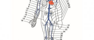
Types of hypoplasia of the left side and the mechanism of blood movement
The first variant of the anomaly is a slit-like left ventricle, not exceeding 1 mm, with fusion of the openings of the aorta and mitral valve . This is the most severe type of defect, in which blood from the left atrium cannot pass into the ventricle; it is directed through the open opening of the septum to the right half and mixes with the venous one.
The mixed flow goes to the lungs, and partly through the Botallian duct passes into the aorta, some blood enters the coronary arteries.
The second type (more common) - the valve openings are narrowed, but function, and the ventricular capacity is 2 - 5 ml. With it, the blood movement is similar, but it is more compatible with life. Both types of hypoplasia are characterized by the presence of:
- dilated ductus Botallova;
- open opening between the atria;
- hypertrophied and expanded right chambers;
- increased diameter of the pulmonary trunk.
Variants of underdevelopment of the right departments
Hypoplasia of the ventricle and its valve apparatus can be of the following types:
- only the ventricle - all its parts are reduced;
- pulmonary stenosis or valve fusion;
- fusion of the tricuspid valve.
The right atrium is dilated and its myocardium is thicker than normal, the left ventricle is hypertrophied, and pulmonary blood flow is reduced.
Venous blood cannot enter the right ventricle, so it passes into the left atrium and arterial network through the foramen ovale. Moreover, until a certain period (several days or years) such a discharge may not detect itself, then, with an increase in the resistance of the right half of the heart, the portion of blood with a low oxygen content in the arteries increases, which is accompanied by cyanosis.
Symptoms of the syndrome in the fetus and newborns
Signs of hypoplasia are most often detected by ultrasound of the fetal heart. Indications for its implementation may include signs of developmental delay or abnormal heart rate. Cerebral and coronary circulation in the prenatal period is provided by the ductus arteriosus, so fetal death does not occur, but a third of children have abnormalities in the structure of the brain.
With left ventricular hypoplasia after childbirth, blood pressure and oxygen levels in the blood drop, and pulmonary hypertension increases. This shows up:
- weak motor activity;
- gray skin color;
- low body temperature;
- rapid breathing and heartbeat;
- increasing cyanosis;
- wheezing in the lungs;
- weakened pulsation of peripheral arteries;
- enlarged liver;
- swelling of the limbs;
- decreased urine output.
The brain and heart muscle suffer most from the lack of blood supply, which leads to ischemia of these organs and death when the functioning of the ductus Botallova stops.
Expert opinion
Alena Ariko
Expert in Cardiology
Cyanosis with underdevelopment of the right ventricle appears in the first hours of life or occurs after a few years (by 14 - 15 years). This is determined by the amount of venous blood discharge. Unlike left ventricular hypoplasia, the period of intrauterine development can proceed without deviations; the entire load falls on the left ventricle, which fully compensates for the failure of the right one.
Children with right-sided hypoplasia are characterized by the following symptoms:
- difficulty breathing during exercise and at rest,
- pain in the heart area,
- attacks of suffocation,
- fingers and nails of a “pulmonary patient” – “drumsticks, watch glasses.”
When palpating and listening to the heart, there may be no changes or a slight noise may occur during the relaxation phase. The course of the disease depends on the severity of circulatory failure - increasing shortness of breath, wheezing in the lungs, cough, hepatomegaly.
Watch the video about congenital heart defects:
Features of manifestations
Hypoplastic right heart syndrome is most often diagnosed in children born at term. With this pathology, blood flow to the lungs is reduced, which is why it is not enriched with oxygen in sufficient quantities. Typically, the deviation does not occur in an isolated form, but is combined with other disorders in the development of internal organs. With such a deviation:
- muscle weakness and a sharp loss of strength are observed;
- the skin takes on a gray tint;
- shallow breathing becomes more frequent;
- heart rate increases;
- body temperature drops below the designated norm;
- wheezing is heard in the lungs;
- A paroxysmal cough occurs, which indicates the development of heart failure.
Immediately after birth, a slight blue discoloration of the skin is observed, but gradually the cyanosis intensifies and concentrates in the lower part of the body. There is a decrease in peripheral pulsation in the extremities, and they are cold to the touch.
The heart cannot function fully from the first days of life. This causes the liver to enlarge and fluid to accumulate in certain areas of the body. The amount of urine excreted also decreases, urine does not enter the bladder, and the acid-base balance shifts toward increased acidity.
A genetic syndrome with hypoplasia of the corpus callosum is possible; ischemia develops in the heart muscle and brain tissue.
All these pathological changes pose a serious danger to the child’s life. Very often the newborn does not have time to save.
Also read: Closure of atrial septal defect - surgery
What is the danger of hypoplasia of the left and right parts of the heart?
Patients with left ventricular dysplasia are called ductus-dependent (ductus-duct). This means that their life expectancy depends entirely on the open ductus arteriosus; when blood flow through it stops, death occurs.
The clinical picture consists of a severe form of cardiac and vascular failure. With underdevelopment of the right half of the heart, congestive heart failure progresses with the accumulation of fluid in the abdominal and thoracic cavity, and with decompensation, pulmonary edema develops.
Diagnosis of the condition
In most cases, fetal echocardiography reveals hypoplasia of parts of the heart - narrowing or fusion of the valve openings, a decrease in the size of the ventricles, stenosis of the aorta or pulmonary arteries. In this case, the birth takes place in neonatal centers, where immediately after the birth of the child, additional examination is carried out.
In case of underdevelopment of the left sections, instrumental diagnostic data are taken into account:
- ECG – deviation of the heart axis to the right, intense hypertrophy of the right half of the heart;
- X-ray – cardiomegaly, heart in the form of a ball, increased pattern in the lung fields;
- EchoCG - narrowing of the aorta, left ventricle, stenosis or fusion of the mitral valve;
- cardiac probing - decreased oxygen content in the blood of the arteries, discharge between the atria (from left to right), high pressure in the right chambers of the heart;
- angiography – open duct, dilated right section.
To confirm the diagnosis of hypoplasia of the right half of the heart, important signs are:
- ECG – axis deviation to the left, high atrial waves in the right leads, combined hypertrophy of both atria and the left ventricle;
- EchoCG - reduction in the size of the right ventricle and narrowing (fusion) of the opening of the tricuspid valve;
- radiography - decreased cardiac pattern, enlarged atria and left ventricle, displacement of the pulmonary trunk;
- catheterization - high pressure in the right atrium and the pressure difference between it and the right ventricle, decreased oxygen content in the aorta;
- angiography - low filling of the right ventricle; when the cavity is reduced to 80% of normal, hypoplasia is considered severe.
Causes
The causes of hypoplastic left heart syndrome is a violation of the embryonic development of the fetus, as a result of which the heart and blood vessels are pathologically altered and cannot function normally. The exact etiological factors that could influence cardiac underdevelopment are not yet clear. It is assumed that, as in the case of other congenital heart defects, teratogenic factors have a negative effect on embryogenesis:
- receiving x-ray and radiation exposure during pregnancy; smoking, drug addiction, drinking alcohol; taking medications that interfere with the normal development of the fetus; past infections during gestation - rubella, influenza, herpes, toxoplasmosis, cytomegalovirus, etc.
It is believed that gene mutations may also play a role in the occurrence of hypoplastic right heart syndrome. In some cases, the role of heredity is also noted: if the mother has any congenital heart disease, this can provoke the development of diseases of this group in the child in approximately 4-5% of cases.
Treatment of the syndrome
A newborn with underdevelopment of the left half of the heart is transferred to intensive care, where Alprostan is immediately administered to prevent the ductus arteriosus from closing, artificial ventilation is given, the acid-base state of the blood is normalized, and heart contractility is supported with Dobutamine.
Subsequently, surgical treatment is indicated. The first stage takes place 13 - 15 days after the birth of the child - a connection is created between the aorta (subclavian artery) and the pulmonary trunk to relieve the right half of the heart. Subsequently (at 3–5 months of age), an anastomosis of the superior venta cava and the pulmonary trunk is established; it initially circulates in two directions, and by the year the blood circulation is completely separated.
In case of underdevelopment of the right ventricle and in a critical condition of the newborn, a temporary measure is to widen the hole in the interatrial septum. Then, up to a year, an aortic-pulmonary connection is created. After five years, an anastomosis of the vena cava and pulmonary artery is formed
.
Treatment Options
If such pathological disorders are detected, the newborn is placed in the intensive care unit, where his condition will be monitored.
To prevent the arterial duct from closing or to achieve its opening, prostaglandin E1 is administered by infusion. It is also impossible to do without artificial ventilation of the lungs. Drugs are prescribed that should normalize the balance of acids and alkalis, remove excess fluid from the body, and strengthen the contractility of the heart muscle.
It is important to undergo surgery if the left side of the heart is not sufficiently developed. This procedure is performed in several stages.
First, they resort to palliative methods, which are used during the first two weeks after birth. This therapy helps reduce the level of stress on the pulmonary artery and ensures normal blood flow into the aorta. This significantly improves the patient's condition.
After this, when the child is three months old or more, a bilateral bidirectional cavapulmonary anastomosis is performed.
To completely eliminate the syndrome of hypoplasia of the left sections, as well as to stabilize the circulatory process, a total cavopulmonary anastomosis is applied after a year. With its help, the blood flow circles are completely disconnected and hemodynamic disturbances are eliminated. This increases your chances of survival.