Infographics for the “bio/mol/text” competition: A clinical blood test is the most common laboratory test prescribed by a doctor when we come to an appointment and complain of feeling unwell. “Blood from a finger, tomorrow from 8:00 to 9:30, on an empty stomach, Nth office,” several generations have invariably heard this phrase. However, blood testing technology has undergone great changes in recent decades and has moved from manual methods to automatic ones. Let's figure out how your grandmother's blood was analyzed and why things are done differently now.
Blood composition
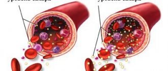
About 60% of blood is plasma, the liquid part. Red blood cells, white blood cells and platelets make up 40%.
The thick viscous liquid (blood plasma) contains substances necessary for the body's functioning. These beneficial substances, moving to organs and tissues, ensure the chemical reaction of the body and the activity of the entire nervous system. Hormones produced by the endocrine glands enter the plasma and are carried through the bloodstream. Plasma also contains enzymes - antibodies that protect the body from infection.
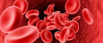
Erythrocytes (red blood cells) are the main mass of blood elements that determine its color.
The structure of the red blood cell resembles the thinnest sponge, the pores of which are clogged with hemoglobin. Each red blood cell carries 267 million molecules of this substance. The main property of hemoglobin is to freely absorb oxygen and carbon dioxide, combining with them, and, if necessary, freeing itself from them.
What will we explore?
Blood is a liquid connective tissue of the body, consisting of plasma and three types of formed elements: red blood cells, platelets and leukocytes. Leukocytes, in turn, come with granules in the cytoplasm - these are neutrophils, eosinophils and basophils - and without granules - lymphocytes and monocytes. In order to distinguish pathology from the norm, you need to know what the concentration of blood cells is, what they look like and what function they perform. It's time to remember who we are dealing with.
Figure 1. Formed elements of blood [1–3]
Figure 2. The figure shows: the concentrations of blood cells are normal; leukocyte formula - the percentage of different types of leukocytes in the blood; erythrocyte sedimentation rate; hemoglobin concentration; hematocrit is normal [4].
So, experts analyze the relative and absolute content of cells, their morphological characteristics, distribution over blood volume and many other parameters. These indicators can tell whether cells are able to fully perform their functions, and if not, then indicate the reason for their “inoperability” and serve as the basis for making a diagnosis.
Figure 3. Blood testing: then and now
Platelets
Blood platelets running near the walls of blood vessels. They act as if in the form of permanent repair teams that monitor the serviceability of the vessel walls. There are more than 500 thousand of these repairmen in every cubic millimeter. And in total there are more than one and a half trillion in the body.
The lifespan of a certain group of blood cells is strictly limited. For example, red blood cells live for about 100 days. The life of leukocytes ranges from several days to several decades. Platelets live the shortest. They only exist for 4-7 days.
Together with the blood flow, all these elements move freely throughout the circulatory system. Where the body keeps a measured blood flow in reserve - this is in the liver, spleen and subcutaneous tissue, these elements can stay here longer.
Each of these travelers has their own specific start and finish. They cannot avoid these two stops under any circumstances. The beginning of their journey is also where the cell dies.
It is known that a larger number of blood elements begin their journey leaving the bone marrow, some begin in the spleen or lymph nodes. They end their journey in the liver, some in the bone marrow or spleen.
Within a second, about 10 million red blood cells are born, and the same amount falls on dead cells. This means that construction work in the circulatory system of our body does not stop for a second.
The number of such red blood cells can reach up to 200 billion per day. In this case, the substances that make up the dying cells are processed and used again when recreating new cells.
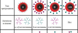
Blood groups
Transfusing blood from an animal to a higher being, from person to person, scientists observed such a pattern that very often the patient to whom the blood is transfused dies or severe complications appear.
With the discovery of blood groups by the Viennese physician K. Landsteiner, it became clear why in some cases blood transfusion is successful, but in others it leads to dire consequences. A Viennese doctor first discovered that the plasma of some people is capable of gluing together the red blood cells of other people. This phenomenon is called isohemagglutination.
It is based on the presence of antigens, named in Latin capital letters AB, and in plasma (natural antibodies) called a b. Agglutination of red blood cells is observed only when A and a, B and b meet.
It is known that natural antibodies have two connection centers, therefore one agglutinin molecule can create a bridge between two red blood cells. While an individual red blood cell, with the help of agglutinins, can stick together with a neighboring red blood cell, resulting in the formation of a conglomerate of red blood cells.
It is not possible to have the same number of aglutinogens and agglutinins in the blood of one person, since in this case there would be massive clumping of red blood cells. This is in no way compatible with life. Only 4 blood groups are possible, that is, four compounds where the same agglutinins and agglutinogens do not intersect: I - ab, II - AB, III - Ba, IV-AB.
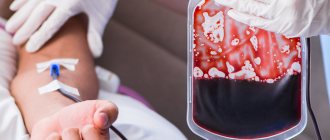
In order to make a blood transfusion from a donor to a patient, it is necessary to use this rule: the patient’s environment must be suitable for the existence of the donor’s red blood cells (the person giving the blood). This medium is called plasma. That is, in order to check the compatibility of the blood of the donor and the patient, it is necessary to combine the blood with the serum.
The first blood group is compatible with all blood groups. Therefore, a person with this blood type is a universal donor. At the same time, a person with the rarest blood type (fourth) cannot be a donor. It is called the universal recipient.
In everyday practice, doctors use another rule: blood transfusions only based on blood group compatibility. In other cases, if this blood group is not available, a transfusion of another blood group can be performed in a very small amount so that the blood can take root in the patient’s body.
Times change
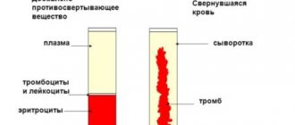
Changes are visible already at the stage of blood sampling: if previously the doctor collected blood in several test tubes with reagents, a glass capillary and made a smear on the glass, now very small volumes are used - from 12 to 150 μl [15] of blood is enough to examine it for all parameters.
Let's take a look into a modern hematology laboratory. Wow! Everything is cluttered with equipment, and the laboratory assistant is nowhere to be seen... Maybe he went off to make himself some coffee? He won't make it in time! The blood test will be ready in a minute, and the device will give the result in the form of a paper tape with numbers and abbreviations, behind which all sorts of parameters are hidden.
Modern hemoanalyzers are divided into several classes, depending on what they can do. Each subsequent class is a new stage of evolution - faster, more accurate, more perfect. The use of a combination of technologies works wonders: if the first analyzers could determine eight blood parameters and did not distinguish between types of leukocytes [16], then the latest devices are able to differentiate up to seven populations of leukocytes [17] and in total examine more than 40 blood characteristics.
As Arthur C. Clarke said, “Any sufficiently advanced technology is indistinguishable from magic.” Indeed, the most detailed results in such a short period of time cannot but surprise. But all magic is based on physical laws. And although names like electrical impedance, light scattering and photometry are a little intimidating at first glance, now we will understand what principles underlie each analysis technology.
Population census
In the middle of the last century, Wallace Coulter made a revolution by patenting the technology of automatic cell counting. One of the leaders in the production of hematology analyzers, the Beckman Coulter company, is named after him [18]. The aperture-impedance method (or Coulter method) is based on the registration and analysis of impulses that occur when a cell passes through an aperture from one container to another, each of which contains an electrode. When there is no cell in the hole, current flows freely through the electrolyte between the electrodes under the influence of an electric field. To direct the cells to the aperture, a pump is used that pumps out liquid from one container, and the formed elements rush into it. Passing through the aperture, the cell displaces a volume of electrolyte equal to its volume from one container to another. In this case, a pulsed change in resistance (impedance) occurs - the cell membrane creates an obstacle to the free flow of current. At the same time, the current strength recorded by the meter also changes. The number of generated impulses corresponds to the number of formed elements, and the height of the impulse is proportional to the volume of the cell [19]. Using information about the number and volume of formed elements, the device can calculate hematocrit, the average concentration of hemoglobin in an erythrocyte, the width of the distribution of cells by volume and many other parameters [15].
Divide and rule
Differentiation of leukocytes into populations can be carried out using a Coulter counter, but a problem arises - different types of leukocytes are similar in volume and similar pulse amplitude does not always allow one to accurately determine the cell type. What should I do? To solve this problem, combinations of reagents are selected that change the size of cells to varying degrees so that it becomes possible to separate them [15].
But the most common method of differentiation is flow cytometry [20]. The method works as follows: cells in the flow are alternately irradiated with a laser, and the resulting light scattering and fluorescence signals are recorded by detectors and analyzed. In order to correctly determine membership in a population, several parameters are examined at once. Thus, light scattering at a small angle provides information about the relative size of cells, and light scattering at a right angle allows you to “look” inside the cell and study its internal structure - the presence of granules and the shape of the nucleus. Another parameter - fluorescence - can tell about the number of antigens and their appearance on the surface of cells - this cannot be accurately determined by eye. Unlike manual differentiation methods, not 100–200 cells are analyzed, but tens of thousands per second! And each leukocyte receives an individual approach: hydrodynamic focusing ensures that the cells line up in a row and are irradiated one by one in the flow cell. The result of the count appears on the screen in the form of scatter diagrams, where cells with similar properties form clusters.
Precipitated: 2.0
Modern instruments can measure ESR in two fundamentally different ways. The first is the modified Westergren method. The principle of operation has not changed since your grandmother's time, but due to automation it has become faster and more accurate. The second is measuring the kinetics of erythrocyte aggregation using an optical method [21]. It happens like this: an anticoagulant is added to the blood, the tubes with blood are placed in a rotor, where automatic mixing occurs. After this, the analyzer takes part of the blood into a microcapillary, where it accelerates and abruptly stops (the so-called “stopped stream” method). The stop causes aggregation of red blood cells, and at this moment the optical density of the blood is determined using a photometer - the denser the red blood cells are located, the less light will pass through the sample. The device uses the data obtained and builds a sedimentation curve - its analysis will allow you to present the result in the usual units of ESR measurement [22], [23].
Photo for memory
To determine hemoglobin concentration, the International Committee for Standardization in Hematology recommends the methemoglobin-cyanide method. However, a different test that does not use toxic cyanide is now widely used. Meet the SLS method. It is named after the main reagent - sodium laurithyl sulfate. SLS destroys red blood cell membranes, after which it binds to heme groups and forms stable complex compounds. They are analyzed photometrically - laser light is passed through the blood sample. Complex compounds absorb part of the light, as a result of which the intensity of the output light flux weakens. Attenuation is measured using a photosensor and the resulting data is converted into hemoglobin concentration units [24].
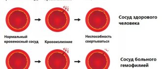
Rh factor
Famous doctors K. Landsteiner and A. Winner, during an experiment on monkeys, discovered an antigen in her, which today is called the Rh factor. Upon further research, it turned out that this antigen is found in the majority of people of the white race, that is, more than 85%.
Such people are noted to be Rh positive (Rh+). Almost 15% of people are Rhesus negative (Rh-).
The Rh system does not have agglutinins of the same name, but they can appear if a person with a negative factor is transfused with Rh-positive blood.
The Rh factor is determined by inheritance. If a woman with a positive Rh factor gives birth to a man with a negative Rh factor, then the child will receive 90% of the father's Rh factor. In this case, the Rh incompatibility of mother and fetus is 100%.
Such incompatibility can lead to complications in pregnancy. In this case, not only the mother suffers, but also the fetus. In such cases, premature birth and miscarriages are not uncommon.
Competition "bio/mol/text"-2019
This work was published in the category “Visually about the beloved” of the “bio/mol/text” competition-2019.
The general sponsor of the competition and partner of the Skoltech nomination is the Skoltech Center for Life Sciences.
Competition sponsor: the largest supplier of equipment, reagents and consumables for biological research and production.
The audience award was sponsored by BioVitrum.
"Book" sponsor of the competition - "Alpina Non-Fiction"
Morbidity by blood type
People with different blood groups are susceptible to certain diseases. For example, a person with the first blood group is susceptible to peptic ulcers of the stomach and duodenum, gastritis, and bile disease.
Diabetes mellitus is very often and more difficult for individuals with the second blood group. In such people, blood clotting is significantly increased, which leads to myocardial infarction and stroke. If you follow the statistics, such people experience genital cancer and stomach cancer.
Persons with the third blood group suffer more than others from colon cancer. Moreover, people with the first and fourth blood groups have a hard time with smallpox, but are less susceptible to plague pathogens.
Blood of a different color in various living creatures
Not all living organisms have red blood.
The protein that gives this color in humans is hemoglobin, contained in hemoglobin. Other living beings have other fat-containing proteins instead of hemoglobin.
The most common shades besides red are:
- Blue. Crustaceans, spiders, mollusks, octopuses and squids boast this color. And blue blood is of great importance for these creatures, as it is filled with important elements. Instead of hemoglobin, it contains hemocyanin, which contains copper.
- Violet. This color is found in marine invertebrates and some mollusks. Typically, such blood is not only purple, but also slightly pink. The blood of young invertebrate organisms is pink. In this case, the protein is hemerythrin.
- Green. Found in annelids and leeches. The protein chlorocruorin is close to hemoglobin. However, iron in this case is not oxide, but ferrous.
The color of blood varies depending on the protein it contains. Whatever the color of blood, it contains a huge amount of useful substances necessary for a living organism. Pigment is important for every organism, despite its diversity.