Disorders of the hematopoietic system are accompanied by critically dangerous symptoms. The overwhelming majority of these disorders pose a mortal threat to the patient and, without timely treatment, take their lives quickly and without any chance of help.
These individuals are treated by hematology specialists. It is possible to attract related doctors (vascular surgeons) as needed.
Hemorrhagic syndrome is a generalized name for a group of symptoms typical of pathologies of the hematopoietic system, accompanied by the formation of small red hemorrhage spots on the skin and in its structures. They represent massive or minor hemorrhages: internal, nasal and others.
This is not an independent diagnosis, but a group of manifestations, that is, a syndrome. Therefore, it is necessary to fight not only and not so much with hemorrhagic manifestations, but with the main pathological process.
The problem is solved strictly in a hospital setting. Mild cases can be treated on an outpatient basis, but are less common. The prognosis depends on the quality and timeliness of the therapy.
Development mechanism
The formation of a pathological process is based on a group of provoking factors. Generally speaking, the following points can be mentioned:
- Genetic abnormalities. They occur relatively often. They account for almost 25% of the total clinical situations. The classic version is hemophilia: an incurable disease in which normal blood clotting is disrupted.
It makes sense to examine the patient for genetic abnormalities, although this is not always possible due to the complexity of diagnosis. Treatment in this case comes down to correction of symptoms; etiotropic effects are impossible for obvious reasons.
- Coagulation factor disorders. There is a whole group of pathologies that are typically characterized by a disruption in the production of special substances responsible for coagulation. These include von Willebrand's disease, disseminated intravascular coagulation syndrome and other similar disorders, including the above-mentioned hemophilia.
Attention:
Diseases of this kind are difficult to correct or cannot be corrected at all. Symptoms are also difficult to relieve.
- Pathologies and inferiority of the vessels themselves, capillaries and larger structures. Increased permeability, fragility. Occurs as a result of congenital hereditary anomalies or acquired problems. For example, with a lack of vitamins B and C, the normal structure of blood vessels is disrupted, and massive bleeding occurs, including fatal ones.
- Other factors that are brought to life by the patient himself or the environment. As a rule, they are transient and can be easily corrected on their own. If there is an understanding of what was the immediate cause of the problem.
Mixed variants of pathogenesis are also often found. In this case, you need to influence all provoking factors in parallel.
Violations of normal coagulation or problems with the structure of blood vessels, their integrity, strength, elasticity, physical properties lead to a pathological disorder of hemostasis.
The blood does not stop on its own, or the capillaries or other structures begin to collapse in large quantities, causing the formation of a specific rash and the release of liquid connective tissue into the joints. Severe disability, emergency conditions, and death are possible.
The pathogenesis of the hemorrhagic process is multiple, caused by congenital or acquired disorders of coagulation and vascular structure.
If you detect at least some symptoms reminiscent of hemorrhagic syndrome, you should immediately consult a doctor.
Hemorrhagic diathesis in children
Due to a lack of vitamin K, hemorrhagic syndrome of newborns can develop, among the signs of which are: hemorrhagic skin rashes, umbilical bleeding. Intestinal bleeding or intracerebral hemorrhage may occur. Doctors name the following reasons for the occurrence of hemorrhages in newborns: during pregnancy, the mother took phenobarbitals, salicylates or antibiotics. Hemorrhagic disease in children occurs when:
- neoplastic lesions of connective tissue;
- thrombocytopenia;
- coagulopathies;
- vasopathy;
- hemophilia.
Classification
The division is carried out according to a group of bases. The first and most important thing that is used in clinical practice is the dynamics of the disorder.
Accordingly they are called:
- Angiomatous form. Accompanied by severe bleeding at the site of the lesion. A typical feature of this type of disease is focal destruction of blood vessels, without involvement of neighboring areas. However, the nature of the leakage of liquid connective tissue makes angiomatous hemorrhagic syndrome deadly, since massive internal bleeding is likely (if a large vessel is affected).
- Hematoma variant. Reminiscent of the previous one, however, much more formidable in terms of prognosis. Develops against the background of DIC syndrome, in the hypocoagulation phase, also hemophilia, in the acute period. Symptoms include the development of massive, profuse hemorrhages in the tissue.
Joints, organs, and brain suffer. Without systematic treatment aimed at preventing such a scenario, a person’s death from complications is likely. It is the matter of time.
- Vasculitic purpuric form. Accompanied by the formation of small hemorrhages on the skin of a purple or dark red hue. The petechial rash itself, passing on its own, leaves pigmented areas and cosmetic defects.
This form does not pose any great danger. However, it develops as a result of infectious inflammatory processes affecting blood vessels. Therefore, it is quite possible for hemorrhage to transform into disseminated intravascular coagulation syndrome; this is already a deadly disorder.
- Petechial type. The classic form of the pathological process. Accompanied by massive rashes in the form of small circles 2-5 mm in diameter. Dark purple or deep red.
Recovery is not possible because a number of clotting factors are involved. As part of symptomatic correction, replacement drugs are used.
This form of pathology behaves unpredictably, therefore it is extremely difficult to predict its course.
- Mixed type. For him, the development of a group of manifestations is pathognomonic. On the other hand, there is also a feature characteristic only of this form. Despite hemorrhages in organs, tissues, and the formation of a rash, the joints are practically not involved.
There are also atypical, rarely encountered species. For example, thrombohemorrhagic syndrome, which is characterized by alternating episodes of hypocoagulation and active, excessive production of coagulation factors with the formation of clots blocking blood vessels (thrombi).
Another way of classification is by the origin of the disease.
- Congenital appearance. Occurs from the moment a child is born. It has hereditary, genetic roots. It is practically not subject to restoration, not counting the most successful cases.
- Acquired form. For some pathologies: von Willebrand, disseminated intravascular coagulation and others. The prospects for therapy are different; anything can be said for sure after assessing the specific clinical situation.
According to the nature of the flow:
- Sharp look. A critical hemorrhagic condition occurs with severe hemorrhages, the formation of a skin rash, symptoms of general intoxication of the body, weakness, muscle and joint pain.
- Chronic form. It proceeds sluggishly, gives a minimum of clinical manifestations, but at any moment it can go into a critical phase. It is poorly diagnosed; as a rule, the patient does not see a doctor until the last moment. Possible accidental detection during a routine examination.
More detailed classifications exist, but they are used less frequently in clinical practice or are not considered generally accepted in medicine.
Types of bleeding
Based on the type of bleeding, a diagnosis can be made and treatment for the disease can be chosen. Therefore, it is customary to carefully consider external manifestations and the localization of skin symptoms.
Microcirculatory type (capillary, petechial-bruising) - looks like small pinpoint rashes (petechiae), larger ecchymoses, bruises on the skin and mucous membranes. Combined with nosebleeds and menstrual bleeding. Rarely, complications associated with hemorrhagic stroke and severe consequences such as paralysis are possible. Most typical type for:
- von Willebrand's disease;
- thrombocytopenia and thrombocytopathies (hereditary pathology often manifests itself as bruises rather than a rash);
- deficiency of factors II, V, VII, X;
- altered fibrinogen content;
- overdose of anticoagulants.
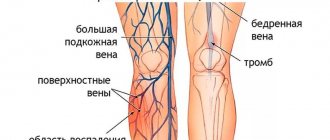
Hematoma - hemorrhages are more abundant, localized in the joint capsule, muscles, subcutaneous tissue, and peritoneum. Hematomas are always painful because they compress the nerve plexuses. During their development, they destroy cartilage and bone tissue, cause contracture of the limbs, and disrupt a person’s motor capabilities.
Possible manifestation of bleeding in the urine, from the stomach and intestines. They are characterized by prolonged bleeding during tooth extraction, cuts, bruises, and surgical operations. Observed when:
- hemophilia;
- acquired coagulopathies with the presence of clotting inhibitor factors in the blood;
- in case of overdose of anticoagulants;
- severe hereditary pathologies.
Mixed or capillary-hematoma - possible in the above conditions; clinical manifestations are also accompanied by damage to the mucous membranes, skin, joints and internal organs.
The vasculitic purpuric type of hemorrhage develops based on inflammation of the vascular wall. Accompanied by damage to the renal glomeruli and intestinal bleeding. Typical of viral fevers, immune vasculitis.
Angiomatous - in its localization it is distinguished by its attachment to the vascular wall. More often observed with:
- angiomas,
- telangiectasia,
- arteriovenous shunts.
Symptoms
The clinic depends on the specific form of hemorrhagic syndrome. It must be assessed accordingly.
Angiomatous variety
- Pain at the site of bleeding. Insignificant in character, weak. Aching, pulling. With a massive process, the phenomenon can be much stronger. Unstable in terms of current. They become pronounced when moving, changing body position, or physical activity.
- An outpouring of fluid connective tissue in a clearly localized area. This is a characteristic distinguishing feature of the angiomatous form from others. There is a delimited area of damage. However, it is not always possible to detect it by eye. Diagnosis required. The symptom is not felt subjectively.
- Swelling of the affected area. With a significant amount of blood released, an enlargement of the localized area occurs. How strong depends on the nature and intensity of the process.
- Weakness, drowsiness, lethargy.
With prolonged course, manifestations of anemia are possible. Skin problems, brittle hair, shortness of breath, heart disorders, exercise intolerance. The result of iron loss.
Hematoma variant
Much more dangerous. Can cause rapid death without treatment.
- Massive hemorrhages in the joints (hemarthrosis). The calling card of this type of violation. It is accompanied by a significant increase in the size of the cavity, compression of the cartilage and bursa, and problems with providing local structures with nutrients.
Motor activity disappears, as does the ability to use the affected limb. As a rule, hematoma hemorrhage involves large joints: knees, elbows, hips. Variations are possible.
- Severe pain at the site of the lesion. It intensifies when palpation is attempted. Movement, changing the position of the arm or leg is impossible due to intense discomfort and swelling. A puncture is required, drainage of the joint cavity and restoration of its anatomical shape and functions.
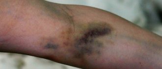
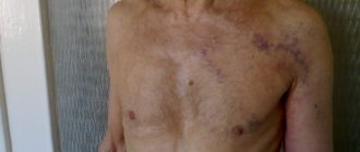
- Formation of large hematomas. On large areas of fabric. They appear as purple or violet, bluish, red areas under the skin. They protrude strongly above the surface of the dermis and change the external properties of the body. Urgent medical attention is required.
- Weakness, drowsiness. As a result of a decrease in hemoglobin concentration. With the development of bleeding in the area of internal organs, focal disorders are possible. For example, problems in the digestive tract, neurological deficits.
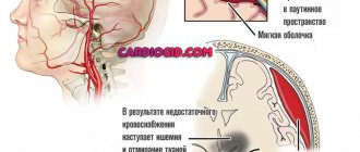
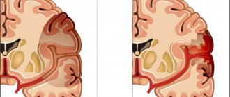
- Necrosis with long-term preservation of blood volume in tissues. As a result of ischemia (malnutrition) and compression of surrounding structures. The most dangerous complication. Possible regardless of the location of the process. In particular, with a cerebral hemorrhage (for example, subarachnoid), symptoms reminiscent of a major stroke cannot be avoided. Provided that the elimination of the consequences is not started in a timely manner.
Vasculitic purpuric form
- Massive rashes all over the body. They look like small spots of violet, lilac shades, possibly turning blue. They rarely exceed 1 cm in diameter. They are completely painless, not accompanied by itching, burning, or other discomfort, which distinguishes them from rashes due to dermatitis and skin diseases themselves. They pass independently without any deviations or special events.
- Weakness, weakness.
- Massive bleeding is possible: uterine, intestinal, pulmonary. Depends on the reason that brought the pathological process to life.
Dangerous complications are typical for this form. The likelihood of such happening is difficult to predict in advance. It all depends on the duration of the process, the main reason, and other factors.
Petechial type
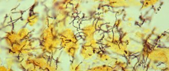
This variety is characterized by the development of a massive rash throughout the body, small red or scarlet spots. Up to 3 mm. Also, in addition to the dermal layers, they affect the mucous membranes. They can form, including on internal organs.
Danger to health and life is relatively rare. However, everything depends on the primary disease.
Mixed view
It manifests itself as hemorrhages in the tissue, as well as cutaneous hemorrhagic syndrome, the formation of spots, petechiae, and purpura. As stated, the development of articular lesions is atypical.
Forms of hemorrhages in newborns
The frequency of detection of hemorrhagic symptoms in newborns in the CIS is 0.25–1.5% of all children born. The manifestations are associated with the low activity of vitamin K, as an activator of other coagulation factors. This is confirmed by the effective administration of the vitamin to all children abroad. There, hemorrhagic disease is found in only 0.01% of births.
According to its form, it is customary to divide the disease into:
- primary - vitamin K penetrates weakly through the placenta in the fetus, in a newborn it begins to be produced by intestinal flora from 3-5 days, with a low content in breast milk, hemorrhages are possible;
- secondary - the blood clotting process is disrupted as a result of malabsorption syndrome (lack of absorption of nutrients in the small intestine), liver diseases, long-term parenteral (through solutions injected into a vein) nutrition.
According to the timing of the occurrence of hemorrhages, they are distinguished:
- early form - on the first or second day of life;
- the classic course is on the third to fifth day.
Late hemorrhagic disease of newborns occurs in infants aged from one to four months (more often in two months). It is observed much less frequently than early - from 5 to 20 cases per 100,000 births.
Causes
If we summarize the possible provoking factors, the following picture emerges:
- Disorders of coagulation processes. Congenital (much more often) and acquired. Hemophilia, other pathological conditions.
- Changes in the concentration of formed cells or their inferiority and inability to perform their own functions (thrombocytopenia - pathies). They occur quite often and can be transient, temporary phenomena.
- Vasculitis, inflammatory processes of blood vessels. Infectious or autoimmune forms.
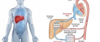
- Disorders of the liver and kidneys. Especially cirrhosis, hepatitis, failure. In this case, hemorrhagic syndrome is considered secondary.
- Tumors of various localizations. Regardless of the type. Typically malignant.
- Genetic abnormalities.
- Mixed diseases such as von Willebrand pathology, DIC syndrome. They have a multiple nature and are accompanied by unstable symptoms from both coagulation factors and the structure of blood vessels.
Types of hemorrhagic syndromes
Depending on the pathogenesis of bleeding, there are:
- Vasopathies accompanied by the secondary development of platelet disorders and coagulation disorders;
- Thrombocytopenia and thrombocytopathy;
- Coagulopathies associated with blood clotting disorders;
- Hemorrhagic diathesis caused by complex disorders of various parts of the hemostatic system;
- Hemorrhagic diathesis provoked by the patients themselves (artificial bleeding).
All vasopathies, coagulopathy, thrombocytopenia and thrombocytopathies, in turn, are divided into hereditary and acquired.
Diagnostics
The examination is carried out within the walls of the hospital, especially in acute cases. The main specialist is a hematologist.
Typical activities:
- Oral interview with the patient. It is necessary to identify and record all complaints for their further assessment and analysis.
- Anamnesis collection. As part of the initial examination, it is necessary to determine the likely origin of the anomalous process. The technique allows you to detect the nature of hemorrhagic syndrome.
- General blood test, biochemistry. Provides information on the composition and rheological properties of the fabric.
- A coagulogram is mandatory. Coagulation rate study. Provides an opportunity to recognize the existence of a problem.
- Ultrasound of internal organs, joints. As part of assessing the condition of anatomical structures, excluding latent hemorrhages.
It is possible to conduct genetic tests and analyzes as needed. If the doctor deems it necessary.
The list of events is incomplete, however, they are held most frequently.
Treatment
Therapy is designed to solve two problems: combating the root cause, correcting symptoms.
Symptomatic supervision. The key direction is implemented through procedures and the use of drugs:
- Glucocorticoids. Stop inflammatory processes, if present. Excellent for the treatment of a number of forms of thrombocytopathy.
- Immunosuppressants. As part of eliminating complex autoimmune conditions. In particular, vasculitis and others.
- Saline solutions. To restore water balance and electrolyte concentration.
- Heparins as needed. For blood thinning during thrombotic phases of DIC syndrome.
- Iron preparations. To eliminate anemia.
- Vitamin complexes. Ascorutin, medicines based on C, B.
If necessary, donor coagulation factor preparations are used, plasma and red blood cells are transfused.
Surgical treatment is performed if necessary. Drainage of hematomas, pumping out blood. These are symptomatic measures. Manifestations are eliminated with the help of replacement agents, also iron-based products. There are many options.
In the future, systematic examinations by a hematologist are indicated, at least 2 times a year. Some disorders do not respond to effective treatment. For example, hemophilia and a number of others.
Prevention
Prevention of vitamin K deficiency involves early breastfeeding, intramuscular or subcutaneous administration of Vikasol to premature infants and orally to full-term infants. In addition, preventive measures include the deliberate prescription of drugs that reduce the activity of the blood coagulation system (for example, heparin) to pregnant women, children and adults, which should be monitored by laboratory diagnostic data. One of the directions for the prevention and treatment of acute conditions, for example, with hemophilia, is to maintain the concentration of plasma factor at least 5%, which is achieved by periodic administration of a concentrated drug.
Similar syndromes: