Lymphangioma is a benign tumor of the lymphatic vessels in children aged 1-4 years. In the medical literature there are other names for this pathology - lymphohemangioma, lymphatic malformation. This disease rarely affects adults. Lymphangioma appears most often during fetal development. This is a collection of lymph nodes that appears as a result of improper arrangement of blood vessels. In other words, an anomaly of embryogenesis. A similar pathology begins to develop from the second month of fetal development. The diagnosis can be determined later - during the first, sometimes third year of life. The tumor can appear on the leg, arm, neck, and mucous membranes.
Types of disease
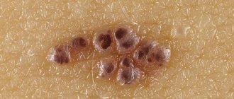
Based on their consistency, doctors distinguish between capillary, cystic and cavernous lymphangiomas.
Capillary or simple - pale nodules located under the skin.
They have a glassy coating. Such lymphangiomas are soft, located on the face, and are often accompanied by bleeding. Cystic lymphangioma is one or more chambered neoplasms, located mainly in the neck, armpits, and mediastinum. It is characterized by clear contours and slow growth. But it is dangerous because as it grows it compresses blood vessels, neighboring organs and nerves.
Cavernous is the most common tumor.
Consists of several cavities filled with liquid. Has a soft consistency. It is unpleasant because swelling forms at the site of the tumor and a cosmetic defect is created.
Lymphangioma in an adult
Lymphangioma is a congenital benign neoplasm that begins its growth from lymphatic vessels and is an elastic cavity with thick walls.
More often, this tumor is detected in childhood, and develops with equal frequency in both girls and boys aged 1-4 years. Sometimes these formations occur in adults. According to statistics, lymphangiomas account for 25% of all vascular neoplasms of childhood. This tumor itself is not dangerous, since it grows slowly and rarely malignizes. However, when it reaches a large size, the formation begins to compress the surrounding tissues and organs, causing disturbances in their functioning.
Why do lymphangiomas occur? What are they? What are the symptoms of this tumor? How is it detected and treated? The answers to these questions can be found in this article.
Causes
The formation of lymphangioma usually begins in the second month of intrauterine development, and its detection usually occurs by the end of the child’s first year of life, when the tumor increases in size.
So far, scientists cannot figure out the exact causes of the development of lymphangiomas. There are two speculative theories about the origin of the cancer in question.
A number of researchers suggest that the main cause of the formation of lymphangiomas is congenital pathologies in the development of blood and lymphatic vessels.
Improper formation of walls and accumulation of fluid in such altered lymphatic vessels leads to their overstretching and expansion. As a result, cavities are formed in their place, which are prone to growth.
This theory about the causes of congenital lymphangiomas is supported by data that with these formations, other congenital developmental anomalies are often detected in the child.
Sometimes a primary tumor can be detected even during ultrasound, which is routinely performed during pregnancy. Such tumors are usually located in the neck or in the fetal mediastinum.
If they are detected, the woman is recommended to conduct research to detect other malformations in the unborn child. If the lymphangioma is solitary and there are no other defects, then dynamic monitoring of the tumor is recommended for the expectant mother.
However, if multiple anomalies are detected, the doctor may raise the question of the need to terminate the pregnancy.
Other scientists suggest that lymphangiomas are true tumors. This theory arose on the basis of data on the staged course of the disease, when tumor cells first divide, and then growth factors and receptors for them appear on them. These processes are not typical for congenital developmental anomalies, and it is likely that secondary lymphangiomas in adults are formed precisely in this way.
In such cases, due to the presence of infection, inflammation or other neoplasm, after surgery or radiation, the flow of lymph in the lymphatic vessels is disrupted. As a result of its stagnation, the cells of the vessel begin to divide and form a conglomerate of dilated vessels or cavities filled with lymph.
Classification
Depending on their size, lymphangiomas are divided into:
- microcystic - no more than 5 cm;
- macrocystic - more than 5 cm.
Depending on the structure, experts distinguish the following types of lymphangiomas:
- Cavernous is a cavity that consists of connective tissue and is filled with lymph. These formations grow slowly, are soft in consistency and do not have clear outlines. When you press on the cavity, the tumor collapses, and after the pressure stops, it restores its shape. These lymphangiomas are usually located on the hand or forearm and appear as fluid-filled blisters that coalesce into large nodules. At the site of their appearance, swelling of the skin occurs. Cavernous lymphangiomas create a cosmetic defect and lead to deformation of the affected area of the body. This form of tumor is detected more often than other types of lymphangiomas.
- Capillary - looks like a swelling consisting of large or small glassy nodules. These lymphangiomas with blurred contours are localized on the face, have a soft consistency and are located in the soft tissues at different depths. The neoplasm consists of connective tissue and inclusions of lymphoid cells. The skin over them does not change or has a bluish or purple color. Neoplasms are prone to lymphorrhea and bleeding.
- Cystic - is a single- or multi-chamber cystic cavity with fluid. These tumors grow slowly, have clear contours, elastic walls, soft consistency and hemispherical shape. They do not adhere to the skin and do not cause pain. Through the stretched and thinned skin above them, blood vessels are visible. When palpating the neoplasm, fluctuation is determined. As lymphangiomas grow, they compress nearby vessels, nerves and organs. Most often they are located in the armpits, mediastinum and neck.
Symptoms
The nature and severity of lymphangiomas depends on their type, size and location.
Superficially located small tumors are usually only a cosmetic defect. Large formations can become a significant defect in appearance and lead to disruption of the functions of neighboring organs.
Lymphangiomas can be detected in different parts of the body: lips, face, tongue, behind the ears, armpits, mediastinum, abdominal cavity, etc. They are defined as a swelling over which the skin does not change color or becomes bluish (sometimes purple).
As the tumor grows, it compresses the surrounding tissues and the patient experiences corresponding symptoms. For example, if located close to the larynx, a large lymphangioma can cause dysphonia, difficulty breathing, shortness of breath, and swallowing problems.
In most cases, lymphangiomas grow slowly and may regress. Rapid growth of the tumor is caused by injury or infection of the tumor.
When lymphangioma tissue is damaged by infection, the neoplasm becomes dense and increases in size. Inflammation of these tumors occurs especially often in children 3-7 years old.
As a rule, such complications occur in the fall or spring.
They are caused by ARVI, tonsillitis, pulpitis, periodontitis, inflammatory processes in the intestines, stomach, or functional disorders of the digestive system.
The patient's temperature rises and signs of intoxication caused by suppuration appear (weakness, loss of appetite, etc.). The skin over the tumor turns red, and touching the tumor causes pain. Bleeding or lymphorrhea may occur due to injury.
Attempts to puncture and evacuate the purulent contents of lymphangiomas do not always lead to the elimination of suppuration, since not all tumor cavities can be drained, and the inflammatory process can recur.
In such cases, only surgical removal of the tumor helps to eliminate repeated suppuration.
Lymphangiomas of the upper lip
This type of lymphangioma has the appearance of a swelling without clear boundaries. They do not cause pain, lead to changes in the shape of the lips, widening of the red border of the lips and drooping of the corners of the mouth. Due to the spread to the nasolabial groove, the tumor smoothes it out.
Source: //venyvarikoz.ru/limfangioma-u-vzroslogo/
Leading clinics in Israel
Assuta
Israel, Tel Aviv
Ikhilov
Israel, Tel Aviv
Hadassah
Israel, Jerusalem
There is also mixed capillary-cavernous lymphangioma . It contains elements of various tissues: vascular, lymph, nervous, connective. Angioneuroma, angiofibroma, hemlymphangioma and others are neoplasms of a mixed type. Often diagnosed in adults.
Since lymphangioma is located on different parts of the body, therefore, the tumors differ in location. Let's look at some of them:
- Lymphangioma of the upper lip . Doesn't hurt when pressed, looks like a swelling, soft to the touch. The main sign of the presence of a tumor is the appearance of pus on the oral mucosa and along the edges of the lips.
- Lymphangioma in the orbital area . Characterized by swelling, then closing of the palpebral fissure. A large tumor leads to poor vision and blindness.
- Lymphangioma of the eye - often discovered when there is bleeding from it. The first signs of the disease are pain and inflammation of the conjunctiva.
- Lymphangioma of the brain . The growth of the tumor leads to compression of neighboring tissues and blood vessels. As a result, neurological symptoms appear - epilepsy, tinnitus, deterioration of vision and smell, etc.
- Lymphangioma of the abdominal cavity . It manifests itself as acute pain in the abdomen, a change in its size. When taking a general blood test, the tumor does not appear.
- Intestinal lymphangiomas . There are 3 stages: lymphangiomas, primary lymphangiectasia, lymphatic cysts. The most common form is lymphangiectasia. Characteristic signs are stagnation of lymph, increased blood pressure.
- Lymphangioma or mediastinal hemangioma is a tumor of the middle part of the thoracic cavity. It is least common in both adults and children.
- Lymphangioma of the neck . Differs in large sizes. Compresses breathing, swallowing, blood circulation.
- Lymphangioma of the tongue , leading to macroglossia, requires special attention This causes the tongue to become enlarged, leading to slurred speech and problems with chewing and swallowing.
Do not confuse lymphangiomas with swelling in the periorbicular area, i.e. on the face. This phenomenon occurs mainly in adults and the reason for this is fluid retention or a simple Botox injection.
Based on the time of appearance, lymphangiomas are divided into:
- Primary . Develop in the womb.
- Secondary . They are a consequence of past infections, surgeries, and radiation.
Both types increase as the child grows.
Types of lymphangioma by size:
- Microcystic - less than 50 mm.
- Macrocystic - from 50 mm and more.
Causes
Doctors do not know exactly why lymphangioma develops. Scientists are inclined to believe that an intrauterine defect in the formation of blood and lymphatic vessels causes the disease. This provokes an abnormal structure of the walls of blood vessels in which fluid accumulates. The vessels dilate and lymphangioma nodules form in them. This theory is confirmed by the primacy of the disease and intrauterine development together with other diseases.
There are a number of other factors that can cause the disease:
- Viral infections that affect a person for a long time;
- Previous surgery;
- Exposure to radioactive rays on the body in high concentrations;
- The presence of previous stagnation of lymph in the vessels - lymphostasis.
A primary pathology develops in a child in the womb. It is difficult to determine the presence of a tumor in a child’s body in the early stages. After many years of research, scientists have found that lymphangioma nodules appear in the first trimester of pregnancy. The disease can only be detected after the first year of life.
Symptoms
Signs of the presence of lymphangioma depend on the consistency of the neoplasm:
- A capillary tumor manifests itself as lumpy compactions in the affected area. Their depth may vary. Some tumors are located just under the top layer of skin, others can lie in the upper layers of muscle. With capillary lymphangioma, lymphorrhea occurs - the release of fluid due to rupture of blood vessels.
- The cavernous neoplasm has a soft composition. When pressing on the tumor, fluctuation of the internal fluid is noticeable, i.e. sensation of a wave upon palpation.
- In cystic lymphangioma, fluctuation is especially pronounced. The skin in the affected area is stretched and thinned. Often, with a cystic tumor, compression of nerves and blood vessels occurs.
Classification
The basis for the classification of lymphatic malformation is the nature of the clinical picture, appearance and structural features of the tumors. Taking into account the combination of all these factors, lymphangiomas are divided into capillary, cystic, cavernous.
Capillary (simple) have the appearance of pale colored small and large nodules with a glassy surface. The epidermis affected by the tumor resembles an orange peel due to its uneven granularity. The structure of capillary lymphangiomas includes large cavities containing lymph. Their thin inner walls are lined with a layer of endothelium. When pressed, the tumors are easily compressed, but over time the formation of connective tissue makes them dense.
Cystic lymphangiomas in children and adults consist of one or more chambers (cysts). They are filled with lymphatic fluid and do not communicate with each other. All cystic tumors are particularly dense. With small formations, the skin is not changed. With large ones, it becomes thinner, small vessels become visible.
The structure of cavernous type lymphangioma is characterized by the presence of several cavities. Each of them is filled with lymphatic fluid, and unevenly: some are overflowing, others remain almost empty. The base of the cavities is connective tissue, spongy, consisting of bundles of smooth muscle fibers, small lymphatic vessels and an elastic frame. The boundaries of cavernous lymphangiomas are clearly defined. If the neoplasm tissues are characterized by diffusion growth, their edges are blurred. When pressed, the tumor decreases in size. After the compression stops, it fills the cavity again.
Depending on the size, lymphangiomas are macrocystic (over 5 cm) and microcystic (not exceeding 5 cm). Based on the degree of growth, diffuse and limited types of tumors are distinguished.
Diagnostics. Prevention and treatment
The presence of lymphangioma is checked by palpating and examining the location of the tumor and the patient’s symptoms. If in doubt, the following diagnostic methods are prescribed: ultrasound, MRI, CT, x-ray lymphography. For a complete diagnosis, tumor tissue histology is used.
The classic treatment for lymphangioma remains surgical. If possible, the tumor is excised completely, sometimes partially. Indications for surgical intervention will be:
- Rapid tumor growth.
- Close proximity to important organs.
- Tumors affecting activities of daily living.
- Presence of branches.
Surgery can be laser, electrocoagulation, radio wave, cryosurgical.
The operation may be complicated by lymphorrhea. In this case, repeated surgery is performed. According to statistics, for about 75% of patients one operation is enough. Due to the low success rates of surgery, scientists are still looking for more effective ways to treat lymphangioma. Among them is sclerotherapy. Consists of administering a streptococcal antibody. Sclerosing agents - ethyl alcohol, reduced quinine - are injected into the affected area. The drugs connect blood vessels to each other and form connective tissue. In most cases, patients make a full recovery. Sometimes cosmetic surgery is required to remove the remaining scar.
Another modern treatment method is the drug Picibanil (OK-432). Has the least number of side effects. The administration of the drug significantly reduces the tumor. Treatment does not require a long hospital stay. Several injections are usually required to completely remove the tumor. The big disadvantage of pinitsibal is that it is not yet registered in many countries. Therefore, for treatment you have to go to foreign clinics.
Microwave therapy - heating the lymph to 42 degrees. In this case, tissue sclerosis and subsequent destruction of the tumor occur. To enhance the effect, a sclerosing drug is introduced. After surgery, cosmetic corrections are performed to remove the scar.
Would you like to receive an estimate for treatment?
*Only upon receipt of data on the patient’s disease, a representative of the clinic will be able to calculate an accurate estimate for treatment.
What is lymphangioma?
Lymphangiomatosis is quite rare. The disease manifests itself as a benign tumor. The nature of the disease is a failure in the formation of the lymphatic system. Lymphangioma develops against the background of impaired outflow of lymph and its accumulation. As a result, the vessels dilate and form cavities.
Capillary
Capillary (or simple) lymphangioma looks like a swelling. It can reach both small and large sizes.
The neoplasm has the following characteristics:
- pale colored;
- the surface resembles glass;
- blurred contours;
- easy to press during palpation;
- becomes dense over time (due to the presence of connective tissue in the tumor);
- resembles an orange peel (due to the layering of vessels overflowing with lymphatic fluid);
- has cavities in the structure;
- affects various levels of soft tissue;
- may cause lymphorrhea and bleeding.
Cystic
Cystic lymphangioma is a tumor-like neoplasm, which has the following features:
- tumor size fluctuates;
- cysts with lymph are connected to each other or separated;
- clearly marked tumor boundaries;
- elasticity of walls;
- softness of structure;
- hemispherical shape;
- slow development;
- reduced growth rate;
- no pain;
- the presence of translucent skin (vessels are visible);
- the presence of fluctuations (indicates lymph in a separate cavity with elastic walls).
Cavernous
Cavernous lymphangioma is considered the most common type. The pathology is presented in the form of a cavity with lymph. It is formed by connective tissue fibers.
This type of tumor is characterized by:
- simultaneous presence of several cavities;
- selective filling of chambers with lymph;
- slow tumor growth;
- tumor decline during palpation and recovery after mechanical action;
- soft structure;
- absence of discernible contours against the background of tumor growth (relevant in the case of multiple cell divisions; often the boundaries are clearly defined);
- the presence of swelling of the skin;
- modification of the affected area.
Tumor localization
Based on their location, it is customary to distinguish the following groups of neoplasms:
- lymphangioma of the face, brain;
- lymphangioma of the neck;
- tongue, upper lip, cheek;
- eye sockets, eyelids and conjunctiva;
- lymphangioma of the abdominal cavity, chest wall, armpit area;
- lymphangioma of the spleen.
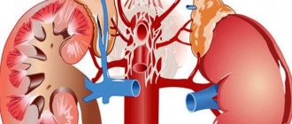
Damage to the kidneys, liver, spleen and foot is quite rare. There are unique cases of neoplasms in the groin, under the knee, and retroperitoneal space.
A tumor on the face is present in the form of local tissue enlargement. A clear liquid may be released.
A neoplasm in the neck area is characterized by an impressive size. Vessels, trachea, and esophagus may be compressed. There is respiratory dysfunction, inability to swallow, and disruption of the blood circulation.
The swelling of the upper lip looks like a lump. It is accompanied by:
- smoothing the nasolabial furrow;
- painlessness;
- lack of contours;
- lip enlargement;
- thickening of the labial edging;
- changing the shape of the mouth.
The neoplasm in the cheek area swells so much that it appears on the surface. The mucous membrane is covered with bubbles.
A tumor in the area of the right or left orbit is characterized by thickening of the eyelids, swelling, reduction or closure of the eye slit. When the size is large, the eye protrudes. The parameters of the eyeball are preserved, but visual function worsens.
A neoplasm on the tongue provokes the appearance of many bubbles. They are able to merge. As a result, it forms a knot. The size of the tongue increases. It practically does not fit in the oral cavity. There are problems with chewing, swallowing and coherent speech.
A tumor in part of the brain causes neurological disorders. Paralysis, paresis, seizures, loss of coordination, nystagmus, ataxia, and tinnitus may occur.
A neoplasm in the abdominal cavity is marked by pain and an asymmetrical appearance of the abdomen.
Reasons for development
According to the nature of formation, lymphangiomatosis is:
- primary (or congenital);
- secondary.
The causes of the congenital appearance lie in the pathological connection of lymphatic and blood vessels. The disease develops in the prenatal period. In this way, lymphangioma is diagnosed in newborns. Its appearance is influenced by hereditary predisposition and adverse external effects that occur during pregnancy.
The secondary type develops as a result of previously suffered infectious diseases or an inflammatory process. Neoplasms arise as a consequence of lymphostasis. This lymphangioma is often present in adult patients.
Symptoms of the disease
Lymphangiomatosis is characterized by the following symptoms:
- compaction in the area of skin lesions;
- there is pronounced swelling;
- contours and boundaries are visible (may be blurred);
- there is no significant change in the skin over the affected area (the cover may slightly stretch and become thinner);
- there is a varying degree of penetration depth;
- soft structure of the tumor (may become denser);
- Bleeding and discharge of lymphatic fluid may occur.
The symptoms of the disease are determined by the location and type of tumor.
Diagnostic measures
Diagnosis of a disease such as lymphangiomatosis includes:
- visual examination of the patient;
- study of medical history and anamnesis data;
- palpation;
- instrumental research.
The instrumental technique involves:
- ultrasound examination (ultrasound);
- magnetic resonance imaging (MRI);
- computed tomography (CT);
- X-ray lymphography (the anatomy of the neoplasm and the nuances of its location are determined);
- histological examination of tumor tissue (carried out after tumor removal; helps to definitively diagnose the disease);
- tumor puncture.
Based on the contents of the neoplasm, the following is revealed:
- form of pathology;
- presence or absence of an inflammatory process;
- damage to borders.
Lymphangioma in children is additionally examined using echography. This method determines the intensity of blood flow.
Treatment in adults and children
Identified lymphangioma requires immediate treatment.
Among the existing methods of influencing lymphangiomatosis there are:
- surgical intervention;
- puncture of the tumor and suction of the contents;
- electrocoagulation procedure;
- sclerosing therapy (medicines are administered that provoke gluing of vascular walls);
- immunotherapy (the use of drugs to stimulate lymphatic drainage and level out tumor growth);
- combined treatment;
- chemotherapy with cytostatics;
- radiation therapy (carried out using close-focus radiotherapy);
- laser excision.
Surgery
Often, lymphangioma diagnosed by ultrasound is subject to surgical intervention. Indications for the operation are:
- tumors that depress the patient’s vital activity;
- dangerous location of the tumor;
- the presence of branched tumors without their involution;
- rapid development of the tumor, dominating the development of the child.
Basic surgical techniques include:
- electrocoagulation;
- cryotherapy;
- laser exposure;
- radio wave treatment.
The purpose of the operation is to eliminate pathogenic tissues.
Conservative treatment of tumors with inflammation
The following categories of medications are used for medicinal purposes:
- non-steroidal drugs that have an anti-inflammatory effect;
- enzymatic agents;
- detoxification medications;
- antimicrobial agents;
- drugs with a desensitizing principle of action;
- restorative medications.
For local action, bandages soaked in ointment are used. The oral cavity is treated with oils and antiseptics over a 10-day period.
Folk remedies
Folk remedies can be used to eliminate the disease.
These methods include:
- “Monastic” collection (includes 16 medicinal herbs);
- Walnut juice (must reach the stage of milky ripeness; a tenth of the liquid is diluted in 500 ml of water; take 3-4 times a day);
- Collection “Anti-cancer” (consists of 25 elements).
If symptoms of the disease are detected, you should consult a specialist. Lymphangiomatosis in a child requires special attention.
Source: //limfosistema.ru/bolezni-limfouzlov/drugie-zabolevaniia/limfangioma
How are cystic lymphangiomas usually treated?
Such tumors require a different approach to treatment. First, the cavity is cleared of lymph. To determine the location of the tumor, its size and shape, a radiopaque liquid is injected into the cavity. At the last stage, microwave therapy and sclerotherapy are performed. This procedure lasts about a week. Removal of the contents in combination with sclerosis leads to the cessation of lymphorrhea and scarring of the cavities.
Drugs used to detect lymphangioma:
- Anti-inflammatory - Diclofenac, Indomethacin, Ibuprofen;
- Antimicrobial - macrolide, cephalosporin, penicillin, aminoglycoside antibiotics;
- Anti-intoxication - glucose, intravenous hemodez;
- LCD enzymes - Mezim, Creon;
- Vitamins, biostimulants, neuroprotectors.
All of the above treatment methods are carried out on an inpatient basis, excluding urgent puncture of the cyst for diagnostic or therapeutic purposes. Newborns are recommended to undergo similar manipulations until 6 months of age. Lymphangioma is a benign tumor, any treatment ends positively.
Prevention
There are no specific preventive anti-lymphangioma measures. They are of a general nature.
Pregnant women should:
- Get tested for TORCH;
- Do not drink alcohol, do not smoke;
- Eat a balanced diet;
- Take vitamin complexes for women;
- Visit your doctor on time.
For children:
- Promptly treat sources of infections - caries, sinusitis, otitis media;
- Accustom to a healthy lifestyle from the cradle;
- Avoid complications in the presence of infections;
- Strengthen immunity, harden;
- If a lymphangioma is detected, register it with a clinic or dispensary.
Feedback from our readers
Anonymous, Vladivostok . It all started for us when we went to kindergarten. My daughter was often sick. Colds, chickenpox, in general, the whole set. To top it all off, there was a lump behind the ear. The doctor said that most likely it was inflammation of the lymph nodes. We decided to watch.
The next day the lump got bigger. We got worried and went to the doctor. Tested - normal. He prescribed tavegil and let me go. On the third day, the lump increased again. We went to the surgeon and sent him for sonography. Nothing showed up there. They sent us for magnetic resonance imaging - it turned out to be cystic lymphangioma. My daughter was prescribed Picibanil (OK-432). At home now. We are preparing for the next injection in February. When it starts to shrink, we will cut it out.
Anonymous, Moscow . Good afternoon My son is 10 months old. Recently a lump appeared in the abdominal cavity with a blue tint, like a bruise. We did an ultrasound and it turned out to be a lymphangioma (((. We made an appointment for the operation at the end of January. Now it has become smaller. We are going to the operation, I am terribly worried about how the baby will tolerate anesthesia, the child is on guard duty, but you can’t feed him before the operation. Just fear.