Inflammation of the walls of the aorta may have a specific etiology. When Treponema pallidum enters the body, syphilitic aortitis develops, also known as aortic syphilis and mesaortitis. The progression of the disease largely depends on treatment, which must be timely and strictly specific. Otherwise, serious complications may occur.
Syphilitic aortitis (SA) is an inflammatory disease caused by Treponema pallidum, which affects the walls of the aorta. The pathology is a complication of the underlying disease - syphilis. Due to the increase in the number of patients with syphilis, cases of aortitis have become more frequent.
The incidence of syphilis is comparatively higher in developing countries. For example, in 1999, about 12 million were infected, of which 90% were in developing countries.
Stamm LV (February 2010). Global Challenge of Antibiotic-Resistant Treponema pallidum. Antimicrob. Agents Chemother. 54(2). With. 583–9.
Syphilis is detected quite often in pregnant women - from 700 thousand to 1.6 million cases are recorded annually. In this situation, spontaneous abortions, stillbirths and congenital syphilis often develop. Syphilitic aortitis is also quite common, which further complicates the course of the disease.
Video Aortic aneurysm and syphilitic aortitis
What is syphilitic aortitis?
The first description of the disease dates back to 1876. Before this time, syphilis had been diagnosed in various parts of the world for 400 years or more. During the development of this disease, not only the aorta is affected, but also internal organs, the brain, and the osteoarticular system. Syphilis is characterized by a chronic course that can last 3-5 years or more.
Syphilitic aortitis occurs in various countries and accounts for 0.2%-0.7%. Until antibiotics were discovered and they began to be widely used, up to 40 thousand people of different ages died every year from SA.
Features of syphilitic aortitis:
- It develops late, approximately 4-20 years after infection.
- The first signs of aortitis may appear at 40-50 years of age.
- SA is more often detected in men.
In its development, SA often concerns the ascending aorta. The specificity of the lesion concerns the aortic wall in the sinus valsalvae area. This section of the aorta is the opening of the coronary vessels of the heart. In some cases, the lesion spreads to the nearby aortic valve, as a result of which its cusps are deformed. The coronary vessels themselves do not become infected, but as the disease progresses, a narrowing of their mouth is observed, which causes angina attacks, and in the worst case, acute heart failure.
Classification of aortitis
The course of aortitis and treatment of the artery depends on how far the inflammatory process has spread. Cardiologists distinguish the following forms of the disease:
- Periaortitis is damage to the outer layer of the artery.
- Mesaortitis is an inflammation of the middle layer.
- Endaortitis is damage to the inner layer.
- Panortitis is the most severe form, in which all layers of the aorta are involved in the inflammatory process.
Aortitis can spread to any part of the vessel - affecting the thoracic or abdominal part of the artery. The disease can be acute, subacute or chronic. At the same time, acute forms include purulent and necrotic pathologies, and chronic forms include productive and granulomatous ones. This classification describes the nature of the vessel damage and is important for choosing the right therapy.
Symptoms of aortitis depend on the cause of the disease, as well as the extent of the pathology. In the inflamed area, the lumen of the artery narrows, which leads to impaired blood circulation and hypoxia - oxygen starvation of tissues. Therefore, patients often complain of fatigue, shortness of breath, headaches, a feeling of cold, they are unable to withstand physical activity, and they also have problems concentrating. Against the background of other symptoms, a person may experience a paroxysmal cough, sometimes similar in intensity to whooping cough. In addition, with aortitis, pain appears in the affected area of the vessel. If the inflammation has affected the thoracic aorta, then discomfort will be felt in the chest, neck, and may resemble angina attacks. When the abdominal region is affected, pain appears in the abdomen and lower back.
If the cause of aortitis is an infection, a person develops symptoms associated primarily with intoxication of the body:
- Fever.
- General malaise.
- Headache.
- Loss of appetite.
- Drowsiness or insomnia.
- Aches and pain in muscles.
With allergic aortitis, the walls of the aorta thicken and fibrosis develops - the replacement of the elastic tissue of the vessel walls with connective tissue. This leads to severe hypoxia and constant pain. The patient may have a fever for no apparent reason, but not above 38 degrees Celsius. Pericarditis also develops - damage to the outer lining of the heart, which over time leads to heart failure.
Causes
Aortitis develops against the background of visceral syphilis due to infection of the patient with a specific pathogen - treponema pallidum, or spirochete. It is characterized by a spiral shape, a high degree of mobility, and residence inside the host cells. It is well transmitted through mucus and various biological fluids.
Syphilitic mesaortitis (m. syphilitica; synonym mesocardiaortitis) M., usually of the ascending aorta, developing in the tertiary period of syphilis and characterized by focal necrosis and disintegration of collagen and elastic fibers, deposition of salts and the formation of infiltrates containing lymphocytes, plasma cells and histiocytes.
Large medical dictionary. 2000.
The disease spreads only between people, transmitted mainly through promiscuous sex, as well as during pregnancy. The spirochete is transmitted directly from a sick person to a healthy person, while without a host it can live no more than a few days. The reason for this is the small genome of the pathogen, which cannot encode metabolic pathways.
Recently, the virulence of the spirochete has decreased. This is due to the availability of quality treatment for many patients. Also, the widespread use of antibiotics of varying strengths made it possible to significantly reduce the prevalence of syphilis in the twentieth century, and with it, syphilitic aortitis.
Causes
There are two types of aortitis:
- infectious;
- allergic.
The reasons of the first type are associated with the fact that an infectious pathogen enters the aortic wall. Penetration is carried out by lymphogenous or hematogenous route. In addition, inflammation can occur due to the occurrence of inflammation in the adjacent tissues of the aorta. Often the development of the disease is associated with tuberculosis, syphilis, and rheumatic fever.
Inflammation of the aorta can occur against the background of syphilis
Allergic aortitis can occur due to autoimmune diseases. Medicine knows cases of the development of this disease also in people suffering from ankylosing spondylitis, keratitis and thromboangiitis. Sometimes aortitis can be an element of Cogan's syndrome.
to the point ↑
Pathogenesis
Syphilitic aortitis develops in the later stages of the underlying disease - syphilis. Associated with increased reproduction of the pathogen and its spread through the hematogenous and lymphogenous route throughout the body. The spirochete penetrates into the intima of the vessel (aorta), as a result of which obliterating endarteritis develops. All walls of the vessel may be affected, causing panortitis. In other cases, only individual layers of the vessel become inflamed, then they speak of endoaortitis, mesaortitis and periaortitis.
Syphilitic aortitis can be:
- ascending aorta;
- aortic arch;
- descending aorta;
- abdominal aorta.
Aortitis is accompanied by expansion of the lumen of the aorta and deformation of its wall. Its uneven thickening and thinning is observed. The inner wall becomes rough and becomes like shagreen leather. By reducing the elasticity and firmness of the aorta, the risk of aneurysm or dissection increases.
Late stages of the disease are characterized by the formation of syphilitic gummas, which are located on the wall of the aorta. Also, a long course of aortitis of the ascending part is accompanied by sclerosis and calcification of the vessel, which leads to aortic valve insufficiency of varying severity. In addition, against the background of SA, atherosclerosis begins to progress, which negatively affects the course of the disease.
Pathological anatomy and pathogenesis
Syphilitic mesaortitis: a - changes in the inner lining of the ascending aorta
Aortitis is characterized by an inflammatory process that covers individual layers (endaortitis, mesaortitis, periaortitis) or the entire wall of the aorta (panaortitis).
The routes of penetration of pathogens into the aortic wall are different: primarily, hematogenously from the lumen of the aorta, along the vasa vasorum, lymphogenously through the outer lining of the aorta, or secondarily when inflammation spreads from neighboring organs.
Depending on the predominance of purulent, necrotic, productive, granulomatous processes, the corresponding forms of aortitis are distinguished. The first two forms occur acutely or subacutely, the rest are chronic. Many of them are accompanied by mural thrombosis.
Syphilitic aortitis (aortitis syphilitica) is manifested by severe damage to the aorta. The inner shell looks wrinkled with scar retractions, cartilaginous folds that have a radiant arrangement, which gives it the appearance of shagreen leather or tree bark (colored fig. a). The changes involve a section of the aorta of several centimeters or are located circularly, more often in the ascending, less often in other sections, ending abruptly at the level of the diaphragm or the mouths of the renal arteries.
Syphilitic mesaortitis: b - inflammatory infiltrates of plasma cells and lymphocytes in the middle and outer membranes; atherosclerotic changes in the inner membrane (hematoxylin-eosin staining; x 80) Syphilitic mesaortitis: c - rupture of elastic fibers in areas of inflammatory infiltration (orcein staining; x 80).
The orifices of the coronary arteries are involved in the process, which leads to their narrowing, but the arteries themselves are not affected. Inflammation spreads to the wall of the aortic sinuses, the area of attachment of the semilunar valve flaps to the aorta. The resulting tension and roller-like thickening of the valve edges with simultaneous ectasia of the aortic mouth with a naturally developing aneurysm of its ascending section lead to aortic valve insufficiency. In the late period of aortitis, diffuse or saccular aneurysms are formed, and the associated atherosclerosis, as a rule, significantly distorts the changes characteristic of mesaortitis. Microscopy reveals chronic productive inflammation, mainly of the middle tunic of the aorta, from which the name comes - mesaortitis productiva syphilitica. In the middle and outer membranes of the aorta along the vasa vasorum, less often in the inner membrane, there are infiltrates of lymphocytes, plasma cells (color Fig. b), sometimes with the presence of giant multinucleated and epithelioid cells. Rarely, infiltrates acquire the character of miliary or large gummas, which makes it possible to distinguish the gummous form of aortitis. (aortitis gummosa). The inner shell is always sclerotic. Localization of infiltrates around the vasa vasorum is accompanied by thickening of the inner membrane and narrowing of its lumen (obliterating endarteritis), which, together with scarring of the infiltrates, leads to lysis of elastic fibers, revealed by staining for elastin (color Fig. c), death of muscle cells and the resulting formation of an aneurysm. Rarely, pale treponemas are detected in the aortic wall using the Levaditi silvering method.
Purulent aortitis develops when inflammation spreads to the aortic wall from the surrounding tissue or neighboring organs, less often as metastatic to the vasa vasorum or as a result of parietal septic thrombosis. Sometimes it has the character of phlegmon or abscess and leads to melting of the aortic wall, the formation of an aneurysm and perforation.
Necrotizing ulcerative aortitis with polypous thrombi with sepsis lenta occurs when moving from the valve or with systemic damage to the endocardium and blood vessels. Mycotic (septic) aneurysms develop. Isolated damage to the aorta is possible. Inflammatory-necrotic, cicatricial processes give the inner membrane a wrinkled appearance, reminiscent of syphilitic mesaortitis.
Tuberculous aortitis occurs during the transition of inflammation from caseous lymph nodes of the mediastinum, retroperitoneal region, paravertebral leak abscess with spondylitis, from the lungs, with pericarditis. The development of specific granulations with foci of caseous necrosis leads to wall thickening, ulceration, aneurysm and perforation. With hematogenous generalization, miliary tubercles or their conglomerates in the form of polypous foci with caseous phenomena can develop on the inner membrane.
In rheumatism, foci of tissue disorganization are found in all layers of the aorta with the sequential development of mucoid edema, fibrinoid swelling and transition to granulomatosis and sclerosis. The connection with rheumatism of foci of accumulation of mucoid substances sometimes found in the tunica media in the absence of elastic fibers and an inflammatory reaction (medionecrosis idiopathica cystica) is debated. In adult patients, the proliferative component predominates with the presence of rheumatic granulomas in the middle shell along the vasa vasorum (rheumatic mes-, periaortitis). When the process worsens, the phenomena of sclerosis are combined with acute tissue disorganization.
Further scarring with destruction of elastic fibers in the middle shell, lymphocytic infiltrates in the outer layer create a picture reminiscent of syphilitic mesaortitis. The changes are localized mainly in the abdominal aorta, giving a tuberous relief to the intima and promoting the development of atherosclerosis [rheumatic “arteriosclerosis” according to Klinge (F. Klinge)]. An aneurysm rarely develops.
Forms
Against the background of the development of visceral syphilis, aortitis can occur in various forms:
- purulent;
- necrotic;
- granulomatous.
The acute course of the disease is characteristic of purulent and necrotizing aortitis. The granulomatous form is characterized by a chronic course.
Purulent aortitis occurs as an abscess or phlegmon. Under the influence of purulent formations, the walls of the aorta seem to melt, which leads to the formation of perforations and aneurysms. The formation of a similar form of syphilitic aortitis most often occurs with inflammation of the tissues surrounding the vessel.
Necrotizing aortitis is more typical for septic conditions, when systemic damage to the vascular system and endocardium occurs. Subsequently, a septic aneurysm develops. In SA, necrotizing aortitis rarely develops.
Classification of aortitis
Depending on the type of pathological changes, different forms of aortitis are identified in the walls of the aorta. In acute inflammation, the disease can occur in the form of a purulent or necrotic process, and in chronic inflammation, it can occur as a granulomatous or productive process. Among infectious aortitis, syphilitic aortitis is singled out as a separate clinical form as the most common. In connective tissue diseases and collagenoses, aortitis occurs as an immunoallergic type.
Syphilitic
The inner shell, under the influence of the inflammatory process, becomes wrinkled, with rough folds consisting of connective tissue. It becomes like the bark of a tree. Syphilis affects the mouth of the coronary arteries, as well as the aortic valves.
Therefore, the clinical picture of the disease is similar to angina pectoris or aortic insufficiency. With prolonged aortitis, sac-like or spindle-shaped aneurysms form in the walls, which can lead to massive and fatal bleeding if they rupture.
The reasons why thickening of the walls of the aorta and its root may be detected may lie in inflammatory and atherosclerotic processes. Symptoms can be easily confused with other diseases. Only the doctor chooses the treatment; folk remedies are used only in combination after agreement.
The aorta is the largest artery in the human body; it is through it that oxygenated blood from the heart enters all the vessels that nourish the body. Therefore, any pathology of this artery always affects the general condition of the patient. Aortitis, inflammation of the walls of the aorta, also negatively affects health and can lead to serious complications. MedAboutMe figured out why the disease occurs and how to get rid of it.
Clinic
During syphilitic aortitis, three clinical variants are distinguished:
- In the clinic, coronary insufficiency is more pronounced, which is associated with narrowing of the mouths of the coronary vessels. The severity of symptoms depends on the degree and progression of arterial stenosis. With this variant of the course of SA, the following symptoms may be observed:
a. The absence of obvious signs, which is explained by the slow development of the pathological process.
b. Angina attacks, which are accompanied by pain in the heart and are relieved with nitrates.
c. Heart failure, more typical of severe forms of SA, manifested by shortness of breath, edema, arrhythmias.
- The disease manifests itself with signs characteristic of aortic insufficiency. Symptoms of coronary insufficiency are often added. During auscultation, it is determined by the characteristic noise above the aortic valve (systolic and protodiastolic). This variant of the course is diagnosed in almost half of patients with SA aged 40-50 years.
- The pathology manifests itself as aortalgia , but is often asymptomatic. The pain is characterized by the absence of irradiation into the arm, nitroglycerin also does not help and there is no visible connection with physical activity. This pain lasts longer than during an angina attack. In some cases, a whooping cough and asthma attacks are noted.
At an early stage of the development of the disease, the pulse on the two radial arteries may differ when compared. This is due to compression of one of the carotid arteries by the enlarged part of the aorta.
Symptoms
Aortitis does not have specific symptoms. The clinical picture of inflammation of the aortic walls consists of the symptoms of the underlying disease - syphilitic or tuberculosis infection, rheumatism, endocarditis. The acute form of the pathology is manifested by severe intoxication: fever, chills, weakness and general malaise, hyperhidrosis, insomnia, loss of appetite.
The symptoms of aortitis are caused by ischemia of organs that are supplied with blood through the branches of the aorta:
- Brain damage is manifested by headache, blurred vision, presyncope,
- Inflammation of the kidneys – development of malignant hypertension,
- Myocardial hypoxia - cardialgia, arrhythmia,
- Intestinal ischemia – paroxysmal abdominal pain.
Aortitis manifests itself as pain. When the thoracic aorta is inflamed, pain occurs in the affected area. In patients it has a pressing, burning, cutting character. Unbearable and constant pain radiates to the upper limbs, back of the head, shoulder blades, and epigastrium. Aortitis of the thoracic aorta is accompanied by shortness of breath, dry and painful cough, and tachycardia. These symptoms are caused by compression of the trachea by an inflamed vessel.
Inflammation of the abdominal aorta manifests itself as pain in the abdomen or lower back. It subsides periodically or is constant. Experts detect an enlarged aorta by palpation. In severe cases, a picture of an acute abdomen develops.
Asymmetry of the pulse in the peripheral arteries is the main factor of the disease. With aortitis, the pulse becomes asymmetrical or disappears completely on one side.
Allergic aortitis is clinically manifested by signs of pericarditis. Patients experience chest pain, low-grade fever, fatigue, tachycardia, and heart murmurs are heard.
Syphilitic mesaortitis - features of the course
Syphilitic mesaortitis is a special form of pathology, characterized by a long asymptomatic course and the development of severe complications. Vasculitis is a late manifestation of tertiary syphilis. Clinical signs of pathology appear 5-10 years after infection. Dull, pressing and aching pain is localized behind the sternum. It occurs after stress, mental and physical strain. Then signs of heart failure appear: arrhythmia, shortness of breath, whooping cough, asthma attacks. Over time, collateral circulation develops.
The uncomplicated form of the pathology is often asymptomatic, objective data are scanty or absent. In some patients, the boundaries of the aorta expand. There are no changes on the ECG.
Aortitis of syphilitic origin usually develops in the ascending aorta. In more rare cases, the aortic arch or descending aorta becomes inflamed. Patients' body temperature rises abruptly throughout the day.
Diagnostics
It begins with a physical examination of the patient. During the inspection, the following changes are determined:
- Syphilitic chancre and other manifestations characteristic of the disease are visible on the skin.
- Signs of aortic insufficiency (visible pulsation of large arteries, low diastolic and high systolic pressure).
Next, auscultation of the heart is performed. During its implementation, one of the characteristic signs is determined - the accent of the second tone at the place where the aortic valve is heard. Other changes in the form of noise in the diastole and systole phase may also be noted.
The following instrumental research methods are used:
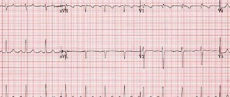
- Electrocardiography - helps to see left ventricular hypertrophy, which leads to aortic valve insufficiency. There are no other characteristic signs.
- Ultrasound - expansion of the aorta is visible; when using Doppler ultrasound, aortic insufficiency becomes noticeable.
- X-ray diagnostics can also confirm the expansion of the aorta; in some cases, calcifications are detected in the vascular wall.
- Serological studies are carried out to determine specific antibodies to the pathogen.
Medical encyclopedia - aortitis
Aortitis is inflammation of the aortic wall. Most often, aortitis develops with syphilis, streptococcal infection, rheumatism, allergic processes, and systemic collagenosis. The disease is chronic, manifested by pain behind the sternum and dilation of the aorta. Prevention and treatment come down to active treatment of the underlying disease.
Aortitis is an inflammation of the aorta, often of infectious origin. The main cause of aortitis is syphilitic infection; Streptococcal, rheumatic, septic, and tuberculous aortitis are less common. Sometimes the aorta is involved in the process due to inflammation of neighboring organs (pulmonary tuberculosis, mediastinitis). Isolated cases of aortitis with systemic thrombangitis have been described. The listed forms of aortitis do not have a clear clinical picture. A. is suspected when dilatation of the aorta is detected against the background of rheumatic, septic or other diseases.
A. refers to late manifestations of syphilis. The first clinical symptoms of A. occur several years after infection, much more often in men. The main clinical symptom of aortitis is pain. Patients usually complain of prolonged dull, pressing and aching pain behind the sternum, which intensifies with physical activity and excitement. With damage to the ostia of the coronary arteries and insufficiency of the aortic valves, the pain can take on a severe anginal character.
Syphilitic aortitis is divided into uncomplicated and complicated (narrowing of the mouths of the coronary arteries, aortic insufficiency, aneurysm). In uncomplicated syphilitic A., objective data are scarce. Sometimes it is possible to note increased pulsation of the aorta in the jugular fossa, and with percussion in the upper part of the sternum - expansion of the aorta. When listening in the second intercostal space to the right of the sternum, a change in the second tone is characteristic, acquiring a ringing metallic character. Often a soft systolic murmur is heard in the same place, in contrast to the rough systolic murmur with aortic stenosis. Often, systolic murmur in syphilitic A. occurs or intensifies when raising the arms (Sprotinin's symptom). When large vessels extending from the aortic arch are affected, a difference occurs in the intensity of pulsation of both carotid arteries, pulse rate and blood pressure in the right and left arms. The electrocardiogram is usually unchanged. A positive Wasserman reaction is observed in 74-95% of patients with syphilitic aortitis. The most important thing for diagnosing syphilitic A. is a thorough X-ray examination (fluoroscopy, teleradiography, X-ray kymography and electrokymography, contrast aortography). Characterized by expansion of the aorta, an increase in the amplitude of its pulsation, uneven contours and an increase in the shadow of the aorta.
When syphilitic aortitis is complicated by narrowing of the coronary artery ostia or aortic insufficiency, a picture of slowly progressive chronic coronary insufficiency develops, followed by cardiosclerosis and circulatory failure. Differential diagnosis is carried out with atherosclerosis of the aorta (see Atherosclerosis), atherosclerotic cardiosclerosis (see), subacute septic endocarditis (see), mediastinal tumors (see).
The prognosis for syphilitic aortitis depends on the activity and extent of the process, and the presence of complications. Prevention and treatment come down to active, comprehensive therapy for syphilis. Treatment is carried out in a hospital setting, starting with bismuth, mercury, iodine and active penicillin therapy (see Syphilis, treatment). In case of severe coronary insufficiency, heart failure, antisyphilitic treatment must be carried out more carefully, combining it with effective coronary dilation therapy, the administration of cardiac glycosides, saliuretics, oxygen therapy [see. Angina pectoris, Blood circulation (insufficiency)]. V. Soloviev.
Pathological anatomy . The term “aortitis” refers to both an inflammatory process in the wall of the aorta, predominantly of an infectious nature, and reactive changes in the aorta of an immunoallergic nature, whose morphological picture resembles inflammation. Based on the localization of the process, endaortitis, mesaortitis, periaortitis and panaortitis are distinguished, but isolated damage to the intima or adventitia is extremely rare (with brucellosis, rheumatism). According to the distribution, aortitis can be diffuse, ascending and descending.
Infectious aortitis is a particular manifestation of the underlying disease (syphilis, rheumatism, sepsis, malaria, brucellosis, gonorrhea, etc.). Mesaortitis and panaortitis are more common. In acute infectious A. (septic, streptococcal, gonorrheal, rickettsial, malarial), the aorta is swollen and poorly elastic. Microscopically, its membranes are infiltrated with polymorphonuclear leukocytes. In chronic infectious aortitis (syphilitic, rheumatic, tuberculous), the aortic wall is compacted, brittle, with calcifications. The intima is thickened, wrinkled, with abundant deposits of lime (syphilitic A.), sometimes with the formation of folds like “valve” (rheumatic A.). The adventitia is spotted and sharply plethoric. Microscopically, in the intima of rheumatic aortitis, foci of mucoid swelling and fibrinoid necrosis are detected: in the media - phenomena of metachromatic edema, sometimes rheumatic granulomas are found. Syphilitic A. is characterized by multiple foci of necrosis with ruptures of elastic fibers, infiltrates of lymphoid, plasma, and histiocytic cells and extensive fields of sclerosis. The microscopic picture of brucellosis and chronic fibrous rheumatic aortitis differs from that indicated by the absence of plasma cells in the infiltrates. Characteristic of tuberculous, syphilitic and actinomycotic A. is the presence of specific granulomas in the adventitia.
Immunoallergic diseases include the so-called juvenile and giant cell aortitis. The first is observed in young people, more often in women. The pathogenesis and etiology of the disease are not clear; the term “aortitis” is purely arbitrary here. The process is characterized by a predominant lesion of the ascending thoracic aorta. The aortic wall is diffusely thickened, inelastic, sometimes with calcified dense adventitia. Microscopically - uneven development of connective tissue under the endothelium, swelling and fragmentation of elastic membranes with the deposition of lipoids and infiltrates of lymphoid and plasma cells. Necrosis of the type of microinfarction is often observed in the aortic wall. In the adventitia against the background of sclerosis, there is an abundance of vasa vasorum, either obliterated throughout, or with extensive proliferation of their endothelium. The lesion from the ascending aorta spreads to its branches with the development of the clinical picture of the “pulseless” disease (Takayasu’s disease).
Giant cell “aortitis”, or idiopathic necrosis of the aortic media, is accompanied by aneurysmal dilatations and ruptures of its wall. The process begins, like rheumatic aortitis, with focal lymphoid cell infiltration of the adventitia with an admixture of Langhans-type giant cells. Infiltrates penetrate into the media, where foci of necrosis appear, surrounded by giant cells. The final stage of the process is fibrosis of all membranes of the aorta and the development of secondary atherosclerosis.
Treatment
Syphilitic aortitis is treated with the participation of a dermatovenerologist.
After diagnosis, patients are sent to specialized departments located on the territory of infectious diseases hospitals, or to the venereology department.
Patients are subject to mandatory hospitalization:
- over 60 years old;
- with a complicated course of syphilitic aortitis;
- with intolerance to penicillin drugs;
- at a late stage of the disease;
- with a burdened somatic history.
Treatment of syphilitic aortitis is identical to the treatment of visceral syphilis. The only thing is that antibiotic therapy must be carried out carefully, since after its initiation the pathological process is often activated, which can lead to coronary insufficiency.
Standard therapy for visceral syphilis includes:
- Antibiotic therapy using cephalosporin or penicillin drugs.
- Symptomatic treatment, which for SA may include beta-blockers, diuretics, vasoprotectors, etc.
In difficult cases, when aortitis is complicated by an aneurysm, surgical intervention is performed if the patient’s health is acceptable.
Forecast
Depends on the course of the underlying disease. If syphilis was identified at an early stage of development, after which it was possible to carry out appropriate treatment, then the prognostic conclusion is relatively favorable.
Severe syphilis does not allow the patient’s health to be so improved that clinical signs do not appear. Medicines can relieve symptoms, but despite this, a chronic course of the disease is observed, with periods of exacerbation and subsidence of the pathological process.
It is important to understand that with syphilitic aortitis, complete recovery is impossible, since the largest vessel in the human body was subject to serious changes and deformations.
The most unfavorable prognosis is for an aneurysm that develops against the background of syphilitic aortitis. When it appears, the prognostic value is influenced by the shape of the formation, its size and location.
Treatment of aortitis
Treatment of aortitis is inextricably linked with active therapy of the underlying disease. For infectious aortitis, antibiotics are the first-line drugs; for allergic aortitis - glucocorticoids, NSAIDs, immunosuppressants; for syphilitic aortitis - preparations of bismuth, iodine, penicillin antibiotics. The effectiveness of therapy is monitored by the dynamics of clinical and laboratory parameters.
The presence of an aortic aneurysm, especially signs of its dissection, is the basis for consultation with a vascular surgeon and angiosurgical treatment - resection of the aneurysm followed by aortic replacement. If aortic stenosis develops, balloon dilatation, stenting, or bypass surgery may be required.
Prevention
The main way to prevent the development of syphilitic aortitis is to treat syphilis at an early stage of development, then there will be no prerequisites for the occurrence of SA.
There is no specific prevention of syphilis, that is, there is no vaccine that would protect a person with a 100% guarantee against the pathogen. Therefore, in order to avoid contracting such a serious infection, one should adhere to high moral values.
Video Treatment of syphilis. Consequences, complications and prevention of syphilis
4.80 avg. rating ( 94 % score) – 5 votes – ratings