Pulmonary hemorrhage - causes
The causes of pulmonary hemorrhage can be very different, and it is often difficult to establish the true cause. Treatment of a symptom (pulmonary hemorrhage) should always begin with the main one - eliminating the cause (the disease itself, which gives such a dangerous symptom).
Background states
What diseases can cause pulmonary hemorrhage?
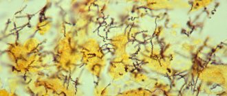
- Infections are the most common and extensive group of causes. This includes diseases such as: pulmonary tuberculosis, pneumonia of various origins, purulent lung diseases (abscess, gangrene), fungus, parasitic infections, bronchiectasis, bronchitis, etc.
- Tumor processes (lung tumor, metastases in the respiratory tract (lungs, bronchi)).
- Trauma to the chest area (bruises, wounds, gunshot injuries, lung rupture, etc.).
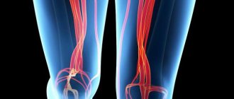
- Vascular causes (PE - pulmonary embolism, aortic aneurysm, congenital vascular features (developmental defects), hypertension (increased blood pressure), pulmonary infarction.
- Coagulopathies are diseases characterized by a defect in the blood coagulation system. Hemophilia, thrombocytopenia, von Willebrand disease, DIC syndrome.
- Other reasons. Pulmonary bleeding can occur during certain medical procedures - puncture, bronchoscopy, postoperative complications, etc.
Immediate causes of pulmonary hemorrhage
The immediate causes of pulmonary hemorrhage most often lie in a defect in the vessels themselves due to inflammatory or sclerotic changes in the lung tissue. There is another group of pulmonary hemorrhages, which is called idiopathic (that is, the cause has not been established), a case when it is impossible to determine what caused the symptom (bleeding).
Basically, it is impossible to single out any specific type of pulmonary hemorrhage that would occur without a background (without an underlying disease).
Mechanisms occurring in the lungs during bleeding
What happens in the lungs during bleeding?
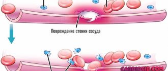
- First, why might bleeding occur? The vascular wall, in the presence of any changes (inflammation, infection, tumors, etc.), changes its properties, becomes thinner, permeable or brittle, which significantly increases the risk of developing this symptom.
- Secondly, a change in pressure in the pulmonary circulation (increased pressure), affecting the damaged vessel, leads to even more increased bleeding.
- And defects in the blood coagulation system prevent the formation of a full-fledged blood clot so that it can “patch” the damaged vessel. Thus, if all three components are present, they aggravate each other.
The entire pathological process occurs mainly in the lungs, but, of course, the condition of the body as a whole also suffers. With frequent or intense bleeding, anemia develops (decreased number of red blood cells, decreased hemoglobin level), hypovolemia (decreased total blood volume).
Additional symptoms
It is important to pay attention to those symptoms that are common to any type of bleeding. Their significance is that they help determine the estimated degree of blood loss and the severity of the patient’s condition in general. Therefore the following should be assessed:
- Pale and grayish-sallow skin tone;
- Shortness of breath and respiratory rate (more than 18-20/min);
- The patient’s consciousness and behavior;
- Ability to move independently;
- Frequent heartbeat (tachycardia - increased heart rate more than 90 beats/min);
- Degree of reduction in blood pressure (below 100/60).
The more severe the bleeding and blood loss, the more they will be determined. Providing assistance to the patient depends on this.
Lung cancer is the most common cause of pulmonary hemorrhage
Classification
The most important classification is based on the volume of blood lost. Here it is presented in a more simplified form:
- lung (streaks of blood in the sputum, total blood loss up to 300 ml);
- moderate severity (up to 1000 ml of lost blood);
- severe (more than 1000 ml per day).
Bleeding is also classified as single or multiple.
Etiology and pathogenesis of hemoptoea
The incidence of LC is about 1-4% of the total number of bleedings. Many people mistakenly do not consult a doctor for this pathology.
Medical care is urgently needed for the patient if signs of pulmonary hemorrhage appear, since the mortality rate for this complication of pulmonary diseases reaches 50-80%.
Massive aspiration of blood is found in both lungs. This is the penetration of biological fluid into the tissues of the respiratory organ during inhalation.
Symptoms indicating pulmonary hemorrhage
General symptoms and signs of pulmonary hemorrhage are similar in various diseases:
- discharge of blood through the mouth and (or) nose (may be intense, may be in the form of streaks);
- foamy sputum with blood (not a constant symptom);
- paroxysmal cough;
- burning sensation in the chest, discomfort;
- feeling of fear;
- dizziness;
- drop in blood pressure;
- fainting (collapse).
The last three symptoms are present with significant blood loss of more than 300 - 400 ml.
Symptomatic manifestations and varieties
First aid for pulmonary hemorrhage begins to be provided if the following symptoms of pathological changes are observed:
- The beginning of the attack is hemoptysis;
- Blood clots of a rich color (bright or dark red) released with coughing;
- If, in addition to bloody sputum, blood begins to be released through the nose, its appearance is different: a foam-like mass appears that does not have clots;
- The cough is paroxysmal in nature, and at first it is dry, and then bloody sputum appears;
- The patient feels discomfort in the throat, while the sounds of “gurgling” are clearly audible;
- A strong burning sensation appears on the side of the affected lung;
- The skin of the face turns pale;
- Blood pressure levels decrease;
- The whole body becomes covered with sweat;
- A rapid heartbeat is detected;
- Dizziness;
- Convulsions;
- Dyspnea;
- Noise in ears;
- General weakness;
- The person temporarily loses vision;
- After severe blood loss - asphyxia.
With prolonged bleeding without appropriate therapy, aspiration pneumonia develops.
Interesting fact!
Symptoms are most often recorded in older men.
In order for medical care to successfully stop the manifestations of bleeding, you need to know their degrees and forms of manifestation.
Diagnostic methods
Diagnosis of bleeding is very important for further tactics of patient management, since without establishing the cause of bleeding, it is not possible to eliminate it. It is also very important to establish whether this is indeed pulmonary bleeding (differentiated from gastrointestinal). With a complete and correct examination, the doctor is able to determine the true cause and draw up a treatment plan.
Every doctor, when caring for such a patient, should be wary of oncology, since pulmonary hemorrhages often accompany cancer.
Physical data
When examining the patient, pallor of the skin is revealed; the general condition can be severe or satisfactory. Blood pressure is reduced. These symptoms are detected with prolonged chronic bleeding or with one, but intense bleeding with a large loss of blood (500 ml or more). During the physical examination, auscultation (listening) of the lungs and heart is also performed.
Often, during a routine examination, especially when the bleeding is not so intense, it is not possible to make an accurate diagnosis; a more thorough examination is required using instrumental and laboratory methods.
Instrumental methods
The main method is x-ray diagnostics; x-rays can be used to identify pneumonia, tumors, purulent diseases (abscess, gangrene), pulmonary embolism, etc. Due to its simplicity, speed and relatively inexpensive cost, this method is used more often than others. In unclear situations, CT and MRI studies are used, which are more expensive procedures.
Fiberoptic bronchoscopy should be performed in all patients with pulmonary hemorrhage. This procedure allows you to determine the source of bleeding, bronchial patency, and identify any changes or neoplasms in the bronchial tree.
Laboratory diagnostics
CBC (complete blood count) detects anemia. OAM (general urinalysis) generally does not provide any data. Blood is used to determine tumor markers (in the diagnosis of tumors), to confirm diagnoses of coagulopathies, pulmonary embolism, etc.
Differential diagnosis
It is first necessary to differentiate pulmonary bleeding from gastrointestinal bleeding. With pulmonary hemorrhage, the blood is most often foamy and bright red in color, while with gastrointestinal hemorrhage it is dark red. Also, in the presence of gastrointestinal bleeding, melena (black coloration of stool) occurs.
A tumor is often differentiated from a tuberculoma or a lung abscess - all these structures have a round shape, and it is not always possible to make a correct diagnosis using just one radiograph. In rare cases, even a puncture of the lung (biopsy) is required.
Diagnostics
The patient must be taken to the emergency department, where, using additional research methods, the doctor makes a diagnosis. If the patient's condition is severe, he is transferred to the intensive care unit before the diagnosis begins, then the diagnosis is made in the intensive care unit.
For diagnosis, the doctor uses the following methods:
- Physical examination. It includes inspection, percussion and auscultation. An important step is to examine the nasal cavity and nasal passages, as well as the pharynx. Sometimes bleeding from these areas can mimic pulmonary bleeding. When percussed in a place where the alveoli are filled with blood, a clear pulmonary sound is replaced by a dull one. On auscultation, moist rales are heard in places where blood accumulates.
- Laboratory research. These include a general blood test, which allows you to see the degree of blood loss and determine the level of platelets, as well as a coagulogram, which evaluates the state of the blood coagulation system.
- Radiography. A very simple and quick method that allows you to see the place where the lungs are filled with blood. Sometimes it is possible to determine the source of the bleeding, for example, if a tumor is visible.
- Selective angiography of the vessels of the bronchial tree. Contrast is injected into the blood vessels of the lungs. It passes sequentially through arteries, capillaries and veins. In the vessel that caused the bleeding, the contrast leaks into the lung parenchyma.
- CT scan. More accurate than conventional radiography. CT allows you to look at the chest cavity in volumetric sections. In this way, even a small accumulation of blood in the parenchyma can be seen and the source of bleeding can be determined.
- Bronchoscopy. The most accurate method that allows you to localize bleeding. In some cases, bronchoscopy can turn from a diagnostic method into a treatment method, for example, if a vessel is coagulated through a bronchoscope.
Differential diagnosis
Nosebleeds can be quite massive. Blood is secreted to a greater extent from the nasal cavity and to a lesser extent from the mouth in the form of spitting. You can also see how it flows down the back of the throat. The discharge is not foamy, the blood is scarlet, and may be mixed with mucus.
Bleeding from the oral cavity can be massive when the tongue is injured. It is important to dry the mouth and carefully examine it for wounds, carious teeth, and gum disease. With such bleeding, the discharge is not foamy, scarlet or dark, mixed with saliva.
When bleeding from the esophagus, the blood is often dark in color, since in most cases its source is the venous plexuses. The discharge flows out in a trickle or in massive portions “mouth full”. There are no impurities in the blood.
When bleeding from the stomach or duodenum, the discharge is dark. The blood mixes with hydrochloric acid in the stomach and becomes the color of “coffee grounds.” Blood is released in portions, which is preceded by jerky movements of the chest.
Stages of treatment
Chronic bleeding with a small volume of lost blood can also begin in an outpatient setting; emergency situations with massive blood loss require urgent action and further treatment in a hospital. Almost all causes of pulmonary hemorrhage require treatment in a hospital setting.
First aid for pulmonary hemorrhage
Help with pulmonary hemorrhage is provided by the person who is nearby; to perform simple actions you do not need deep knowledge of medicine, you just need to remember a few rules:
- unfasten the first buttons on clothes, belts, locks, etc.;
- lay on your back or side (in case of massive bleeding) and be sure to raise your head!!!;
- Do not give the patient water or food under any circumstances.
Upon arrival of the ambulance, the medical worker examines the patient and assesses his condition. In case of massive blood loss, the respiratory tract is cleared of blood and clots, then oxygen is supplied and catheters are installed for transfusion of solutions to replenish the volume of lost blood. All these actions should be carried out on the way to the hospital.
Further inpatient treatment
In the hospital, laboratory and instrumental studies are carried out to make a correct diagnosis. But the first priority in this case is to stop the bleeding.
Solutions are used to replenish the BCC (circulating blood volume) - crystalloids and colloids (saline solution, Ringer's solution, Dissol, etc.), with a large loss of blood (more than 21% of the total volume of all blood), red blood cells, fresh frozen plasma and other blood products.
When bleeding does not stop, invasive methods are used - fibrobronchoscopy (for the purpose of removing blood clots from the bronchi and irrigation with various drugs), a special hemostatic sponge can also be used, which tampons the bleeding site. In case of extensive bleeding (for example, due to injury), surgical operations are performed on the chest.
Along with priority measures, an accurate diagnosis is established. Then, after stopping the bleeding, treatment of the disease itself begins. For pneumonia, abscesses - prescribing antibacterial drugs, tuberculosis - anti-tuberculosis drugs, tumors and metastases - chemotherapy, radiation therapy, surgery.
The doctor determines the further treatment plan, either continues the treatment himself if the disease is within his competence, or refers the patient to a specialized medical institution.
Rehabilitation
Rehabilitation of patients depends on the underlying disease. Rehabilitation measures must be prescribed by the attending physician (usually recommendations for further treatment are given to patients along with discharge from the hospital, or there are specialists such as rehabilitation specialists who give recommendations taking into account all the characteristics and diseases).
Rehabilitation consists of correcting lifestyle, first of all, giving up bad habits, possibly changing work, climate, living conditions, work and rest schedule, etc. In fact, rehabilitation predetermines the patient’s further comfortable state, so there is no need to be skeptical about the recommendations and still try to follow them.
Clinical examination of patients after pulmonary hemorrhage
Medical examination depends on the underlying disease. That is, after treatment in a hospital or outpatient setting, the patient must be registered with a specific doctor, with whom he will be observed in the future (oncologist, phthisiatrician, hematologist, pulmonologist, etc.).
Typically, in the first year after the disease, the number of examinations is reduced to 2–6, or maybe more, in the second year – 2–4, then, after stable remission, examinations can be carried out once a year. It all depends on the specific disease and condition of the patient.
Emergency care and treatment for pulmonary hemorrhage
First aid for internal pulmonary hemorrhage is very limited. Patients are urgently hospitalized in the department of pulmonology or surgery. Transportation is carried out in a sitting or semi-sitting position with legs down.
Emergency care consists of removing blood from the respiratory tract with a special aspirator, administering hemostatic drugs and antibiotics, transfusion of blood components, restoring CTCs, performing therapeutic bronchoscopy and surgical treatment.
The treatment algorithm for patients includes general recommendations: swallowing pieces of ice, drinking cold water in small portions, applying a cold compress to the chest. Patients need to be reassured and explained the need to cough up sputum. Excessive emotional stress can aggravate the situation.
In the department, patients are placed on the sore side, oxygen and the necessary medications are administered by inhalation. Bronchoscopy is performed and, if necessary, the optimal extent of surgical intervention is determined: lung resection or pneumonectomy.
There are temporary and permanent ways to stop pulmonary hemorrhage. The first include: drug-induced hypotension, hemostatic drugs, endobronchial methods of hemostasis. The second group includes most operations: lung resection, vascular ligation.
Conservative treatment
Treatment of pulmonary hemorrhage is aimed at eliminating the underlying disease. Currently, medications are used only for minor and moderate forms of pulmonary hemorrhage.
Medicines prescribed to patients:
- Hemostatic drugs - “Vikasol”, “Sodium etamsylate”, “Gordox”, “Kontrikal”;
- Antihypertensive drugs - “Pentamine”, “Benzohexonium”, “Arfonad”, “Clonidine”;
- Immunosuppressants and glucocorticoids - “Cyclophosphamide” for the treatment of systemic diseases;
- Painkillers – “Analgin”, “Ketorol”, some narcotic analgesics;
- “Codeine”, “Dionine”, “Promedol” to suppress a painful cough;
- Cardiotonic drugs – “Strofanthin”, “Korglikon”;
- Desensitizing drugs – “Pipolfen”, “Diphenhydramine”,
- Diuretics – “Lasix”;
- Oxygen therapy.
Red blood cell replacement therapy for significant blood loss: patients are administered native plasma, Reopoliglyukin, Polyglyukin, saline and colloidal solutions - isotonic sodium chloride solution, Ringer, Trisol. To relieve bronchospasm, patients are administered inhaled m-anticholinergics – “Atropine Sulfate” or b-adrenergic agonists – “Alupent”, “Salbutamol”, “Berotek”.
Endoscopic methods
If conservative therapy is ineffective, they proceed to bronchoscopy, during which pulmonary hemorrhage is stopped in various ways. To do this, they use applications with drugs, install a hemostatic sponge, coagulate the vessels at the site of the lesion, obstruct the bronchi with fillings, and embolize the arteries. But these methods bring only temporary relief.
X-ray endovascular occlusion of a bleeding vessel is performed by experienced radiologists who are fluent in angiography techniques. Arteriography allows you to determine the source of bleeding. Polyvinyl alcohol is used for vessel embolization. This method of treating pulmonary hemorrhage is highly effective, but causes a number of complications: ischemia of the myocardium, brain or spinal cord.
Surgery
Main types of operations:
- Palliative - collapse therapy, thoracoplasty, extrapleural filling, pulmonary artery ligation, pneumotomy;
- Radical - partial lung resection, marginal resection, segmentectomy, lobectomy, bilobectomy, pneumonectomy.
Death in patients with massive pulmonary hemorrhage most often occurs from asphyxia rather than from blood loss. Ensuring airway patency is a primary and vital task in the treatment of such patients.
Causes of the disease
Pulmonary hemorrhage most often occurs in middle-aged and elderly men.
The risk group includes:
- children with recurrent pneumonia;
- patients taking steroid hormones for a long time;
- convicts serving sentences;
- external migrants;
- women bearing a child;
- diabetic patients;
- patients with pulmonary tuberculosis;
- elderly people;
- patients with acute pneumonia.
LC is a manifestation of many ailments.
Pulmonary hemorrhages are caused by the following factors:
- destructive pathological processes in the respiratory tract;
- irradiation;
- pulmonary heart;
- infectious diseases;
- psycho-emotional stress;
- oncological processes;
- injuries of the lungs and bronchi;
- lung abscess;
- diseases of the esophagus;
- venous stagnation;
- vitamin deficiencies;
- systemic illnesses;
- blood diseases;
- development of heart disease;
- mediastinal diseases.
With chronic atrophic bronchitis, bronchiectasis, tumors and pulmonary tuberculosis, this dangerous syndrome occurs most often. Up to 90% of blood loss cases are due to tuberculosis.