Recently, cancer is being diagnosed more and more often. Leukemia is a dangerous malignant neoplasm that affects the hematopoietic organs. In the early stages, the disease is asymptomatic, which complicates the diagnostic process. Symptoms of the disease appear in later stages. Doctors and scientists have long been looking for options for detecting leukemia in the early stages. A blood test for leukemia will immediately show any deviations in the indicators. Doctors recommend donating blood annually. This will allow the disease to be identified at an early stage.
Types of tests to determine leukemia
Leukemia has specific symptoms that appear in adults and children. The disease also has enough unusual symptoms that it is difficult to make a correct diagnosis. Symptoms are often similar to other diseases. To clarify, you need to undergo diagnostics. Based on the existing symptoms, the doctor will decide on the type of tests and quantity. Basically, doctors prescribe the following types of diagnostics:
- A complete blood count (CBC) will help determine the level of basic indicators - the number of leukocytes, red blood cells, basophils, ESR and other elements. The indicators of these elements are among the most important in determining abnormalities in the body.
- Blood biochemistry shows disturbances in the functioning of internal organs. Based on the results of the analysis, a decision is made on the method of therapy to improve the patient’s well-being.
- Leukemia is also diagnosed by performing a bone marrow puncture with lymph nodes. Ultrasound examination, tomography, radiography and copy are considered clarifying techniques for a preliminary diagnosis based on a general blood test.
It is possible to clarify the existing signs of leukemia using a blood test in a short time - indicators of the main elements in the blood that go beyond the normal limits will indicate the degree of damage to the body.
Blood cells at 1000x magnification
How is leukemia determined?
The first symptoms of leukemia in adults are not noticeable from the very beginning of the disease.
However, here are the signs:
- weakness;
- severe fatigue;
- frequent infectious diseases;
- loss of appetite;
- joint pain;
- bleeding from the nose, gums;
- anemic shortness of breath;
- hemophilia.
With myelo- and monoblastic leukemia, the temperature often rises. The size of the spleen and kidneys increases, while the liver cannot be palpated.
With lymphoblastic leukemia, the inguinal and axillary lymph nodes become enlarged. Sometimes one of the testicles increases in size. Even if there is no pain, you should urgently do a blood test. Often swollen lymph nodes are accompanied by a dry cough and shortness of breath.
In a quarter of cases, leukemic meningitis is diagnosed. Its signs: vomiting, weakness, headache, convulsions, inadequate perception of reality, irritability, seizures, fainting. Deterioration of hearing and vision is possible. In the cerebrospinal fluid, the rates of cytosis and blast cells increase.
The skin turns red or brown in advanced stages of leukemia.
General blood analysis
A general analysis can determine the presence of many diseases, inflammations and abnormalities in the functioning of internal organs and body systems. If the picture of the main elements in the analysis does not change, then the body works without deviations or disturbances. The following elements are taken into account for diagnosis:
- An increased ESR is observed - this indicates the presence of possible pathologies. To confirm blood cancer, you need to compare other indicators.
- White blood cells may be higher than normal or lower. In acute or chronic leukemia, the level depends on the stage of the process and the form of the pathology. Acute leukemia is characterized by aggressive cell division, which is expressed in an increase in the rate from normal. In a preschool child and a teenager, there is often a difference in the readings of leukocyte levels.
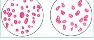
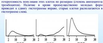
- Identification of cells with different sizes indicates the presence of anisocytosis.
- With leukemia, platelets are reduced by 10-15 times (the norm is 180-320). The initial stage may show normal levels.
- A decrease in red blood cells to 1-2*109/l indicates an oncological process in the body. The cell is responsible for transporting oxygen to tissues and organs. A lack of transport provokes shortness of breath and headaches in humans. The malignant process at an early stage does not affect the number of red blood cells in the blood.
- Reticulocytes are the germ cells of red blood cells. A low rate is present at an early stage of the tumor.
- Hemoglobin decreases in later stages of the disease. A reading of 50 or 60 g/l is considered the main sign of leukemia. But for this, the doctor excludes the reasons for the decrease associated with other diseases - deficiency of vitamin B12 with iron and severe bleeding.
- The absence of basophils and eosinophils indicates the presence of oncology that has affected the body’s hematopoiesis.
Clinical analysis is carried out in the same way for patients, regardless of age. This type of diagnosis is a mandatory event in the anamnesis collection program. In children, acute leukemia is lymphoblastic in nature, in adults it is mainly myeloblastic. Chronic leukemia occurs in people over 45 years of age.
Leukocytes
White blood cells are cells responsible for protecting the body from infectious and viral substances in the body. During the disease, the shape and structure of leukocytes undergo changes that affect their functionality. The disease can increase or decrease the number of cells. A blood test with bone marrow puncture allows you to detect the extent of damage to the body.
Any changes in indicators indicate the presence of the disease. Lymphocytes are heterogeneous in structure and differ in appearance. Low or elevated levels (leukocytosis) not only with leukemia. This is possible during other diseases - internal inflammation or viral damage. There is an example of the course of the disease against the background of a normal number of lymphocytes. But the leukocyte system shifts towards agranulocytes or granulocytes. Granulocytes mainly increase during an infectious disease.
Platelets
Platelets in the blood circulation perform an important function - they stop bleeding. When a vessel is injured, the system quickly forms a blood clot to stop bleeding. Normally, they should be contained from 180 to 360 in men and women. Leukemia affects differently - an increase (thrombocytosis) or a decrease (thrombocytopenia) is possible. A dangerous diagnosis is thrombocytopenia - decreased clotting function (DIC syndrome).
A decrease in platelets is possible with leukemia, hepatitis and systemic lupus erythematosus. Increased levels occur with erythremia, cancer of the pancreas, and after surgery.
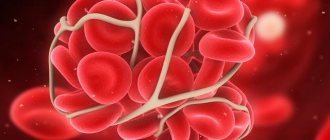
Platelets in the blood
Red blood cells and hemoglobin
Red blood cells contain hemoglobin to transport oxygen to human tissues and organs. The level of red blood cells depends on the amount of hemoglobin. Normally there should be 4-5*1012/l. In the last stages of leukemia, the rate drops to 1-2*1012/l.
A decrease in hemoglobin causes an acute state of anemia, which is characterized by the following symptoms:
- You feel very tired and have decreased performance.
- The skin becomes dry and pale.
- Nail plates become brittle and thin, hair falls out.
- Light physical activity is accompanied by rapid heart rate and shortness of breath.
- There is a change in taste preferences.
- There are extraneous noises in the ears, attacks of nausea and dizziness.
- Unstable emotional background.
These symptoms worsen the patient’s condition, complicating the treatment process.
Hematocrit
Hematocrit is the ratio of red blood cell volume to plasma. The indicator is directly related to the number of red blood cells. The disease affects the entire hematopoietic composition.
Hematocrit helps detect the degree of anemia. The development of anemia reduces the amount to 25% and below. Severe blood loss with blood transfusion does not take into account hematocrit data due to the late reaction to the change of basic elements. You also need to take the test correctly, since the value changes depending on the position of the body.
ESR during illness
ESR is the value of the erythrocyte sedimentation process. The speed of this process is taken into account. Against the background of cancer, the picture does not look the same as in a normal state. Usually the speed increases. This is due to a disruption in the functioning of the immune system and the presence of a secondary infectious lesion. Based on this value, the doctor can understand the patient's condition.
Device for blood ESR analysis
Leukocyte formula
Diagnosing a patient with leukemia also includes assessing the leukocyte count. It is not the number of leukocytes that is taken into account here, but it is important to identify a qualitative change. The quantity often remains unchanged, but there is a qualitative imbalance. The analysis shows mature and young forms, without the presence of intermediate forms.
Oncology provokes a decrease in reticulocytes. The peculiarity of the leukocyte picture depends on the type of leukemia - an increase or decrease is possible.
Analysis in children
Children are susceptible to acute forms of the disease. The age group at risk is from 2 to 5 years. In the chronic type, symptoms are often not detected, but the disease can be determined by the values of the main blood elements. The development of a malignant tumor can be recognized by the following signs:
- A sharp decrease in the level of hemoglobin with red blood cells;
- Red blood cells settle at a high rate;
- The number of reticulocytes decreases;
- Unstable leukocyte count;
- Platelets shift towards decrease.
Doctors recommend taking an annual blood test to determine the clinical picture and tumor markers.
Why is regular examination necessary?
Sometimes it is impossible to determine even a cold in a child based on the general condition and symptoms. In addition, obvious symptoms of anemia are already evidence of body dysfunction. It is much more reliable and safer to detect leukemia using laboratory methods - by donating blood for a general analysis. This must be done at least once a year as part of a general medical examination. In addition to leukemia, such an examination can detect a host of other diseases at an early stage of development.
If it is known that the patient has a predisposition to blood diseases (oncological diseases in relatives, a history of cancer, autoimmune diseases), then it is necessary to take blood tests every six months and whenever there is a deterioration in health, which could indicate the onset of the disease.
Today it is much easier to prevent a serious illness than to cure it. Monitoring your health allows you to detect cancer in its early stages. Some types of cancer can be predicted even before they start using genetic analysis. Therefore, diagnostic measures should not be ignored.
In the vast majority of cases, children with acute leukemia can be cured and relapses can be avoided in the future. In adults, unfortunately, the chance of relapse is slightly higher, but careful medical monitoring and timely assistance can prolong life for as long as possible. It also happens that leukemia itself is a consequence of another cancer.
Myeloid leukemia
With myeloid leukemia, it is possible to detect an increase in leukocytes and a decrease in platelets - this is characteristic of the chronic type of the disease. Anemic syndrome is actively developing with a decrease in erythrocyte level. At the initial stage of tumor development, eosinophilia, basophilia and an increase in ESR are detected.
Further formation of a cancerous tumor leads to an abnormal change in the shape and size of the main elements - poikilocytosis with anisocytosis. Biochemistry often reveals a decrease or absence of alkaline phosphatase.
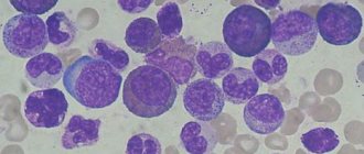
Lymphoblastic crisis under a microscope
Lymphoblastic crisis is characterized by an acute form of anemia with critically increased values of blast cells and minimal values of neutrophils. Acute myeloid leukemia is determined by leukocytosis, young genes.
Description of the disease
With leukemia, healthy cells in the blood are replaced by pathological ones, which disrupts the reactions occurring in the body.
There is a lack of various necessary cells in the circulatory system, and blood cancer develops.
Leukemia is divided into the following types, each of which occurs separately:
- acute - develops rapidly , can be detected in the initial stages;
- chronic - the course of the disease is slow, it cannot always be detected immediately.
Acute form
Expert opinion
Kovaleva Elena Anatolyevna
Doctor-Laboratory Assistant. 14 years of experience in clinical diagnostic services.
Ask a question to an expert
This type of disease is characterized by sudden development. With the disease, patients experience an increase in the size of organs and excessive bleeding. With timely diagnosis and prompt initiation of therapy, the disease can be completely cured.
The disease has the following symptoms:
- joint pain;
- frequency of occurrence of various infectious diseases;
- nausea;
- weakness;
- bone pain;
- increased fatigue;
- vomit;
- temperature increase.
Acute forms of the disease:
- Myeloblastic leukemia - during the disease, a change occurs in the myelocytes formed in the bone marrow. The disease is characterized by rapid development. If granulocytopenia develops, ulcers form on the mucous membranes. Thrombocytopenic hemorrhagic syndrome and body intoxication are not uncommon in the disease. and specific infiltration of the uterus, kidneys, skin
- Lymphocytic leukemia is a disease characterized by the degeneration of blood cells into leukemic ones. In the first stages of the disease, the following appear: intoxication, pronounced enlargement of lymph nodes, anemia, and hemorrhagic syndrome.
Biochemical analysis
The prognosis for leukemia is also made based on a biochemical blood test. The doctor draws up a clinical picture of the functioning of organs and the internal state of tissues. Leukemia is characterized by the following values:
- The level of urea with uric acid increases - disturbances in kidney function.
- An increased level of gamma globulins indicates abnormalities in the functioning of the digestive tract.
- A malfunction of the liver cells indicates an increase in bilirubin levels, AST with ALT and LDH.
- A decrease in sugar values characterizes improper functioning of the pancreas.
- Severe damage to liver tissue is detected by a decrease in albumin and fibrinogen, but sometimes their values remain normal.
Leukemia: mechanism of development
Hematopoiesis occurs in the red bone marrow, which in adults is located in the sternum, vertebral bodies, pelvic bones, skull bones, and ribs. It is here that blood cells form and mature before entering the vascular bed. Leukemia begins with a mutation in a single hematopoietic precursor cell, which stops responding to the body’s regulatory signals, loses the ability to die naturally, and begins to divide uncontrollably. A huge mass of leukemia cells - and in three months the number can reach a trillion, 1012 - clones of the very first, mutated one.
Of course, such a load on the bone marrow is not in vain: all resources are taken over by the leukemic clone, the production of normal cells decreases, and this applies to all hematopoietic germs. This is how the blood picture in leukemia is formed: many pathologically altered cells and a reduced number of all others.
If leukemia cells are presented in mature forms - they have time to form before entering the blood - leukemia is called chronic. If these are young, unformed blast cells, leukemia is called acute. It is difficult to talk about which of these forms is more favorable - much depends on the age of the patients, genetic changes in the cells and other related factors. The principle “the less differentiated the cell, the more malignant the tumor and the worse the prognosis”, applicable in oncology, does not work for leukemia. For example, acute lymphoblastic leukemia in children, from the point of view of modern pediatrics, is curable, and chronic lymphocytic leukemia in adults (cases of the disease in children are unknown), according to clinical recommendations, in principle cannot be treated, you can only slow down the development.
In addition to the predominance of blast forms, a blood test for acute leukemia (more common in children) differs from that for chronic leukemia (more often in adults) by the so-called leukemic failure: the absence of transitional forms between blast and mature cells.
How to distinguish between acute and chronic leukemia using a blood test
Acute leukemia is characterized by:
- An increase in the level of immature leukocytes and blasts - immature erythrocytes, promyelocytes with myelocytes and lymphocytes. The level of immature pathogens exceeds the number of mature pathogens.
- The absence of an intermediate link of leukocytes or a minimal volume is revealed.
- The main blood cells decrease.
Chronic leukemia is defined by:
- The increase in leukocyte values is due to mature forms. Present in the tissues of the liver, spleen and lymph nodes. Blasts are presented in a reduced volume.
- The remaining cells are characterized by minimal volume.
Symptoms of leukemia in adults
The onset of the disease is characterized by an increase in the number of infectious diseases, increased fatigue, constant chills and fever. All this occurs as a result of a decrease in white blood cells, which are replaced and displaced by diseased cells.
The accumulation of integrated cells leads to a decrease in platelets, hemorrhages under the skin and bleeding.
In acute leukemia , vomiting, pain are noted, lymph nodes become larger and their palpation is painful, convulsions, decreased muscle tone and lack of control over the movements of the legs and arms. The disease affects the internal body from the inside - the kidneys, organs of the genitourinary system, lungs and others.
In chronic leukemia, in addition to the above symptoms, damage to the spleen, excessively rapid saturation with food, and sufficient weight loss for no reason are added.
Acute leukemia: principles of diagnosis and treatment
Acute leukemia is a severe malignant lesion of the hematopoietic system, which is based on the formation and rapid division of immature cellular elements, which in a short period of time displace hematopoietic sprouts.
The general principles for diagnosing acute leukemia include several informative methods, among which are:
- Hematological tests, including sternal puncture and laboratory blood tests. When examining peripheral blood, the development of a malignant process is indicated by the presence of blast elements, a decrease in the number of platelets, anemia, leukocytosis and an increase in ESR. In a laboratory study of bone marrow fragments, the ratio of cellular elements is assessed (myelogram). An important criterion is the ratio of erythrocyte and leukocyte cells. With the development of leukemia, this ratio changes in favor of cells of the leukocyte series.
- Cytogenetic study. During diagnosis, chromosomal mutations and genomic abnormalities are determined. In 90% of patients with the acute form of this disease, one of the disorders is determined at the chromosomal and gene level.
- Immunological analysis using monoclonal antibodies. The essence of the technique is to treat blood cells with monoclonal antibodies with a fluorescent label and introduce them into a blood vessel, which is illuminated by a laser. The study evaluates the amount of antigens expressed by the labeled cells.
General principles of treatment of acute leukemia
Treatment of acute forms of leukemia is carried out in the oncohematology department. Such patients are prescribed courses of chemotherapy, immunotherapy and sessions of exposure to ionizing radiation. The entire treatment process consists of the following sequential stages:
- The induction stage is aimed at achieving a state of remission of the disease.
- The consolidation stage, the purpose of which is to consolidate the achieved result.
- Stage of maintaining a state of remission.
These stages are implemented in accordance with polychemotherapy regimens, which are selected individually for each patient, taking into account the morphological characteristics of the disease. It may take 4 to 6 weeks of intensive treatment to achieve remission of leukemia. Next, to consolidate the result obtained, at least 2-3 courses of chemotherapy are carried out. To maintain the result, the patient receives anti-relapse therapy for 3 years.
To prevent the development of DIC syndrome, agranulocytosis, neuroleukemia and infectious complications, patients are prescribed a course of antibiotic therapy, transfusion of fresh frozen blood plasma, platelets and red blood cells. A radical method of treatment and prevention of relapse of the acute form of the disease is bone marrow transplantation. Before the transplant is performed, the patient undergoes a course of radiation and chemotherapy, which allows the remnants of pathologically altered cells to be destroyed. Also, measures are taken to suppress the immune system and prevent transplant rejection.
Patients with acute leukemia require comprehensive and ongoing care, including the following measures:
- Hygienic treatment of the oral cavity.
- Prevention of bedsores.
- Toilet the external genitalia after each urination and defecation.
The diet of such patients should be balanced and fortified.
We can talk about a complete recovery if, within 5 years after the end of complex treatment, the patient has not experienced a relapse of the disease. In the process of complex treatment, not only pathological cells, but also healthy cellular elements are exposed to the harmful effects of ionizing radiation and chemotherapy drugs. Cells with accelerated division are the first to be attacked. Chemotherapy and radiation therapy damage intestinal epithelial cells, oral mucosa and hair follicles.
In addition, chemotherapy sessions lead to nausea and vomiting, temporary alopecia and appetite suppression. To combat nausea and vomiting, antiemetic medications are prescribed in combination with chemotherapy.
Treatment of complications
A decrease in the number of red blood cells and platelets in the peripheral blood leads to the development of complications of acute leukemia such as bleeding and severe anemia. In addition, the body of such patients becomes susceptible to the development of infectious diseases. To treat infectious complications, empirical antibiotic therapy is used, including 3rd and 4th generation cephalosporins.
To combat bleeding, platelet transfusions are performed. Treatment of severe anemia involves transfusion of red blood cells. Another common complication is tumor disintegration (lysis) syndrome. Treatment includes taking antiarrhythmic drugs, performing cleansing enemas (for chronic constipation), and correcting the water-salt balance using drip administration of physiological solutions. If necessary, anti-inflammatory and analgesic drugs may be prescribed. In severe tumor decay syndrome, hemodialysis is performed.
Forecast
Modern chemotherapy drugs can achieve stable remission in 65-80% of patients with a similar diagnosis. Of this number, at least 20% of patients achieve complete recovery. Prognosis for survival and recovery is most favorable in acute lymphoblastic leukemia. A less favorable prognosis concerns myeloblastic leukemia.
Still have questions? You can call us or leave a request on our website and experienced coordinator doctors will answer all your questions about the best specialists, clinics and prices for treatment!