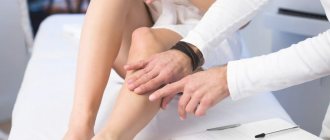
Many serious pathologies originate in seemingly small, insignificant manifestations, which rarely anyone pays attention to. Even mild vascular changes can subsequently lead to stroke, heart attack, or amputation of limbs. Vascular diseases occur for various reasons; these can be psycho-emotional stress, unbalanced nutrition, or a stressful rhythm of life. The earlier an unstable condition and the onset of the disease are detected, the more competent and adequate the therapy is selected, and this is an important aspect for maintaining health. In medicine, preference is given to non-invasive research methods that are more informative, one of them is rheovasography of the lower extremities (RLG). Let's take a closer look at this method: what is the purpose of rheovasography, who is it indicated for, and how the examination results are interpreted.
The concept of rheovasography
Rheovasography is one of the diagnostic methods, which examines blood circulation in the lower and upper extremities of the patient. Blood circulation studied in this way is called hemodynamics. It reveals the picture of the condition at the time of examination of the cardiovascular system, and also allows you to assess vascular tone. RVG shows the condition of arteries and veins in a selected area of the legs and arms, and makes it possible to determine the changes occurring in the walls of blood vessels. When deciphering, it is clear whether there is partial narrowing of the vessels or whether there is complete obstruction.
In human life, the main load falls on the lower extremities, so most often the study is prescribed for this part, this allows you to assess the blood flow of the vessels of the legs and prescribe the correct treatment.
It is possible to undergo a revision scan not only with a doctor’s direction, but also independently. This study is carried out in many specialized clinics at an affordable cost. If you need rheovasography of the lower extremities in Moscow, you can contact the following addresses:
- 2nd Tverskoy-Yamskoy lane, building 10 - JSC "Medicine".
- Banny Lane, building 2 - SVSR.
- Microdistrict Novogorsk in Khimki - KB No. 119.
- St. Novozavodskaya, building 14a – clinic No. 2.
- St. Lyapidevskogo, 14/1 - “Dobromed”.
- St. Budaiskaya, building 2 – JSC Russian Railways, Central Design Bureau No. 2.
Depending on the institution in which the procedure is performed, its reputation, location, and the quality of the equipment used to perform rheovasography of the lower extremities, the price may fluctuate. Cost - from 800 to 2500 rubles.
Rheovasography is one of the non-invasive examination methods that does not require penetration into the body. High frequency current is used for conduction. This study is not used for the vessels of the head; in this case, the diagnosis is called rheoencephalography.
Rheovasography: what is it, types, how is the study carried out?
To examine various pathologies associated with the vessels of the limbs and blood supply, rheovasography is often prescribed. The technique is available in clinics of any level. Often, thanks to this method of research, diseases are discovered that other techniques are not able to detect.
RVG is one of the newest modern non-invasive diagnostic methods. It is used to examine the blood vessels of the legs and arms.
The technique helps to identify areas of arterial blockage that lead to inflammation. It is also possible to examine the vessels and identify disorders associated with their operation.
Principle of operation
The essence of the method is to detect the resistance of a skin area through which a harmless electric current is passed.
In the sensors attached to the body, a certain force and frequency of charge oscillations are set.
If the blood flow is not intense enough, then the resistance of the skin and tissues becomes higher, and vice versa.
The device’s readings are displayed on the monitor and then on a paper tape. Where all changes are recorded in the form of a curved line.
The process of performing the examination and providing information about the results is similar to an ECG. Based on these data, the doctor makes a diagnosis regarding the area being examined.
Kinds
There are two types of rheovasography:
- Detection of blood flow pathologies in the legs.
- Study of the vascular channels of the hands.
Based on disease statistics, diseases of the lower extremities are the most common, therefore rheovasography of this type is used more often.
Main indications
The disease can be identified and a procedure prescribed based on a person’s complaints. The patient must list the symptoms manifested over a certain period of time.
Complaints:
Suspicion of diseases
Some symptoms may indicate obvious diseases that can be identified through examination:
- atherosclerotic lesion due to stenosis or complete occlusion of the supply artery;
- autoimmune pathology with abnormal hemodynamics in the hands;
- diabetes mellitus with complications;
- inflammation of the walls of arteries from the inside;
- varicose veins
In addition, using RVG it is possible to determine the etiology of the disease. Understand what influenced the subsequent development of the pathology, the structure of the canals or reasons caused by lifestyle.
Contraindications
Speaking about contraindications to rheovasography, we can only name relative ones, since there are no absolute ones. Namely:
- In the presence of chronic diseases during the period of their exacerbation, this research technique cannot be carried out.
- Existing infectious diseases.
- If a person takes medications on a continuous basis that affect vascular tone, for example hemostatic agents or psychostimulants.
How to prepare
The procedure itself is quick and does not require special preparation.
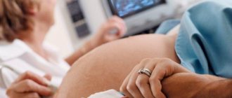
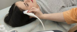
The patient should sit on the couch, on his back. The doctor attaches sensors in the form of suction cups to the area being examined. Then observes the indicators for 15 minutes.
Before performing RVG it is recommended:
- Before starting the examination, you need to relax your muscles and lie down for 20-30 minutes;
- stop taking medications that affect blood flow and blood vessels at least one day before the test;
- do not drink alcoholic beverages several days before the procedure;
- those who smoke should abstain for a couple of hours before rheovasography;
- on the day of the examination, do not overwork and try to avoid emotional stress.
This preparation guarantees the most accurate result of the readings obtained during rheovasography.
If you ignore the rules of preliminary organization, then a single ailment can be confused with a serious pathology. The specialist must take this into account.
In case of violation of the rules, an additional test must be carried out. This will allow you to compare the results of the indices and make the correct diagnosis.
How it goes
The room where the procedure itself takes place must be quite warm, since the patient is forced to take off his clothes, freeing his limbs for examination.
In order for the fixed electrodes to show accurate data, the person must lie in a motionless position. For convenience, a soft couch is available.
Before fixing the sensors, the surface fatty layer is removed from the skin. They are installed clearly according to the principle of longitudinal or transverse placement. This depends on the limbs being examined.
If the mechanical activity of the vessels of the arms is examined, then the sensors are placed in four places throughout the limb. On the legs these are the feet, legs and thighs.
Decoding the results
Obtaining research results does not take much time. Within 30 minutes, a specialist can decipher the received line and display the indices. RVG decoding is output in Ohms.
As mentioned above, RI shows the total volume of filling of blood vessels. With readings of 0.05 Ohm and above, we can say that their condition is normal.
With deviations of one unit, mild deficiency is observed. If RI is sharply reduced, the patient is diagnosed with a blood supply disorder.
Standards for vascular tone data without deviations are calculated from 0.4. Values that do not reach the specified units are already considered violations. The lower the indicator, the lower the tone of the vascular wall.
The outflow index shows the speed of blood movement in the venous canal. RVG should range from 0.2 to 0.5. Anything that exceeds this line indicates serious illness.
The IPI responsible for counteracting vascular channels should be in the range of 0.2-0.45. In case of obvious blood flow pathologies, the index may be higher or lower than the norm.
Additional samples
Violations that were recorded during RVG do not always indicate damage. The resulting index may have a functional nature of deviations.
Often such results are obtained when preliminary preparation is not followed. There are two additional samples. They help determine what is really happening, a temporary spasm or pathology:
- Nitroglycerin. Using the drug, the rheovasography procedure must be completed several times. The first occurs before the drug is used, and the second after its action. The doctor receives data from two studies and conducts an analysis. If comparable results do not differ significantly, then we can conclude that there is a pronounced pathology on the face. In the case where the drug had an effect and the indicators improved, one-time disturbances caused are assumed.
- Compression. The essence of the procedure is to use a cuff that compresses the required area of study. As in the case of the RVG drug, the patient undergoes it twice. Before and after applying the cuff to the limbs. This test helps determine how quickly blood supply is restored when the deep veins are blocked.
The latest rheovasography devices have a large number of positive aspects. They greatly simplify the work of specialists. The system itself calculates the results, which is much faster than routine calculations.
At the same time, the patient does not receive any radiation or other negative effects on his body. The method is more than safe and informative. Due to this, this diagnosis is relevant in medical practice.
Carrying out the procedure
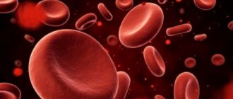
To carry out rheovasography of the vessels of the upper and lower extremities, you need an electrocardiograph (ECG machine), special electrodes and a rheographic attachment. The task of a medical worker is greatly facilitated if computer recording of indicators is used. The study uses high-frequency current, which does not cause any unpleasant sensations. Blood is capable of conducting electrical current, for this reason the resistance changes in different phases of the blood flow. The dependence here is inversely proportional: with greater blood supply, lower tissue resistance. There is a constant fluctuation from high to low indicators. The rheovasogram graphically displays the passage of the pulse wave; the amplitude increases with increasing blood supply and, conversely, decreases with its decrease. The functional diagnostics doctor sees abnormalities on the graph and can draw conclusions about the nature of the blood flow.
Indications for examination
Rheovasography of the vessels of the lower extremities is used both for the initial study of vascular pathologies and for monitoring the development of the disease. The indications are:
- Atherosclerosis, which often contributes to disruption of proper tissue nutrition and narrowing of the lumens in the artery.
- Diabetes. Rheovasography of the upper and lower extremities is performed. This disease can lead to micro- and macroangeopathies, which subsequently develop diabetic foot.
- Raynaud's disease. Neurocirculatory asthenia is characteristic, periodic spasms of peripheral arteries are possible.
- Severe headaches that arise due to disturbances in the outflow of venous blood, as well as spasms of arterial vessels.
- Chronic brain pathologies, intracranial hypertension.
- Neurocirculatory asthenia, which depends on the lability of vascular tone.
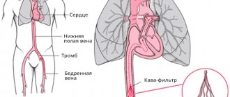
- Diseases of the venous bed, which contribute to disturbances in the outflow of blood, in particular through the veins of the legs.
- Thrombophlebitis, leading to the formation of blood clots on the walls of blood vessels.
- Phlebeurysm.
- Embolism, when there is a blockage in the bloodstream.
- Obliterating endarteritis, which can lead to lameness.
In the absence of pronounced diseases, RVG is recommended in cases of numbness of the extremities, the appearance of blue discoloration and convulsions, loss of sensitivity, changes in skin color, and swelling.
Indications for RVG of extremities
Rheovasography is recommended not only for an existing disease, but also in case of complaints from the patient about decreased temperature sensitivity (chilliness), tingling, cramps, blue discoloration, swelling, pain in the extremities. Below is a list of the most common diseases for which RVG is mandatory to assess the degree of hemodynamic impairment:
- varicose veins of the lower extremities, complicated by thrombophlebitis, phlebitis, ulcerative skin defect, swelling of the legs;
- obliterating endarteritis in long-term smokers or other diseases of the arteries of the lower extremities;
- atherosclerosis with the formation of atherosclerotic plaques of blood vessels, the presence of ischemia;
- Raynaud's syndrome (angiotrophic neurosis, leading to spasm of the small terminal arterioles of the hands, less often the legs);
- diabetic vascular angiopathy in patients with diabetes mellitus;
- some rheumatic diseases (periarteritis nodosa);
- acute embolism, thrombosis;
- vibration disease with damage to the arteries of the hands.
Pros of the examination
Each study has both its advantages and disadvantages. They may be related to the technical side or to the professionalism of the staff. It is important that all negative aspects can be corrected and the diagnostic process can be debugged for the better. The advantages of RVZ are as follows:
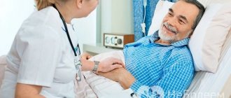
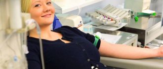
- Non-invasive method. A significant positive point is that the patient is not exposed to unpleasant and painful influences, for example, venous catheterization. The research does not require a secure, specially equipped office; the necessary instruments are sufficient.
- Ease of implementation. A big plus for the staff themselves. A rheovasogram can be taken by a qualified nurse; its task is to apply electrodes and turn on the resograph. The results will be analyzed by a doctor.
- Inexpensive and accessible equipment for research.
- Safety of the procedure. RVG can be performed in children and pregnant women.
- Reliable information. The patency of blood vessels, the quality of blood outflow, pathologies and their level of prevalence are determined with great accuracy.
- It is possible to carry out a differential diagnosis of vascular damage from a functional disorder.
Prices for rheoencephalography (REG) at SM-Clinic
| Name of service | Price, rub.)* |
| Rheoencephalography (REG) | RUB 3,550 |
— This is a painless and non-invasive (bloodless) medical diagnostic procedure that can be repeated as needed from several times a week to several times a month, including during pregnancy. - Conducted in a state of passive wakefulness. - In a sitting or lying position. — If an REG examination is prescribed for a child, parents should explain to the child on the eve of the examination that this is not a scary procedure at all (for example, that it is very similar to a game in which you can imagine yourself as an astronaut). — For a child with a high level of anxiety, the doctor may offer to attend another child’s examination in order to demonstrate to the little patient that the procedure is painless and safe.
— It is not recommended to discontinue antihypertensive drugs on the eve of the REG study due to the high risk of worsening the course of the disease or developing a hypertensive crisis. — The withdrawal of medications on the eve or on the day of the REG is carried out only in exceptional (expert) cases (as a rule, in a 24-hour hospital setting) - this must be agreed upon with your attending physician.
Wash your hair the day before the examination. Do not use gel, foam or hairspray. Before performing REG, hair must be dry.
— When conducting REG, a special gel (electrode paste) or physiological sodium chloride solution will be used. If you have an allergic reaction to the above listed medications, inform your doctor in advance. — Take a waffle towel or cotton napkin with you to dry your hair. — It is recommended to dry your hair with a hairdryer before going outside or before using hats (for example, in the cold season). — In our clinic you will be offered a free hairdryer so that you can dry your hair (when contacting other diagnostic centers, check the availability of such a service in advance). You must use your own (individual) comb.
Eating on the day of the REG should be no earlier and no later than 1.5-2 hours before the study. A light breakfast or afternoon snack (for example, a sandwich with juice or milk) is recommended.
— For children in the first months of life, REG is performed in a state of rest (relaxation) immediately after feeding, in a position in the mother’s arms or lying in a crib. - For children over one year old, the test is carried out no earlier than 1 hour and no later than 2 hours after eating. — On the day of the examination, do not drink tea, coffee, cola, caffeine or taurine-containing soft drinks (for example, “energy drinks” - flash, bern, etc.). — The day before and on the day of the examination, do not drink alcohol or low-alcohol drinks (for example, beer and kvass).
When contacting the clinical neurophysiology office for the purpose of conducting an REG, you must have the following medical documents with you:
— a referral for examination from your attending physician indicating the clinical diagnosis, medications you have been taking over the past month, and their daily (single) dose; — outpatient card at the place of attachment; — medical history with a prescription sheet, if you are being treated in a day hospital or in a 24-hour hospital; results of previous REG examinations.
The duration of the EEG examination is about 10-15 minutes.
— Arrive at the appointed time for the REG examination without delay (preferably 10 minutes before the start), otherwise the date of the examination may be postponed (postponed). — The duration of the study in restless children is increased by 20-25% (compared to the recommended) to achieve relaxation of the child, since tension and anxiety affect the results of the REG. — Plan your work (study) schedule in advance so as not to be nervous during the research. — A certificate can be prepared for you to present at your place of work (study) stating that you were in our Center for examination. If you need this medical document, please inform the doctor at the clinical neurophysiology office immediately upon contact.
Minuses
Rheovasography of the lower extremities has very small disadvantages, these include the following points:
- Human factor. If there are too many patients, the nurse's lack of proper rest can lead to a lack of attentiveness, and this can affect the quality of registration. If the working conditions and rest regime are observed, and the qualifications of the medical staff are high, then this minus is absent.
- Quality of technology. Rheographic attachments can cause interference, which affects the results. The use of computers eliminates such shortcomings in research.
Preparation for rheovasography
Preparation for rheovasography of the lower extremities is not at all difficult, but you must meet all the requirements that are presented before the study.
- 15 minutes before the test, the patient should completely relax and get a preliminary rest.
- Those who smoke should completely eliminate the entry of nicotine into the blood within two hours.
- If you are undergoing treatment with any medications, you must stop taking the medications a day before the RVZ.
- When undergoing rheovasography, the limbs must be freed from clothing.
Conducting research
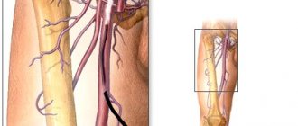
The patient lies on the couch, position - on his back. The skin of the limbs is degreased with alcohol. Sensors are attached to the treated areas. Wires stretch from the device and end with electrodes, which will read the information. The signals are transmitted to the screen, the rheovasogram is recorded, and the main indicators are calculated.
A rheovasogram can be carried out using multichannel rheographs, or the study is carried out sequentially, starting from areas remote from the center ones, ending with those close to them. A characteristic feature when placing conductors is strict symmetry.
A little information about the rheograph
The device is based on a generator that generates current. Using a special attachment, measurements are converted into graphic form.
Using appropriate electrodes attached to certain areas of the body, a rheogram is recorded.
What is rheography? At the beginning of the procedure, an electrophoresis-treated napkin is placed on the patient’s skin, through which the electrode reacts better with the body.
The skin is first wiped with medical alcohol; the presence of a fatty layer worsens the result of the process.
Indicators
Rheovasography of the lower extremities is carried out to study the condition of blood vessels using quantitative indicators. Synchronous waves of readings are equal to the pulse rate. The waves can be used to judge the filling of blood vessels, depending on the phase of cardiac contraction in a certain time period. As a result of the study, RI (rheographic index) is derived. It is calculated by comparing the wave amplitude with the height (calibration pulse). The value of RI directly depends on the filling of the vessels with blood; it can be used to calculate the overall intensity with which the organ is filled with arterial blood. The main indicators also include the following parameters: the amount of outflow; elasticity; peripheral resistance.
Modern diagnostic methods
To make an accurate diagnosis and identify the characteristics of tissue damage, consultation with an endocrinologist is not enough.
Considering that blood vessels in diabetes mellitus can be affected in any part of the body, identifying angiopathy often requires examination by such highly specialized specialists as a vascular surgeon, cardiologist, neurologist, ophthalmologist, dermatologist, etc.
To compile a biochemical blood profile, the following is clarified:
- Glucose level;
- Contents of lipoproteins, cholesterol and triglycerides;
- Platelet count;
- Blood clotting rates.
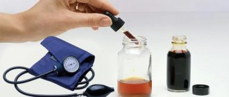
To assess the state of the cardiovascular system and identify angiopathy in this area, the following is prescribed:
- ECG;
- daily monitoring of blood pressure and ECG;
- EchoCG;
- CT angiography;
- coronography;
- Doppler ultrasound of the aorta.
To clarify the extent of vascular damage in the brain, duplex scanning, ultrasound and angiography are performed. When characteristic symptoms of angiopathy of the lower extremities appear in diabetes mellitus, the following is prescribed:
- rheovasography;
- peripheral arteriography;
- duplex scanning;
- capillaroscopy;
- oscillography.
To assess the patency of the arteries of the legs and the speed of blood flow during angiopathy, an ultrasound study of tissue permeability is carried out in them. Ultrasound can quickly detect the presence of atherosclerotic plaques and blood clots.
Rheovasography of the vessels of the lower extremities (interpretation)
After receiving the rheovasogram, it’s time for decoding, which is carried out by a qualified specialist. This is the main point of the study; here all pathologies are detected and the issue of further treatment is decided. Decoding rheovasography of the lower extremities includes the following important points:
- The main emphasis is the study of RI. A value of 0.04 indicates a sharp decrease in the norm, an index of 0.04-0.05 indicates a moderate decrease. An RI greater than 0.05 is considered normal.
- Elasticity index (IE). A sharply reduced rate - less than 0.2. Moderately reduced - 0.2-0.4. Normal - 0.4.
- To determine the outflow of blood in the vessels, the corresponding index is calculated, the norm of which is 0.2-0.5. If the value is lower, the outflow is facilitated; an indicator above 0.5 indicates difficulties in outflow.
- Peripheral resistance indicators: more than 0.55 – overestimated; less than 0.15 – underestimated. The norm is 0.2-0.45.
Results, their interpretation
After deciphering the received rheovasogram, the doctor calculates several indicators, which include:
- Elasticity index - shows the condition of the artery walls (their elasticity). Normally it should be above 0.4. A decrease below normal indicates poor elasticity of the artery walls with a corresponding deterioration in blood flow in them. A decrease in this index below 0.2 is a prognostically unfavorable sign.
- Rheographic index or outflow index - shows the capabilities of the venous bed; normally this indicator is 0.5. An increase indicates a deterioration in venous blood flow.
- The peripheral resistance index normally varies from 0.2 to 0.45; it characterizes the intensity of blood flow in small vessels (microvasculature).
Modern devices for rheovasography calculate the main outcome indicators using a built-in processor, which greatly simplifies the routine work of a doctor.
Additional drug tests may also be performed during this study. Typically, during the procedure, the person being studied is given nitroglycerin, which affects the functional state of the arteries. By its change, the doctor can judge the structural state of its walls. Compression tests are also carried out, which consist of temporarily compressing the main vessels of the limb, followed by their release and studying the rate of resumption of blood flow.
Due to the absence of any negative effects on the body, there are practically no contraindications for rheovasography. It is a safe and informative method of functional diagnostics that has not lost its relevance today.
Source: prof-med.info
Functional tests
Functional pharmacological tests can be used to identify hidden pathologies:
- Nitroglycerin. After the nitroglycerin is absorbed, RVG is performed, after which the result is compared with the usual one. This is necessary in order to distinguish organic contractions from functional spasms.
- Compression. Used in the study of deep vein thrombosis. To compare with conventional RVH indicators, a cuff is placed on the thigh and the indicators are recorded after its removal.
- Cold. It is carried out to diagnose various pathologies in the hands. Cold water is used. The rheogram is taken three times after a certain time, the results of blood flow restoration are compared, and conclusions are drawn. Negative test for RI recovery at the 7th minute. Positive test – slow recovery up to 30 minutes.
Treatment of the disease
Therapy for angiopathy of the lower extremities and vascular lesions in other parts of the body depends on the severity of the manifestations, the age of the patient and the presence of individual characteristics.
When treating angiopathy, medications are often used to eliminate vascular spasm, including in the legs, eyes, kidneys and other target organs. The choice of medications for blood vessels in diabetes mellitus complicated by angiopathy remains with the endocrinologist.
Conservative treatments for leg angiopathy and disruption of the circulatory system due to diabetes mellitus are aimed at reducing glucose, cholesterol and blood viscosity and eliminating arterial hypertension.
With progressive disruption of tissue trophism, surgical interventions can be performed to restore blood flow.
Drug therapy
Statin drugs are included in the treatment regimen. The most commonly used medications for diabetes mellitus include:
- Atorvastatin;
- Simvastatin;
- Lovastatin.
These drugs are especially useful for the blood vessels of the legs and coronary arteries of the heart, as they prevent the formation of cholesterol plaques.
To improve blood microcirculation, blood thinning drugs are prescribed. Commonly used means of this type include:
- Vazaprostan;
- Plestasol;
- Pentoxifylline.