You are here: Blood test -
General blood analysis -
Anisocytosis in general blood test
- Etiology
- Classification
- Symptoms
- Diagnostics
- Treatment
- Prevention and prognosis
Anisocytosis in a general blood test is a sign indicating a change in cell size in a biological fluid. It is detected only during laboratory blood tests. A person of any age, even a child, can experience this disorder.
The reasons for such measurements are often pathological conditions - the course of oncological processes, diseases of the liver and other internal organs.
Most often, such changes occur asymptomatically for humans. Without considering the symptoms of the underlying disease, signs such as sudden increase in heart rate, shortness of breath and weakness may indicate anisocytosis.
Diagnostics aimed at identifying the etiological factor may consist not only of a general clinical blood test, but also of instrumental procedures.
This anomaly is treated using conservative methods. If the root cause of the pathology is not treated, there is a high probability of complications developing.
What is anisocytosis?
The morphology of erythrocytes is varied and is divided mainly based on the size of the element:
- Microcytes,
- Megalocytes,
- Macrocytes,
- Normocytes.
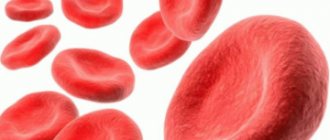
Altered and normal red blood cells
Normocytes are cells with a size of about 7.1-9 microns. Microcytes measure up to 6.9 microns. Macrocytes are represented by cells with a size of 8 microns. And megalocytes have sizes from 12 microns.
With normal red blood cell counts, the blood contains about 70% of the total number of normocytes. Macro- and microcytes occupy a niche of 15%. If the analysis shows an increase or decrease in the content of one or another type of element, then this is already a sign of developing disorders in the body, and therefore the RDW value also increases. This is called anisocytosis.
An increase in RDW can be recorded during the treatment of anemia, even at the initial stages. The picture changes as soon as the first stage of therapy is completed.
That is, if we explain it in simpler terms, then with anisocytosis the qualitative composition of erythrocytes changes significantly, in which normocytes are replaced by micro- and macrocytes. The main difference between the pathology is expressed in the violation of the correct ratio of different sizes of red blood cells. This deviation is determined by a general blood test (CBC).
Even with minor micro-changes in the composition of the blood, it is necessary to carry out a number of additional diagnostic manipulations of laboratory and hardware type (blood, urine, stool tests, MRI, CT, and so on, depending on which diagnosis needs to be clarified) to exclude more serious diseases. Microcytosis is most often associated with anemia, in which the hemoglobin content in all red blood cells is noticeably reduced. Therefore, it is necessary to periodically monitor the condition, which will exclude more serious diseases.
Platelet anisocytosis
The protective function of preventing acute blood loss is performed by blood particles called platelets. They are very important for the human body and are responsible for the ability of blood clotting. In the analysis, the normal indicator of the number of particles of changed sizes should be from 14 to 18%. With platelet anisocytosis, the numbers are different. When examined in the analysis, the platelet index is designated as PDW.
This pathology has its origins and is explained by the presence of various diseases, since it is only an accompanying symptom. The patient feels a physiological change. His health is deteriorating. Changes in the size of platelet cells are provoked by hemorrhoids (anal fissures) and heavy menstruation. Other possible reasons:
- myeloneoplastic processes;
- leukemia;
- liver failure;
- viral infection;
- radiation sickness;
- aplastic anemia;
- lack of biologically active substances;
- DIC syndrome.
RDW standard
A normal body produces red blood cells that are almost identical in volume. Moreover, in a healthy adult, normocytes make up about 70% of the total volume of red cells. With anisocytosis, red cells have different shapes and volumes.
RDW analysis allows us to determine the degree of distribution of red blood cells by volume, that is, their heterogeneity. As a person ages, these cells gradually begin to decrease in volume. The same changes occur with anemia.
The following approximate indicators are considered the RDW norm:
- 13% for adults,
- 16.8% for children under six months,
- 13.2% for children six months and older.
Indicators can vary between 1.5-1.9%. If the transcript shows a deviation from the norm, then it is necessary to examine the patient followed by therapy.
RDW standard
Classification of anisocytosis
Anisocytosis is divided primarily according to the severity of the condition.
- The first stage is characterized by slight to moderate anisocytosis. Red blood cells are 30-50% composed of macro- and microcytes.
- The second stage is characterized by average anisocytosis with a content of micro- and macrocytes in the amount of 50-70%
- The third stage is pronounced, severe anisocytosis, in which micro- or macrocytes occupy more than 70% of the total volume of red blood cells. If the index is so elevated, the reasons should be sought in the body itself - from severe anemia to oncology.
- This stage of pathology development is characterized by complete replacement of normocytes with micro- and macrocytes.
Normocytes are also elevated, as are micro-, macro-, and megalocytes. This is evidence of the development of various pathological processes in the body.
This disease is accordingly divided according to the type of cell predominant in the composition of red blood cells:
- Macrocytosis,
- Microcytosis,
- Mixed type anisocytosis, in which macrocytes and microcytes predominate at the same time.
This division allows you to pay attention to specific deviations in the composition of the blood, as well as what pathologies could cause them. The lower the indicator, the more serious the problem.
Anisochromia is a different degree of cell staining during OAC. It can be either the result of a serious pathological process or a normal reaction of the body to changes in the environment.
Types of Anisocytosis
To identify positive changes in the blood, a complete study should be carried out.
Anisocytosis poses a threat to the body not on its own, but as a cause for the occurrence of more serious diseases. One of them is poikilocytosis.
The disease is diagnosed when red blood cells change significantly in appearance and undergo deformation, and their concentration exceeds a critical value.
The stages of disease formation are determined by the following parameters:
- minor degree – below 30%;
- moderate – 30 – 50%;
- pronounced – 50 – 70%;
- pronounced – more than 70%.
Even with a reduced rate of anisocytosis, especially in children, auxiliary tests should be taken for disorders that have occurred in the body to exclude other serious pathologies.
When the number of healthy blood cells is greatly reduced, metabolic processes slow down. This indicates a delay in the delivery of oxygen to tissue cells, resulting in anemia. Anisocytosis is the first sign of the development of initial stage anemia.
If treatment is not prescribed in a timely manner, the pathology moves into the second stage, at which poikilocytosis develops.
Anisocytosis is determined not only when the size of red blood cells is changed. Disturbed platelet sizes also pose a significant threat to health. These deviations can occur for various reasons.
Symptoms
Any pathological changes in red blood cells adversely affect the human condition. You should consult a doctor and get tested if you experience the following symptoms:
- Fatigue, general malaise.
- Deterioration of breathing, shortness of breath with slight physical exertion.
- Heart rhythm disturbances. Appears quickly and disappears on its own.
- Pale skin.
- Brittle hair and nails.
- Regular bumps on the corners of the lips.
- Dry mucous membranes.
- Difficulty swallowing, feeling of a foreign body in the throat.
- Deterioration in taste.
- Nausea, vomiting.
- Burning, itching on the labia minora and majora in women.
- Headache.
- Dizziness. Fainting conditions.
- During pregnancy - swelling, fetal hypoxia.
Signs
With mild and moderate anisocytosis, the patient's condition slightly worsens. Signs appear that are not typical for this pathology. With severe anisocytosis, the following symptoms are observed:
- muscle weakness that does not disappear even after a long rest or night's sleep;
- decreased physical and mental performance;
- moderate pain localized in the parietal or occipital part of the head;
- daytime sleepiness;
- tremor of the limbs observed after waking up.
With prolonged absence of treatment, the pathology worsens, and more dangerous manifestations, such as cardiac syndrome, are added to the above symptoms. The main signs of severe and critical anisocytosis include:
- increased heart rate;
- increased heart rate;
- breathing problems (shortness of breath, attacks of asphyxia);
- a feeling of shortness of breath that occurs at any time of the day and does not depend on physical activity;
- pallor of the skin, lips and mucous membranes of the oral cavity (in severe forms of anisocytosis, the tissues acquire a bluish tint).
Features of the course in pregnant women
Slight anisocytosis with a predominance of microcytes and macrocytes is often found in pregnant women. This is due to the development of anemia or hypovitaminosis caused by non-compliance with recommendations for proper nutrition and taking nutritional supplements. The clinical picture may include the following signs:
- pallor of the skin and mucous membranes;
- dizziness;
- general weakness, apathy;
- decreased appetite;
- deterioration of hair and nails.
In children
Anisocytosis can be detected in a blood test in a child of any age. In most cases, the cause of this phenomenon is a previous infection or vaccination. Weakening of the body contributes to the development of iron or vitamin B12 deficiency, due to which cell sizes deviate slightly from normal. You should consult a doctor if the pathological condition is accompanied by the following symptoms:
- pallor of the skin and mucous membranes;
- brittle hair and splitting nails;
- dry skin;
- indigestion, in which constipation is replaced by diarrhea;
- frequent occurrence of ulcers on the mucous membranes of the oral cavity;
- increased fatigue, irritability;
- deterioration of dental condition, change in taste perception;
- increased heart rate.
Important information: What do low neutrophils in the blood of an adult indicate (reasons and how to increase)
Causes of pathology
The main reason for the formation of a high level of small red blood cells is considered to be a failure of hemoglobin synthesis. The second reason is diseases of the plasma membranes. A number of diseases also contribute to pathology:
- Iron deficiency anemia - development causes starvation of the body due to an insufficient amount of incoming oxygen. Failures in hemoglobin synthesis occur.
- Microspherocytosis. In most cases (75%), it manifests itself hereditarily, provoked by a gene mutation, a failure in the production of substances responsible for the condition of the membrane - it can be significantly damaged. In such cases, the size of red blood cells decreases, they become round, entering the spleen. In children, the skull becomes deformed, a high palate develops, it is possible that they have extra fingers on the limbs, and the spleen and liver become enlarged.
- Thalassemia. Passed along a hereditary line, otherwise called “Cooley's syndrome,” it is characterized by low production of adult hemoglobin. In a child, the skull may resemble a cube; there is a violation of the shape of the nose and bite. If the pathology was formed in the early stages of life, then a lag in mental and physical development is possible.
- Inflammation, chronic infections.
- Cancer pathologies, which often manifests itself in neoplasms in the lungs, thyroid gland, mammary gland, bone marrow. A child has neuroblastoma.
If microspherocytosis is detected in a general blood test, additional tests are prescribed, thanks to which the presence of hepatitis is accurately determined.
In addition to diseases, the following factors can provoke microcytosis:
- Significant blood loss.
- Lack of iron in food.
- Breastfeeding period during pregnancy.
Laboratory diagnostics. Criteria
The main way to diagnose anisocytosis is a blood test.
Red blood cell distribution width (RDW)
This parameter is used to measure the size and volume of red blood cells. RDW increases in accordance with changes in red blood cell size.
The normal RDW parameters are as follows:
| Adults | 13 % |
| Children under 6 months. | 16,8 % |
| Children from 6 months. | 13,2 % |
If results exceed the normal range, this indicates the presence of anisocytosis.
Doctors often compare the RDW result to the mean cell volume (MCV).
Results may show:
| results | Causes |
| Normal RDW and MCV | Anemia may be caused by a chronic illness or blood loss. |
| Normal RDW and low MCV | Anemia caused by a chronic condition or thalassemia. |
| Normal RDW and high MCV | Liver pathology or alcohol abuse. Use of antiviral and chemotherapeutic drugs. |
| High RDW and normal MCV | Deficiency of iron, B12 or folic acid. Possible chronic liver disease. |
| High RDW and low MCV | Iron deficiency, microcytosis. |
| High RDW and high MCV | Lack of B12 or folic acid, macrocytosis. Possible chronic liver disease. |
Photo: https://pixabay.com/photos/medic-hospital-laboratory-medical-563423/
Color index
Another indicator used in the diagnosis of various types of anisocytosis. They indicate the amount of hemoglobin in a red blood cell.
The normal value is in the range of 0.85 - 1.05.
Hypochromia in a general blood test, when the color index is less than 0.85, may indicate the presence of microcytosis. Hyperchromia (indicator more than 1.0) possibly indicates macrocytosis.
Pathology in children
In the first months of life, an increased level of microcytes is observed, and this is normal. This is explained by the fact that the child’s organs are not fully formed.
This process is completed by about three months. If significant deviations are diagnosed, the child is hospitalized to exclude the development of congenital diseases.
In 1 year, the indicator should be completely normalized, otherwise a thorough examination is required. The primary degree of development indicates a violation of the immune system and requires adjustments in nutrition and the prescription of a vitamin complex.
In severe situations, indicates the development of leukemia.
Minor violations are normally allowed in adolescence, this is associated with hormonal levels and goes away on its own.
Anisocytosis in a child's blood test
Different types of anisocytosis occur in newborns, infants, as well as in preschoolers and schoolchildren. Microcytes in increased content are observed after infectious diseases.
Macrocytosis is normally found as a physiological process in infants, especially in the first two weeks of life. By 60 days of life, this pathology disappears on its own.
When diagnosing any type of anisocytosis in children, it indicates the following diseases:
- neuroblastoma;
- hypochromic anemia;
- chlorosis.
Diagnostics
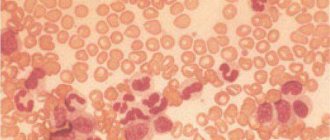
Microcytosis is detected during a blood test. If the presence of anemia is determined, a smear of peripheral material is additionally prescribed.
Thanks to this measure, the initial symptoms of micro- and macrocytosis (the diameter of red blood cells exceeds the norm) or a mixed type of anisocytosis, combining signs of the first and second types, are revealed. Normo-, hyper- or hypochromia are detected in the same way.
A hematologist deals with the disease; it is he who prescribes further diagnostics, determines the cause, and the necessary treatment.
As additional diagnostic measures, tests are prescribed to determine celiac disease, blood and stool tests for the presence of the microorganism Helicobacter pylori.
The specialist clarifies in detail all the signs and symptoms present in the patient; it often becomes necessary to consult a gastroenterologist (if pain appears in the abdominal area). Sometimes the following diagnostic methods are needed:
- Transabdominal ultrasound examination.
- Endoscopy of the stomach and intestines.
- Computed tomography of the abdomen.
If adult women experience heavy periods with pain, it is necessary to exclude the presence of uterine fibrosis and other factors that cause heavy bleeding.
Factors in the development of the disease
Formal components can distort their shape and size under the influence of various kinds of prerequisites. When performing a complete blood test, anisocytosis is detected immediately. A more in-depth analysis is required to detect poikilocytosis.
The main factors causing anisocytosis include a number of reasons:
- Poor nutrition.
- Blood transfusion.
- Myelodysplastic syndrome.
- Oncological diseases.
- Lack of iron, vitamins A, B12.
- Liver diseases.
- Various types of anemia.
- Thyroid gland dysfunction.
In addition, physiological disturbances in the diameter of red blood cells can be detected in an infant from the first days of life, which is not a symptom of any illness.
Factors causing an increase in anisocytosis are:
- microcytosis – iron deficiency anemia;
- megakaryocytes – lack of vitamin B12.
When a complete blood count reveals anisocytosis, nutrition should be reconsidered. The body requires elements involved in the construction of blood cells.
Treatment
Therapy is initially aimed at eliminating the causes of the pathology. For example, a lack of iron requires taking medications with a high iron content, which eliminates anemia. At the same time, it is recommended to use vitamin C, as it promotes better absorption of iron.
When diagnosing serious blood loss (acute or chronic), the clinical picture worsens. Women with heavy periods and iron deficiency are prescribed hormonal treatment.
The development of complications such as heart failure requires a blood transfusion or restoration of the number of red blood cells from a donor.
If nutrient deficiencies are detected, the diet must be reviewed. Such situations are best treated.
In advanced situations, lack of treatment can cause serious complications.
Treatment of anisocytosis
Treatment will depend on the cause of the anisocytosis.
It is important to determine the cause of the problem so that proper treatment can begin.
Anisocytosis is often associated with anemia, and anemia is usually caused by iron or vitamin deficiency.
The usual treatment for iron deficiency is taking iron supplements and changing your diet to increase your iron levels with iron-rich foods.
Iron-rich foods:
- dark green leafy vegetables;
- brown rice;
- beans;
- nuts and seeds;
- meat and fish;
- tofu;
- eggs;
- dried fruits.
Patients can also address vitamin deficiencies by taking supplements and making changes to their diet.
In severe cases of anisocytosis, your doctor may recommend a blood transfusion. This process will replace blood containing abnormal cells with blood containing healthy cells.
Photo: https://pixabay.com/photos/asparagus-steak-veal-steak-veal-2169305/
Consequences
If, with severe microcytosis, the patient does not receive the necessary treatment, the consequences can be serious. This primarily concerns cancer diseases, since their development leads to death.
Microcytic anemia can cause starvation of organs and tissues of the body due to low oxygen supply. This often results from heart failure and lung diseases.
In children, this leads to a decrease in the immune system, disruption of all organs and systems in the body.
Over time, clinical analysis shows that the number of normocytes decreases, this reduces the area of red cell membranes, and as a result, a number of disorders are observed:
- Deterioration of nutrition, protection, and gas transportation by cells.
- Disorders of metabolic processes.
- Decreased vascular tone.
- Low blood pressure.
- Coronary vascular disorders.
- Pulmonary pathologies.
- Shock.
Forms of the disease
Changes caused by pathology appear gradually in children. The table below discusses all stages of anisocytosis and their clinical features.
| Stage | Short description |
| First | The number of pathological cells does not exceed 25%. The condition of the formed elements is normal. |
| Second | The number of modified elements reaches 50%. The patient begins to show the first symptoms of the disease, such as: |
| change in skin color to a paler color; | |
| the appearance of shortness of breath and tachycardia. | |
| Third | The number of pathological cells reaches 70%. The young patient exhibits lethargy in behavior and physical activity decreases. The symptoms described above also appear. |
| Fourth | The number of changed cells reaches 90-100%. This condition is life-threatening for the patient. The patient cannot lead his usual lifestyle. The size of some organs changes. |
It is the form of the disease that is decisive when drawing up a treatment plan. It should be noted that the fourth form of anisocytosis is extremely rarely observed in children. As a rule, its appearance is preceded by cancer.
Prevention
There are the following measures to prevent microcytosis:
- Healthy eating. The diet should include fresh vegetables, fruits, herbs, and lean meat. Avoid eating processed foods and fast food.
- Exercise, walks in the fresh air. Due to them, the supply of oxygen to tissues and organs is improved.
- Refusal of alcoholic beverages and tobacco products.
- Establish a daily routine with eight hours of sleep and sufficient time for rest.
- Regular tests. Timely treatment starts speeds up recovery and avoids any complications.
- Avoid close contact with poisons and chemicals.
Self-medication is extremely unwise, since such measures can aggravate the situation. The specialist will make an accurate diagnosis as soon as possible, find out the cause that provoked the pathology, and prescribe the necessary therapy.
Microcytosis
Click to enlarge
Microcytosis is a condition when the majority (about 30%) of red blood cells change into a minority. Their number has increased, but the standard size differs several times.
Microscopic red blood cells do not transport full-fledged oxygen throughout tissues and organs in the same way as healthy and full-fledged normocytes produce.
With a normal, average diameter of a red blood cell in adults, 6.8 and 7.5 microns are found.
With a regular biconvex disc, the red blood cells are of normal diameter, volume, colors and shapes, this is called a normocyte. In private clinics, the norm is indicated, ranging from 6 to 8.5 microns. A child or teenager, taking into account age, is subject to a norm of 7 to 8.1 microns.
If microcytosis is detected in the analysis, iron deficiency anemia can be further diagnosed.
Symptoms of macrocytosis
At an early stage, symptoms may not appear. Changes in the size of blood cells are often discovered by chance during a routine blood test. Later stages are characterized by the following symptoms:
- fatigue, weakness;
- sudden weight loss;
- varnish tongue (with a smooth, shiny surface);
- pallor or yellowness of the skin;
- fainting;
- dyspnea;
- cardiopalmus;
- tingling in the limbs;
- brittleness of hair and nails;
- flashing “flies” before the eyes;
- dizziness.
All these symptoms are characteristic of the state of anemia, and each symptom separately helps to determine what exactly the body lacks at the moment to restore oxygen metabolism. Brittle nails and hair indicate a lack of iron, a varnished tongue indicates a lack of vitamin B 12.
Symptoms of macrocytosis appear cyclically, that is, the patient notices them periodically, and not constantly. This often leads to the fact that a person simply gets used to it and does not pay attention to changes in his body. Meanwhile, any of these symptoms is a reason to consult a doctor.
Mixed anisocytosis
In this pathology, the level of the overall content of increased and decreased cell sizes decreases. To correctly determine the percentage during the survey, the Price-Jones method is used.
Price-Jones curve: click to enlarge
This type of anisocytosis indicates the predominance of macrocytes. The source of the changes is a lack of vitamin A, B12, which suggests the formation of anemia.
The problem may also relate to kidney failure. If macrocytosis is detected above the established norm, this means iron deficiency.
Causes of macrocytosis
A detailed examination of the body allows us to determine the cause of the pathology.
The main reasons are:
- Lack of cyanocobalamin (vitamin B 12),
- lack of folic acid (vitamin B 9),
- dysfunction of the thyroid gland,
- hepatitis,
- intestinal diseases,
- damage to liver tissue,
- bone marrow pathologies,
- spleen diseases,
- some forms of leukemia,
- use of medications (Chloridine, Allopurinol).
Macrocytosis can also be caused by congenital anomalies. But often it is poor nutrition or alcohol abuse that becomes hostage to disorders.
Treatment of macrocytosis
Abnormally large blood cells are an indicator of the presence of the disease. Therefore, it is necessary to treat the root cause, and not the consequence of the development of the disease. The basis for restoring natural processes in the bloodstream is good nutrition, as well as giving up alcohol and smoking. A diet poor in vitamins provokes disturbances in blood formation, and alcohol-containing drinks lead to impaired liver function. Cigarettes contain poisons that contribute to intoxication of the entire body.
Additionally, depending on the disease, the following may be prescribed:
- course of vitamins,
- immunoglobulins,
- iron-containing preparations,
- blood transfusion,
- therapy with glucocorticoid drugs.
Severe forms of disease require mandatory hospitalization of the patient.
Prevention and prognosis
To prevent the problem from developing, it is enough to follow a few simple rules. Preventive recommendations:
- complete renunciation of addictions;
- complete and balanced nutrition;
- frequent exposure to fresh air;
- constant strengthening of the immune system;
- avoiding physical and emotional stress;
- regular comprehensive preventive examination in a medical institution.
Regardless of whether the volume of formed parts of blood is reduced or increased, the prognosis will be dictated by the etiological source. It is necessary to take into account that each basic disease has a number of its own complications.
Prognosis and possible complications
Macrocytosis is a dangerous sign of a number of diseases characterized by a lack of oxygen in the body.
Immune suppression is one of the first consequences of this disorder, which leads to frequent infectious diseases and their complications.
In order to speed up the delivery of hemoglobin to the organs, the heart muscle begins to contract more often. The result is heart failure and tissue wear.
One of the common complications is a disorder of the central nervous system, which is manifested by impaired concentration and memory.
Lack of oxygen also leads to deformation of the structures of the mucous membranes and skin.
In childhood, the impact is on mental and physical development.
A condition in which tissues are not fully saturated with oxygen over a long period gradually leads to their degeneration. Severe forms of the disease lead to heart attacks, cardiogenic shock, and acute renal failure.
Timely treatment and dietary correction allow you to restore the natural circulation of red blood cells. However, in the future you should regularly monitor your health and undergo examinations.
Reasons for the development of anisocytosis
Isolated anisocytosis of red blood cells may be present in mild stages of anemia. It can develop in a woman during her period, especially if for one reason or another it is somewhat delayed. There can be many reasons for this condition, both minor and quite serious.
The most common symptom of anisocytosis is:
- Anemia, hypochromia, chlorosis, post-hemorrhagic anemia, hyperchromic anemia and so on,
- Lack of vitamin B12,
- Iron deficiency
- Lack of vitamin A,
- Lead poisoning
- Malignant neoplasms,
- Received blood transfusion.
Iron deficiency
In the case of the last point, we can say that this is a passing anisocytosis, which will eliminate itself as soon as the body gets used to the “new” blood and replaces the diseased cells with healthy ones. So in this case, you just need to wait time and, if necessary, replenish internal reserves with vitamins, minerals and other useful substances.
Often the state of platelets changes in parallel with pathological changes in erythrocytes.
Causes of microcytosis
An increased content of microcytes is typical for different groups of people at different periods of life. Microcytosis can often be observed after an infectious disease in a child. If the level of such red cells is slightly increased, then it can be easily corrected with the right lifestyle and nutrition.
This is a common condition in infants, since the systems are just returning to normal. Therefore, such conditions can go away on their own without treatment. Microanisocytosis also often affects women during pregnancy and lactation due to hormonal changes in the body and high consumption of nutrients.
In other cases, anisocytosis of this type can be caused by:
- Iron deficiency or microcytic anemia,
- Microspherocytosis,
- Cooley's syndrome or thalassemia,
- Chronic infectious and inflammatory diseases,
- Heavy bleeding
- Lack of foods with iron in the diet,
- Oncological lesions.
It is important to remember that some diagnoses require not only clarification and lifestyle adjustments, but also serious treatment. Oncological diseases can develop very quickly, and therefore one should not delay diagnosis.
The deterioration of cell characteristics can lead to the deterioration of red blood cell function.
Causes of macrocytosis
Macrocytosis may also indicate the development of anemia. Third-party factors may also influence this sign. Even a lack of nutrients and vitamins in the diet can significantly change blood composition.
But often an increase in the number of macrocytes is caused by such conditions as:
- Heavy bleeding
- Various types of anemia
- Bone marrow pathologies,
- Oncological lesions, most often blood,
- Thyroid dysfunction
- Chronic alcoholism,
- Diseases of the hematopoietic organs,
- Taking certain medications.
Macrocytosis
Some diseases are quite serious and require immediate intervention. Often, some pathologies end in death in the absence of adequate therapy.
Mixed type
The mixed type of anisocytosis is manifested by the dominance of macrocytes and microcytes over normocytes. Combined pathology with a predominance of macrocytes is characterized by the presence of an impressive number of abnormally large red blood cells. They are detected in blood smears and have a hyperchromatic structure without clearing in the center. The cells look like ovals with a diameter of about 11-12 microns.
Detected when:
- Vitamin B12 deficiency,
- Folic acid deficiency,
- Hypochromic anemia,
- Pernicious anemia,
- For anemia during pregnancy,
- With dyserythropoiesis,
- With helminthic infestation.
This deviation quite often develops when affected by worms, and therefore it is necessary to identify their presence, as well as determine the type of disease. Only in this case will it be possible to recover from pathogenic organisms.
Anisocytosis rate higher than normal can even be due to a simple change in the environment, due to moving.
Development mechanisms
The process is based on a group of factors. Among these are three key points:
Congenital anomalies of the hematopoietic system
They are relatively rare in clinical practice. Such deviations are usually caused by genetic mutations.
They are inherited in a recessive way, that is, for the development of a pathological process, a combination of altered biomaterial of the father and mother is required.
Changes cannot be corrected qualitatively, since the problem lies deep in the body. The only thing that specialists can do is to fight the symptoms, maintain well-being at an adequate, acceptable level, and prevent deadly complications.
Bone marrow changes
For example, damage due to malignant neoplasms, myeloproliferative diagnoses, disorders due to radiation sickness, intoxication with toxic substances. Be it lead, mercury, arsenic, vapors and salts of heavy metals. There can be many options.
All of these factors directly affect the nature of hematopoiesis. Erythrocytes (red blood cells) and platelets become too large or inappropriately small.
The coagulation process is disrupted, and it is not possible to provide the body with oxygen and nutrients.
Structural pathologies
Not directly affecting the hematopoietic system, but affecting the processes of production of formed cells and their size.
This includes diseases of the digestive tract, possibly hepatitis, and cirrhosis of the liver. These also include kidney disorders and hormonal imbalances. A common cause of anisocytosis is anemia: iron deficiency and megaloblastic.
Mechanisms for the development of anisocytosis in adults and children occur, including in combination.
Assessment of pathogenesis is necessary to develop a high-quality correction scheme, determine prognosis and prospects for recovery.
Also, specification allows us to better understand the essence of the disorder, which is important within the framework of the general epidemiology of diseases accompanied by changes in the structure of formed blood cells.
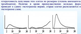
Analysis on RDW
This analysis is carried out using analyzers. They help to accurately count red blood cells of all sizes and shapes in 1 μl of blood. The sampling is carried out on an empty stomach. The material for the study is taken from a vein.
The average capacity of an erythrocyte is determined automatically, and the deviation in the number of normocytes in relation to macro- and microcytes is calculated. The data is displayed in the form of a histogram. If the percentage of RDW falls within the normal range, then the result is recorded as negative.
If the norm is exceeded, a positive result is indicated. For such cases, the procedure is duplicated in order to identify the causes of abnormal indicators.
Sometimes a false positive result occurs, especially if there are a large number of macrocytes in the blood. These results often occur after surgery or blood transfusion. The redistribution or change in the type of cells itself can change frequently, and in a short time.
Therefore, it may be necessary to process the data using a statistical analysis method. The standard deviation value is calculated using a number of formulas. Previously, the results were obtained manually, but this is an extremely labor-intensive process, and therefore today it has been practically abandoned in favor of computer data analysis.
Treatment of anisocytosis is directly related to the elimination of the cause of its development. Depending on the type of disease, a treatment regimen is prescribed to eliminate influencing factors. In case of iron deficiency anemia, a course of replenishing the deficiency of the substance in the body with the help of medications is required.
Oncological diseases require a more serious approach in the treatment of anisocytosis using hormonal, chemotherapy, radiation therapy, and so on.
If a general blood test reflects a change in the volume of normocytes according to the RDW index, then there is a high probability of developing anemia. This is one of the first indicators when making this diagnosis. But only this auxiliary criterion in making a diagnosis will not give an accurate picture of the type of pathology and the patient’s state of health.
Therefore, it is necessary to undergo a full diagnosis and begin treatment of the disease.
How to treat anisocytosis?
Complex therapy includes:
- Treatment of the disease that caused the development of anisocytosis.
- Taking special medications that help maintain the number of increased blood cells within normal limits. In some cases, the use of medications is not required. In newborn children, abnormalities are often detected during a general blood test. But this phenomenon is the norm in most cases. After a certain time, these changes disappear on their own.
- Refusal to use medications that reduce iron levels in the body.
The duration of the therapeutic course is determined by the doctor. It depends on the degree of the disease and the patient’s condition.