The mechanism of development of expansion of the borders of the heart
The pathogenesis of cardiomegaly depends on the cause. Most often, left ventricular hypertrophy occurs in people with metabolic syndrome, coronary artery disease, or arterial hypertension.
When oxygen supply is low, the heart muscle contracts more strongly than usual and gradually increases in size. Roughly the same thing happens with hypertension.
In this case, the heart does not have time to pump blood quickly enough due to its high pressure, so the organ requires more effort. The mechanism of development of cardiomegaly differs in stenosis and valve insufficiency.
In the case of these pathologies, the blood does not completely flow into the adjacent chamber or vessel (aorta, pulmonary artery) and causes stretching of one of the parts of the heart. With long-standing defects, both the ventricle and the atrium enlarge.
In some cases, hypertrophy of the entire organ may occur. Right ventricular failure occurs with pulmonary pathologies and liver diseases.
Coronary diseases
Hypertension is the most common cause of heart enlargement.
This is explained by the fact that due to increased blood pressure, the organ is forced to pump large volumes of it and work in enhanced mode.
This causes the heart muscles to enlarge and the organ itself to expand.
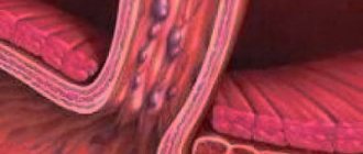
If a person has ischemia, the heart muscle cells constantly do not receive enough nutrients, as a result of which they degenerate, and connective tissue appears in their place.
The latter, unlike muscle tissue, is not capable of contraction; as a result, the organ cavities become deformed and increase in size.
What to do if an x-ray showed that the organ is enlarged, and the cause of this phenomenon is a disease of the cardiovascular system?
The answer to this question is simple and obvious - treat the root cause and return the organ to normal limits.
If a patient is diagnosed with hypertension, he is usually prescribed pharmaceuticals that lower blood pressure. The latter helps restore the normal size of the organ.
A patient with hypertension or coronary artery disease who has been diagnosed with an enlarged heart must take medications.
The fact is that despite the increased size of the organ, a large heart performs its most important function - pumping blood - much worse, which means that human organs and systems do not receive the nutrients they need - heart failure develops, and the whole body suffers.
Reasons for the formation of pathology
Synchronous cardiac hypertrophy to the right and left is an indicator of ventricular expansion. The main reasons that cause changes in the boundaries of the organ are chronic diseases, heart diseases, drug and alcohol intoxication.
The most important organ that ensures the vital functions of the entire body consists of 4 chambers: two ventricles and two atria. There are two sections of the heart - right and left, each of which includes an atrium and a ventricle.
Normally, the boundaries of the heart change throughout life. People who play sports experience an increase in its size, which is considered a completely natural process and does not cause any cause for concern.
The weight of a man's heart is 332 grams, a woman's - 253 grams. An enlarged heart is observed when the myocardium grows and (or) expands its cavity.
Most often, the organ enlarges to the left, which is observed in many diseases: hypertension, stagnation of blood in the systemic circulation, heart defects. Enlargement of all parts of the organ is dangerous.
This condition is called “bull’s heart”, and only surgical intervention can improve a person’s quality of life.
The reasons influencing the enlargement of the organ are:
- Hypertonic disease. An increase in pressure leads to changes in the cardiovascular system: vascular tone increases, the thickness of the muscle layer increases, and the systemic circulation suffers.
- Coronary heart disease: angina pectoris, myocardial infarction. Oxygen starvation of the organ tissues occurs with the death of their cells and replacement with connective tissue, which leads to an increase in the size of its left section.
- Rheumatic heart disease. It is a consequence of tonsillitis (frequent tonsillitis). Rheumatic disease is manifested by an inflammatory process occurring in the tissues of the organ. As a result, the valves suffer and defects form.
- Myocarditis.
- Renal dysfunction.
- Alcohol abuse. The most common example is alcoholic cardiomyopathy.
- Smoking.
- Acute pericarditis (inflammation of the serous membrane).
- Congenital heart defects.
The weight of an average man’s heart is 332 grams, a woman’s – 253. It is considered normal if the weight of the organ varies within these limits.
As for the sizes, they are usually correlated with a person’s fist. For an organ to function normally, it is very important that all its parts (atria, ventricles) are normal, or more precisely, the thickness of their walls, length and width as a whole.
What to do if fluorography (x-ray, ultrasound) showed that the heart is enlarged and dilated? How dangerous is it to literally have a big heart? And as a result of what can the organ become larger? Let's figure it out in order.
The most important reasons why the heart is larger than normal in a fluorography image include:
- Great physical activity.
- Diseases.
In people who engage in heavy physical labor every day, as well as in professional athletes, the heart also works harder: it is forced to beat more often and pump blood faster.
This leads to the fact that there are often more heart muscle cells and they grow. As a result, the weight of the organ and its size increase.
If physical activity in the future is moderate, an enlarged heart for this reason does not pose a health risk. If a person subjects his body to excessive stress for a long time, then it is possible to develop a pathology such as a hypertrophied heart, which is already fraught with serious complications and even life-threatening.
The reason that the heart is enlarged in size can be diseases of the cardiovascular system (coronary diseases: for example, hypertension, coronary disease) and the heart itself (viral, inflammatory diseases), as well as heart defects.
Enlargement of the heart to the left
Hypertrophy of the ventricular walls means an increase in the heart muscle. The following reasons can lead to pathology:
- vices;
- diabetes mellitus and high blood pressure;
- long-term treatment with antibiotics;
- pregnancy period;
- anemia;
- alcohol consumption;
- renal failure;
- irregular training with enlarged heart muscle in athletes;
- rheumatism;
- myocardial infarction.
People who are constantly forced to do hard work, these include professional athletes, their main vital organ is forced to work more actively. Regular increased exercise leads to the proliferation of muscle cells. Fluorography will definitely show that their heart is enlarged to the left in the picture. Reducing the load to moderate will bring the enlarged organ back to normal. If the amount and strength of the load is not changed for a long time, complications and death may occur.
Changes begin to appear when the load increases regularly. The pathology has no clear signs, but it can manifest itself with the following symptoms:
- shortness of breath and fatigue;
- swelling of the lower extremities;
- feeling of heaviness on the right side under the ribs;
- headache and tinnitus;
- high blood pressure;
- cough for no reason.
Enlargement of the organ to the left is more common, which may indicate diseases such as hypertension, stagnation of blood in the systemic circulation, and various defects. This condition is sometimes called "bull's heart."
Diagnosis and treatment
Today in the world of medicine there are a huge number of methods for diagnosing cardiac pathology. In this situation, the cardiologist prescribes:
- Chest X-ray.
- Electrocardiogram.
- Echocardiography.
- Computed tomography or magnetic resonance imaging (if necessary).
- Blood chemistry.
It should be remembered that a correct diagnosis plays an important role in the treatment of any disease. Therefore, after studying the results obtained, the doctor determines treatment tactics.
Treatment should begin with lifestyle changes. Proper nutrition (exclusion of fatty, spicy, salty foods), sleep for at least 7 hours, absence of bad habits, and exercise will help you get rid of the diagnosis of “heart hypertrophy.”
Treatment consists of prescribing diuretics (removing excess water reduces the load), anticoagulants (reduce the risk of blood clots and, as a result, minimize the manifestations of coronary heart disease). Drugs are also prescribed to help restore cardiac activity.
Surgical treatments are used when a person's life is at risk. The most advanced form is considered to be “bull heart”; in this situation, transplantation of the affected organ is recommended.
If the structure of the valve changes, prosthetics are performed. If changes in heart rhythm are observed, a device is installed under the skin to correct it.
Experts associate changes in the volume of the heart muscle with:
- with the formation of hypertrophic enlargements of the ventricles;
- accumulation of metabolic products in the cavities of the heart;
- with neoplastic processes - the appearance of neoplasms in the tissues of the heart.
The deviation is also recorded in healthy people - professional athletes, pregnant women. The standard volumes of the heart muscle are individual for each person. Experts note that males always have a larger organ than females.
Heart size is related to the patient's body weight. The diagnosis of “cardiomegaly” can be made solely according to diagnostic studies - in some cases, a slight increase in the heart muscle may be included in the normative indicators.
Upon initial visit to a medical facility, the patient is referred for consultation to a cardiology specialist. A presumptive diagnosis is made by collecting anamnestic data:
- complaints of the sick person about changes in the usual state;
- presence of chronic diseases;
- existing bad habits and duration of passion for them;
- previous surgical procedures.
After a physical examination, the patient is sent for instrumental examination:
- X-ray of the thoracic region - the images will help to see the expansion of the heart muscle, existing congestion;
- fluorography is a mandatory method for determining pathological abnormalities in the pulmonary region;
- ECG – will show general data on the work of the heart;
- echocardiography - will determine the physical characteristics of the organ - the size of the heart chambers, necrotic areas and ischemic lesions;
- Ultrasound;
- CT;
- MRI;
- clinical blood tests to determine cholesterol, bilirubin, protein elements, hormones, etc.
After identifying the root causes of the formation of a pathological deviation, the specialist will make a final diagnosis and prescribe symptomatic therapy.
First of all, the doctor must collect the patient’s medical history: find out about the presence of chronic diseases, previous operations, possible bad habits. After which research is carried out. Percussion determines the size and boundaries of the organ, which makes it possible to identify which parts of the heart are enlarged, and then judge the possible causes of the disease.
The laboratory carries out:
- blood chemistry,
- fluorography,
- Ultrasound,
- CT scan.
If a doctor determines that the causes of a large heart are chronic or acute diseases, then treatment of these diseases must be carried out. If it is started on time, the organ decreases in size.
If the cause is a heart defect, then you need to consult a cardiac surgeon and, if necessary, undergo surgery. This will allow you to maintain the functionality of this vital organ for a long time. After surgery, symptomatic treatment is prescribed.
It is necessary to slow down the process of heart enlargement in the patient. If a person moves little, does not watch his diet, or has a number of bad habits, to solve the problem he needs to reconsider his lifestyle. This means starting to exercise in moderation and eating foods high in vitamins and microelements.
If treatment is not started promptly, the consequences can be very serious. That is why you should not neglect the recommendations if the doctor prescribes diet, sports or surgery. For any cause of the disease, drug treatment is prescribed that will last throughout the patient’s life.
Non-coronary diseases
Another fairly common cause of an enlarged heart is inflammatory processes affecting muscle tissue (carditis), primarily rheumatic carditis.
So, if a person has suffered a serious infection such as tonsillitis or scarlet fever, complications (rheumatism) can also affect the most important organ that transports blood.
In this case, the muscle loses its elasticity, and the ventricles are overstretched, as a result of which the size of the organ can increase several times, and its functionality, accordingly, will decrease several times.
In this regard, timely treatment of rheumatic carditis is very important. To date, drugs have been developed to completely eliminate streptococcal infections and prevent overstretching of the heart.
If therapy is not followed, the person may die. In addition, being a carrier of streptococcus, the patient infects others.
Endocarditis is an inflammatory disease that affects the internal cavity of the heart and its valves.
Endocarditis in an advanced stage causes expansion of the organ, loss of muscle elasticity and ability to contract. The disease requires immediate treatment.
Myocarditis is a consequence of viral infections, accompanied by arrhythmia and shortness of breath, and heart failure may occur. Video:
In this regard, a patient with myocarditis needs immediate medical attention and supportive care.
Chronic alcohol consumption can cause cardiomyopathy and cardiac dystrophy, as a result of which the heart cavities expand and the heartbeat rhythm changes significantly.
Also, patients with alcoholism, as a rule, have high blood pressure - another factor that contributes to the modification of the heart muscle.
If a person recovers from alcoholism and stops drinking alcohol, and if he has hypertension, takes blood pressure-lowering medications, after some time the organ will restore its normal size.
Thus, if a fluorography image reveals an increase in the size of the heart, you should immediately contact a specialist, find out the cause of the pathological changes and, if necessary, begin therapy: the problem is solvable in most cases.
Treatment
During therapy, it will be important to detect the focus, and therefore establish the disease or failure that caused the enlarged heart. It is necessary to do fluorography to get a complete picture. After carrying out the necessary diagnostics, based on the image, therapy is prescribed, which is aimed at eliminating this pathological process.
After an x-ray is taken, medications are prescribed as an additional treatment to remove obstacles to the outflow of blood and relieve the intensive functioning of the ventricles. This will make it possible to prevent the occurrence of adverse consequences in the form of myocardial infarction, angina pectoris, shortness of breath and cardiac arrhythmias.
If therapeutic actions do not give the desired result, the specialist prescribes surgery to improve blood flow. But it should be used only in extreme situations.
Signs of growth of the organ shadow within the ventricles and atria are similar to other diseases of the cardiovascular system.
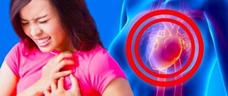
The following symptoms are observed:
- chest pain;
- dyspnea;
- swelling of the legs and arms;
- high fatigue;
- fainting;
- pale skin.
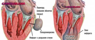
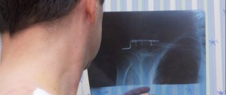
You can find out that the heart has expanded its boundaries during examination using ultrasound, radiography, echocardiography or MRI. For example, Figure No. 1 is presented in the form of a fluorography image. The shadow marked in the image indicates the expansion of the organ's shadow.
Figure No. 2 allows you to visually compare the normal state of the atria and ventricles, and the moment when the organ expanded its boundaries.
The course of treatment with medications depends on the severity of the disease:
- Diuretic medications reduce pressure in blood vessels.
- To avoid the risk of blood clots in the vessels, anticoagulants are prescribed.
- To restore the functioning of the cardiac system, angiotensin prescription blockers are prescribed. The drugs Warfarin and Heparin are effective in treating atrial and ventricular dysfunction.
- Medications of the beta blocker group are designed to normalize the pulse.
The expansion of the organ's shadow leads to various consequences - blood clots, cardiac arrest, rhythm disturbances, and death.
The greatest danger is the enlargement of the boundaries of the ventricle of the left chamber. This pathology most often leads to death.
If conservative treatment methods have no effect and the patient’s condition worsens, then doctors advise surgery. The type of intervention is considered on an individual basis. For example, they may prescribe inserting a defibrillator under the skin to adjust the rhythm.
If conservative therapy does not bring results, and the patient’s well-being continues to deteriorate, the cardiologist recommends surgery. Depending on each specific case, certain surgical intervention will be required:
- cardioverter-defibrillator. A small device is implanted under the skin in the upper chest. With the help of this defibrillator, heart rhythm is corrected;
- valve replacement. The damaged heart valve is replaced with a new one in order to restore its normal functioning. The duration of the procedure is 3-6 hours;
- A heart transplant is a last resort. First, the doctor signs the patient up for a transplant. The average waiting period varies from 200 days to a year. A successful operation makes it possible to live an additional 5-10 years.
When this symptom is detected, patients wonder what to do if the heart is enlarged. Treatment should begin only after a complete examination and clarification of the causes.
If necessary, bronchodilators, antihypertensives, and diuretics are prescribed. In some cases, a combination of these agents is necessary.
Regardless of the cause, it is important to take medications to suppress heart failure. These include drugs “Coronal”, “Propronalol”, “Captopril”, etc. In case of severe heart defects, surgical treatment is necessary.
It is also prescribed for persistent ischemia and acute circulatory failure.
How is hypertension classified by ICD-10 code?
Have you been struggling with HYPERTENSION for many years without success?
Head of the Institute: “You will be amazed at how easy it is to cure hypertension by taking it every day...
Read more "
Any disease has its own code in a special classifier of diseases, which is used in international practice. This article will focus on ICD-10 codes for hypertension.
I10 – primary (essential) hypertension
In 9 out of 10 hypertensive patients, this type of disease is diagnosed. Possible provoking factors include genetic background, obesity and the presence of regular stressful situations.
Symptoms:
- pain and feeling of pressure in the head;
- state of insomnia;
- tachycardia;
- tinnitus, spots before the eyes;
- elevated blood pressure (BP);
- excessive irritability;
- tachycardia;
- dizziness;
- bleeding from the nasal cavity.
If not treated correctly, damage is caused to the brain, kidneys, heart and capillaries. This is also fraught with severe complications (renal failure, cerebral hemorrhage, heart attack) and even death of the patient.
The benign form of the disease develops over a long period of time. In the first stages, the patient’s blood pressure increases only occasionally and does not cause concern. Most often, benign hypertension is detected during clinical examinations.
The disease in its malignant form is especially dangerous. It is difficult to cure and threatens with consequences incompatible with life.
I11 – pathologies associated with hypertensive heart disease
These include:
- I11.0 – when congestive failure develops in the heart;
- I11.9 – the heart is affected, but congestive heart failure does not occur.
Middle-aged and older people are at risk of getting sick. The disease is accompanied by signs of primary hypertension, mainly cardiac symptoms (pain, shortness of breath, angina attacks).
I12 – hypertension, mainly affecting the kidneys
The ICD includes the following types of hypertension:
- I12.0 – in combination with functional renal failure;
- I12.9 – without functional renal impairment.
Due to high blood pressure, the structure of small renal vessels changes.
Primary nephrosclerosis may develop, provoking pathological processes:
- fibrosis;
- deformation of small arteries (loss of elasticity, thickening of their walls);
- the renal glomeruli do not work well, and the renal tubules atrophy.
There are no clear signs of kidney damage in hypertension. Special examinations will help to establish dysfunction.
These include:
- Ultrasound examination;
- urine analysis (albuminuria exceeding 300 mg/day indicates obvious problems);
- blood test;
- study of glomerular filtration rate (alarming symptom - indicator > 60 ml/min/1.73 m2).
If such a violation is detected, the amount of salt in the diet should be reduced. If this measure does not prove effective, medications are prescribed to protect the kidney tissue from deformation. These medications include AP enzyme inhibitors and angiotensin II antagonists.
Why do you feed pharmacies if hypertension is afraid of the usual like fire...
Tabakov has revealed a unique remedy against hypertension! To reduce blood pressure while preserving blood vessels, add to…
I13 – hypertensive disease, mainly damaging the heart and kidneys
Combines malfunctions of the kidneys and/or heart - up to signs of organic or functional failure of these organs.
Included:
- I13.0 – hypertensive process with insufficient cardiac function;
- I13.1 – process with insufficient renal function;
- I13.2 – disease with renal and heart failure;
- I13.9 – unspecified hypertension.
I15 – secondary (or symptomatic) hypertension
These include:
- 0 – increased blood pressure due to low blood supply to the kidneys;
- 1 – in relation to other kidney diseases;
- 2 – secondary to endocrine diseases;
- 8 – other;
- 9 – unspecified nature.
This type of disease accounts for about 5% of all hypertensive conditions. It is caused by diseases of the organs that maintain blood pressure balance. The processes occurring in them lead to the fact that the tonometer begins to record excessively high pressure values.
Symptoms of secondary hypertension:
- the disease develops rapidly;
- there is no positive effect when two (or more) drugs are prescribed;
- patients are usually young people;
- their relatives have never had hypertension;
- the course of the disease worsens despite drug therapy.
It has been established that approximately 70 different ailments can provoke increased blood pressure.
Among them are:
- diseases of the endocrine system (increased or weakened thyroid function, diabetes mellitus);
- kidney pathologies (inflammatory or tumor processes, urolithiasis, polycystic disease, transplantation, connective tissue diseases, etc.);
- diseases of the adrenal glands (pheochromocytoma, Cohn's disease, Itsenko-Cushing's disease);
- cardiovascular disorders (inflammatory process of the aorta);
- neurological pathologies (head injuries, inflammatory processes of the brain).
Secondary high blood pressure, which is difficult to stabilize, can be caused by taking certain medications. For example, contraceptives containing hormones, anti-inflammatory drugs, MAO inhibitors simultaneously with ephedrine.
I60–I69 – disease involving cerebral vessels
Arterial hypertension of this type is classified by ICD-10 under the heading “Brain damage”. It is not assigned a specific code, because it can accompany any brain disorder.
If there is no treatment, increased blood pressure changes the structure of the blood vessels in the brain. Sclerosis forms in small veins and arteries, causing vascular blockages or ruptures with cerebral hemorrhage. Both small and large vessels are deformed. In the latter case, this leads to a stroke. Deterioration of blood flow over a long period of time also causes a lack of nutrients. The consequence is mental disorders.
I27.0 – primary pulmonary hypertension
A rare type of disease with unknown causes. It usually develops by the age of 30.
Signs:
- blood pressure readings in the lungs <25 mm Hg. Art. at rest and <30 during exercise;
- pain in the chest area that is not relieved by antianginal drugs (nitrates);
- heart failure, fainting;
- dry cough during physical exertion;
- bloody discharge when coughing;
- shortness of breath, especially during exertion.
P29.2 – neonatal hypertension
The disease is characterized by a lack of cardiac function in the newborn, as well as an enlarged liver, bluish skin tints, and convulsions. The disease can cause swelling of the brain.
The main factor is usually aortic narrowing or a blood clot in the renal artery. Other reasons include:
- maternal drug use;
- polycystic kidney disease;
- Itsenko-Cushing pathology;
- inflammatory or oncological diseases;
- use of glucocorticosteroids, theophylline, etc. by a pregnant woman.
I20–I25 – hypertension affecting the coronary vessels
Coronary arteries become a target in hypertension. These vessels are the only ones that supply the myocardium. Under the influence of increased pressure, their lumen narrows, which threatens a heart attack.
O10 – pre-existing hypertension accompanying pregnancy, childbirth and the postpartum period
O10.0–O10.9 according to ICD-10 combines all types of hypertension.
O11 – pre-existing hypertension accompanied by proteinuria
Includes this if it was diagnosed before the child’s conception and was observed for at least 1.5 months after birth.
O13 – pregnancy-induced hypertension (without proteinuria)
This section includes:
- hypertension developed during pregnancy (NOS);
- slight degree of preeclampsia.
O14 – hypertension in pregnant women, accompanied by severe proteinuria
This section combines:
- O14.0 – moderate preeclampsia;
- O14.1 – severe preeclampsia;
- O14.9 – preeclampsia of an unspecified nature.
It usually occurs after the fifth month of pregnancy. Characteristic manifestations are severe swelling and high protein content in urine (0.3 g/l and above). It is a serious pathology that requires constant monitoring by a medical specialist.
O15 – eclampsia
This type of arterial hypertension includes the following conditions according to ICD-10:
- O15.0 – observed during pregnancy;
- O15.1 – arising during the birth process;
- O15.2 – appeared immediately after birth;
- O15.9 – pathology, not specified by date.
O16 – maternal eclampsia, unspecified
The pressure has increased to critical limits. There is a threat to the life of the mother and fetus. Hypothetical reasons for development:
- hereditary predisposition;
- infectious pathologies;
- thrombophilia.
Symptoms:
- bluish color of mucous membranes and skin;
- wheezing;
- spasms starting from the facial muscles;
- loss of consciousness;
- eclamptic coma.
Pathology treatment methods
Taking into account the degree of the disease, the doctor selects medications that help improve the functioning of the heart muscle. List of drugs:
- anticoagulants - “Heparin”, “Angioks”;
- diuretics - Trifas, Lizax, Furosemide;
- beta blockers – “Anaprilin”, “Digoxin”;
- angiotensin blockers - Eprosartan, Losartan.
The traditional method of treatment should not be excluded. Freshly prepared juices help well: carrot, cranberry, onion, birch. Beekeeping products work well: propolis, honey.
Enlarged heart: consequences of the disease
In case of increased myocardial activity and its subsequent increase, it is necessary to be observed by an experienced cardiologist and undergo regular examination courses. An important part of drug therapy will be a review of your lifestyle.
In particular, you need:
- Give up bad habits - alcohol and cigarette abuse.
- Get rid of obesity and extra pounds to reduce the load on the heart muscle.
- Reduce the amount of salted, smoked, cholesterol-rich foods you consume.
- Balance the diet, enrich it with microelements and substances to normalize heart function.
- Reduce inadequate stress on the heart.
If measures are not taken to maintain the myocardium in a healthy state, this can lead to the development of stroke, heart attack, heart failure and even death. People with alcohol addiction are at particular risk. Against the background of constant intoxication, the heart of a drinker sometimes reaches very large sizes.
Restoration of myocardial size can only occur if alcohol is completely abstained.
An enlarged heart can entail significant risks for the patient, which depend on the underlying disease that caused the enlarged heart. When the heart becomes too large, some parts of the myocardium are subject to greater pressure and therefore an increased risk of ischemia and dangerous complications such as stroke and heart attack.
However, statisticians note that one can live with the pathology without serious consequences, keeping it under control with the help of appropriate treatment. In Russia, in fact, about 9.4 million people have an enlarged heart, especially in the left ventricle.
Unfortunately, heart failure rarely goes away completely, as it is a chronic, progressive disease. With inadequate therapy or its absence, the consequences can be serious.
In the case of severe cardiomegaly, the patient constantly lacks air, as a result of which all organs suffer. The disease can also lead to myocardial infarction, stroke, thromboembolism of the heart or pulmonary vessels.
Prevention
Lifestyle changes will help not only prevent the development of the disease, but also improve the condition of an already enlarged ventricle. Since hypertrophy is common in people suffering from obesity, maintaining an ideal body mass index will be the best prevention of the disease.
It is also worth limiting the amount of salt in your diet to normalize blood pressure. If hypertrophy is suspected, it is recommended to drink alcohol in moderation, and if treatment is prescribed, then it is better to avoid strong drinks altogether.
Despite the fact that one of the reasons for an enlarged ventricle of the heart is heavy physical activity, you should not give up sports. Regular physical exercise, such as walking, Pilates, yoga, will not only do no harm, but, on the contrary, will strengthen the heart.
If a diagnosis of hypertrophy has already been made, it is necessary to ask a physiotherapist to select the optimal exercise program. 30 minutes of moderate physical activity will strengthen weakened heart muscle and prevent its enlargement. A healthy lifestyle and proper nutrition will allow you to forget about problems with the left ventricle for a long time.
Symptoms of the disease
Manifestations of cardiomegaly are purely individual. In some cases, symptoms do not occur at all, and a person learns about the expansion of the organ’s boundaries by chance, for example, during fluorography or a doctor’s examination. If the pathology makes itself felt, its manifestations are not much different from the symptoms of diseases of the cardiovascular system. But there are cases when the pathological process develops so rapidly that a “bull’s heart” quickly forms.
Signs include:
- Pain behind the sternum.
- Dizziness, loss of consciousness for no reason.
- Fast fatiguability.
- Shortness of breath with slight exertion.
- Arrhythmia (change in rhythm).
- The appearance of edema (especially in the lower extremities in the evening).
- Cough not associated with respiratory diseases.
Forecast
It depends on what exactly caused the enlargement of the heart:
- With arterial hypertension, the prognosis is favorable. If you take the medications prescribed by your doctor on time, your heart will soon return to normal and will no longer enlarge.
- In case of ventricular septal defect – relatively favorable.
- For dilated cardiomyopathy – unfavorable. Full recovery occurs only after transplantation. However, it is not always possible to find a donor for a heart transplant. In addition, the risk of postoperative complications is high.
- In hypertrophic cardiomyopathy – relatively unfavorable. With an asymptomatic course of the disease, patients die before the disease is detected. With proper therapy, the risk of death is reduced.
- Metabolic cardiomyopathy has a favorable prognosis. When metabolism is established, complete recovery occurs.
- With aortic stenosis without treatment, life expectancy ranges from 1 to 4 years from the onset of symptoms. If the operation is performed in a timely manner, the prognosis is relatively favorable.
- If mitral stenosis is left untreated, 50% of patients die within 5 years of the first symptoms appearing. After surgery, the prognosis is relatively favorable.
- In case of Ebstein's anomaly, it is relatively favorable. The risk of sudden death is 3–4%.
- For myocarditis – favorable. Complete recovery occurs after 4–8 weeks in 90% of cases, after a year – in 10% of cases.
- For exudative pericarditis – favorable. All operated patients recover.
- In case of amyloidosis – unfavorable. The maximum life expectancy is 5 years from the date of diagnosis.
class=»fa»>If the operation is not performed in time, there is a risk of developing aortic valve insufficiency, severe rhythm disturbances, left ventricular dysfunction and sudden death. If the patient is operated on, the heart will no longer bother him.
What does an enlarged heart mean on the left side of fluorography?
X-ray diagnostic methods make it possible to identify various pathologies of internal organs. So, it is possible to notice that the heart is enlarged to the left in the fluorography image. Experts advise not to ignore this symptom, but to contact a cardiologist who could correctly identify the disease and prescribe treatment.
What does expansion of the heart to the left mean?
The average weight of this cone-shaped hollow muscular organ in an adult man is approximately 300 grams, in a woman - 250 grams. Its dimensional characteristics are comparable to the size of a clenched fist: longitudinal size - 12 centimeters, transverse - 8-9 centimeters, anteroposterior - 6 centimeters.
An expansion or increase in the volume of an organ is called cardiomegaly. If it is expanded on the left, this is a symptom of transformation of the left ventricle, which means hypertrophy of the heart muscle. This sign means that it has become larger in diameter.
As a rule, the increase in volume occurs on the left side. This may be a manifestation of hypertension (hypertension), blood stagnation in the systemic circulation, and other pathologies. Sometimes this phenomenon is called “bull’s heart”.
Causes of pathology in adults
Provoking factors for cardiomegaly can be:
- diabetes;
- hypertension;
- long-term treatment with antibiotics;
- the onset of pregnancy;
- anemia (anemia) – a decrease in the level of hemoglobin protein in the blood, which is responsible for transporting oxygen in the blood;
- renal failure;
- alcohol abuse;
- rheumatism – inflammation of the connective tissue structures of the cardiovascular and musculoskeletal systems;
- ischemia – impaired blood supply to the myocardium;
- Myocardial infarction – damage to the heart muscle. It develops with an acute lack of oxygen. Occurs due to blockage of a blood artery by a blood clot. Subsequently, the damaged areas die off. The process of their death is called necrosis;
- athletes with enlarged heart muscle undergoing irregular training;
- genetic predisposition;
- cardiomyopathy - thickening of the walls of the ventricles, stretching of the cavities of the heart.
The described pathology does not manifest itself in any way at first, but over time, with increasing loads, the following symptoms may appear:
- a condition characterized by shortness of breath, fatigue;
- swelling in the lower extremities;
- the appearance of a feeling of heaviness on the right under the ribs;
- cephalalgia, tinnitus;
- causeless cough.
Enlarged heart in children
An increased volume of an organ in children can be a congenital pathology, which will subsequently cause serious health problems. It is important to identify such deviations from the norm immediately - from the first days of a child’s life to six months.
Cardiomegaly in children can occur due to the presence of an inflammatory process of connective tissue in the body - rheumatism. This disease, in turn, can appear as a complication after a sore throat, exacerbation of chronic tonsillitis, or with some types of caries. It is acquired and develops regardless of the child’s age.
Signs of the described pathology in children may be:
- weakness;
- dyspnea;
- pain in the sternum area;
- fever 1-2 weeks after recovery from tonsillitis;
- sweating;
- increased fatigue;
- pain in muscles, joints.
The reasons for the appearance of signs of an enlarged heart on a fluorography image may also be:
- hypertrophic cardiomyopathy - manifests itself in thickening of the wall of the ventricle, most often the septum between the ventricles;
- myocarditis is an inflammatory process in the heart muscle. It can be of viral, fungal, bacteriological origin, or may appear due to autoimmune problems or improper use of medications;
- endocarditis – inflammation of the endocardium (inner lining);
- pericarditis is an inflammatory process in the serous membrane.
The last two pathologies in minor patients are quite rare.
If the described symptoms occur, you should consult a doctor; it is highly undesirable to leave this situation without proper attention.
Is it possible to detect on fluorography that the heart is enlarged? How does this manifest itself?
An enlarged heart is almost always noticeable in a fluorography image. The condition manifests itself in the fact that the shadow of this internal organ increases in size and changes shape. It is also visualized that its boundaries have been increased.
Is it possible to make an accurate diagnosis based on a fluorographic image?
An enlarged heart in a fluorography image can only indicate the presence of diseases or pathologies of this internal organ, but this is not enough to make an accurate diagnosis.
The diagnosis is made by a combination of signs, which include cardiomegaly. Moreover, treatment is prescribed not for the symptom itself, but for the disease that caused it.
After proper therapy, the size of the organ returns to its previous parameters.
What additional diagnostic measures are taken?
To make an accurate diagnosis and prescribe appropriate treatment, the following types of examinations are required.
- X-ray of the sternum area. You can notice an expanded shadow of the heart group and determine congestion in the circulatory system.
- Electrocardiography (ECG).
- Echocardiography (EchoCG). Allows you to determine the parameters of the heart muscle, the size of the chambers, identify necrosis (death of cells/tissue areas) or ischemic disease (impaired blood supply to the myocardium due to damage to the arteries).
- Ultrasound examination (ultrasound) of the heart muscle.
- Computed tomography (CT).
- Magnetic resonance imaging (MRI).
- Immunological and biochemical blood tests. They determine the levels of hemoglobin, bilirubin, protein, hormones and urea.
The doctor will be able to prescribe adequate treatment only after making a diagnosis. And to determine the disease, the doctor carefully examines not only the data of hardware examinations, but also the results of clinical tests, as well as the clinical picture.
Treatment
For almost any disease with a symptom of heart enlargement to the left on fluorography, doctors recommend leading a healthier lifestyle, adhering to a special diet, which means avoiding spicy, fatty and salty foods. Patients are advised to give up bad habits and may be prescribed special exercises.
As a rule, medications from the following groups are prescribed for cardiomegaly:
- angiotensin-converting enzyme (ACE) inhibitors;
- angiotensin receptor blockers;
- beta blockers;
- diuretics.
Angiotensin-converting enzyme inhibitors
Such drugs dilate blood vessels and lower blood pressure, reducing the workload on the cardiovascular system. The drugs have the following names: Enalapril, Captopril, Lisinopril. A side effect is often a dry cough.
Angiotensin receptor blockers
Being analogous to ACE inhibitors in terms of pharmacological effect, these drugs do not cause a dry cough as a side effect.
Beta blockers
The use of beta blockers helps reduce heart rate and normalize blood pressure. However, such drugs are only auxiliary in treatment.
Diuretics
Diuretics (diuretics) can improve blood flow to the heart and help lower blood pressure. The names of these drugs: Chlorthalidone, Hydrochlorothiazide.
It is not recommended to resort to self-medication; only a doctor can make a diagnosis and determine an effective drug therapy regimen.
Surgery may also be required. It consists of restoring or completely replacing the arterial valve. In severe cases, a whole organ transplant is necessary.
Prevention
Prevention of cardiomegaly can be a change in lifestyle. One of the main factors in the occurrence of heart pathologies is obesity. It is recommended to maintain a normal body mass index. Losing weight if you are overweight, against the background of an already enlarged heart, helps stabilize the condition.
A special diet is recommended to prevent cardiovascular diseases. It is advisable to consume fish, soybean, flaxseed, corn or olive oil at least twice a week.
To strengthen the heart muscle, you need to eat cranberries, cabbage, viburnum, eggplants, peaches, dried apricots, apples, pomegranates, walnuts, melon. To normalize blood pressure, you need to avoid excessive consumption of salt and salt-containing foods.
In addition, it is advisable to reduce the consumption of alcoholic beverages or completely abandon them.
Although an increase in heart volume may be caused by intense physical activity, in moderate doses, sports activities can only benefit the patient. Thus, yoga, Pilates, and walking will help strengthen the heart.
If hypertrophy has already been identified, it is better to consult a physiotherapist. He will help you choose a set of therapeutic exercises, designed for about half an hour. Regularly performing this set of physical activities will reduce the volume of the heart muscle.
Eight hours of sleep and avoidance of excessive physical and emotional stress are helpful. In addition, regular walks outside are recommended.
Thus, a healthy lifestyle and proper nutrition will help minimize the risk of complications with an enlarged left ventricle.
Risks and possible complications
There are various prognoses that are directly dependent on the diagnosis made when the heart expands to the left.
- For arterial hypertension (hypertension, high blood pressure), the prognosis is considered favorable. After taking the medications prescribed by the doctor, the patient’s condition returns to normal, the heart returns to its normal parameters and does not enlarge again.
- If a ventricular septal defect is observed, surgery is necessary. Otherwise, the pathology will lead to aortic valve insufficiency, serious rhythm disturbances will occur, and left ventricular dysfunction is possible. All of the above can cause death. After the operation, the patient’s condition returns to normal, and he no longer faces such heart problems.
- Dilated cardiomyopathy (stretching of the heart cavities) is characterized by a poor prognosis. Full recovery becomes possible only after a heart transplant.
- Hypertrophic cardiomyopathy (thickening of the walls of the ventricles) - the prognosis is relatively unfavorable, the disease may be asymptomatic. Without proper and timely treatment, there is a risk of death. When undergoing treatment procedures, the likelihood of death becomes lower.
- Metabolic cardiomyopathy is a non-inflammatory myocardial lesion of various etiologies. It can cause insufficiency of cardiac functions, in particular contractile function. The prognosis is favorable. If you establish proper metabolism, complete recovery will occur.
- Myocarditis. Characterized by a favorable prognosis. Complete recovery is possible within 1-2 months in nine out of ten cases, after 1 year in the remaining patients.
- Aortic stenosis (the mouth of the aorta becomes narrow, as a result of which blood flow from the left ventricle to the aorta is impaired) - without treatment, the life expectancy will be from 1 to 4 years from the time the disease is detected. With proper treatment, the prognosis is relatively favorable.
- Exudative pericarditis (inflammation of the serous membrane of the heart) has a favorable prognosis, all patients recover.
Source: https://iDiagnost.ru/issledovaniya/fluorografiya/chto-znachit-rashirnnoe-serdtse-v-levoj-chasti-flyuorografii