Doctors around the world use palpation, percussion and auscultation to diagnose various diseases. During pregnancy, the gynecologist also uses these objective examination methods to determine your baby’s vital signs. You are probably familiar with palpation (from the Latin palpatio - stroking, feeling), and percussion (from the Latin percussio - blow, tapping), and auscultation (from the Latin auscultare - listen, listen) from childhood. But you will only have to encounter auscultation in the form in which it is practiced in gynecology during routine visits to the antenatal clinic. What is this term and what is this method? Of course, you are interested in delving into the essence of all the manipulations carried out during examinations by your attending physician, because now you are responsible not only for yourself, but also for the little person growing and developing in your womb.
- 2 Listening to the belly of a pregnant woman: why is this necessary?
- 3 Main determined indicators
- 4 Listening points
- 5 Preparation for manipulation
- 6 Algorithm for performing auscultation
- 7 Video “Technique for listening to the fetal heartbeat”
Auscultation: what is it?
Auscultation (from the Latin “auscultare”) in translation sounds like listening. In midwifery practice, this is listening to fetal heart sounds
Auscultation (listening) is one of the methods of examining a patient for the purpose of diagnosing diseases. In therapy, surgery, pediatrics and other areas of medicine, auscultation is carried out using a stethoscope - a device invented in 1816 by the French physician and anatomist Dr. Laennec, who was the discoverer of the auscultation method. He also named and described all the main auscultatory phenomena.
In gynecology, auscultation is performed with an obstetric stethoscope. It differs from a conventional stethoscope in the width of the funnel, which the doctor firmly applies to the pregnant woman’s exposed abdomen during auscultation.
In addition, this manipulation can be successfully carried out using a stethophonendoscope - an invariable attribute of all doctors, a device with which we have all been familiar for a long time.
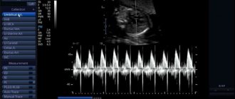
Today, instead of listening to the pregnant woman’s abdomen, a modern analogue of an auscultatory examination, phonocardiography, can be used. But to carry out such diagnostics, it is necessary to have special equipment - an ultrasound machine with the Doppler effect. And this is not available in all antenatal clinics.
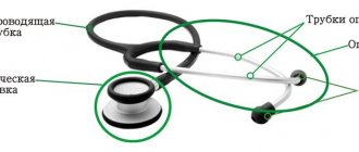
Obstetric stethoscope
How is listening performed?
Auscultation of the fetus is carried out using several techniques. It is also not necessary to have special equipment to listen to your baby’s heartbeat at home.
- Fetal Doppler is a portable device with headphones. With it, heartbeats can be heard already at 8 weeks. Obstetricians advise not to rush and wait until later. It is not recommended to use the device frequently, or to listen to the stomach for more than 10 minutes. This device can be purchased and used at home, but the price is not cheap.
- Regular listening is a free method. You can listen to the belly and hear the beating of the baby’s heart muscle as early as 30 weeks. It is important to know where the head is. It should be borne in mind that the thicker the fat layer on the abdomen, the quieter the sounds will be.
- A stethoscope can be purchased for personal use. In this case, you need to be able to listen through this device. Without the help of a specialist, you may not be able to hear sounds if the period is less than 30 weeks.
Determining a child's heart rate is an important procedure. Auscultation of the fetus helps to detect this or that abnormality and prescribe the necessary treatment in time.
The material was prepared specifically for the website kakrodit.ru, edited by doctor O.V. Klochkova. Specialty: obstetrician-gynecologist of the highest category.
Listening to a pregnant woman's belly: why is it necessary?
What does the doctor hear during auscultation of the expectant mother’s abdomen? And he can hear uterine and intestinal sounds, the beating of the abdominal aorta, which coincides with the woman’s pulse, the noise of the vessels of the fetal umbilical cord, its dull jerking movements in the mother’s womb, and, most importantly, the heart sounds of the fetus. They should be audible well and clearly with a stethoscope. A decrease in sound conductivity can be caused by a large amount of amniotic fluid, the location of the placenta on the anterior wall of the uterus, or significant fat deposits in the abdominal cavity. During multiple pregnancy, the heart sounds of the fetuses are dried in different parts of the uterus.
Auscultation of the fetal heartbeat can tell the doctor a lot about its development and physical condition. In particular, it can be used to determine the presence of congenital heart defects and pathologies of large blood vessels in a child. The baby's muffled heart sounds usually dry out due to hypoxia.
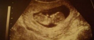
The fetal heartbeat during auscultation becomes clearly visible at the beginning of the second half of pregnancy, but no later than 18–19 weeks. Every week these sounds get better and better. The heart rate of the baby in your belly is normally 120–140 beats per minute.
At earlier stages, it is better to use phonocardiography or fetal cardiotocography to determine the cardiac activity of the embryo. The indicators from these studies will be more accurate.
Fetal heart rate can be used to determine gestational age
How to prepare for the procedure
For an accurate diagnosis, it is better for a woman to undergo this procedure after 20 weeks, or better after 24. Once at the appointment, you can see all the necessary paraphernalia in the doctor’s office. There will be a couch with oilcloth there.
The specialist in the office has a stethoscope, balls and a stopwatch. It is also important for a pregnant woman to remember to bring her individual card with her.
Process description.
- The doctor explains to the pregnant woman the need for the method and its nuances.
- The stethoscope and couch are treated with 0.5% calcium hypochlorite.
- The patient is placed on a clean sheet placed on the couch, on her back. Legs should be in a straight position.
- The doctor conducts a preliminary external therapeutic study - determines the place where the baby’s head is located. It is also important to identify the position of the body.
- The wide part of the stethoscope is placed on the anterior abdominal wall. By moving the stethoscope, the listening point is sought.
- The specialist counts how many times the child’s heart beats per minute. Auscultation of the fetus using an obstetric stethoscope helps assess the clarity, purity and tonality of heartbeats.
- I record the data in the woman’s card. Auscultation of the abdomen is considered normal when, from the 20th week of gestation, 140-160 beats per 60 seconds.
Main determined indicators
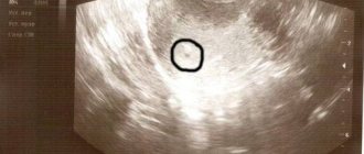
According to the rate of fetal heart rate, the entire period of pregnancy can be divided into several periods. The heart leaf is laid and makes its first tremors in the third week. And on the sixth day it can already be determined using an ultrasound machine.
It is noteworthy that it is in the sixth week of pregnancy that the fetal heart rate completely coincides with the maternal heart rate (plus or minus 3 beats/min).
After the 6th week, your baby's heart begins to beat faster. His heart rate increases by an average of 3 beats per day. Knowing this, determining the age of the fetus histologically is not difficult for the gynecologist.
At the beginning of the ninth week of pregnancy, the embryo's heart beats at a speed of 175 beats/min. This is the peak of fetal cardiac activity. Next, the baby’s heart rate begins to gradually slow down. And, starting from the second trimester, his heart rate is set at 140–160 beats/min.
Any deviations from this norm may indicate oxygen deficiency of the fetus - hypoxia. At its initial stage, the baby is diagnosed with tachycardia, and at a severe stage - bradycardia.
Listening points
Auscultation of the fetus - necessity, main indicators
The medical diagnostic method “listening” helps to identify characteristic sounds in a pregnant woman’s abdomen and use them to judge whether the child has any health problems. Fetal auscultation is an effective way to help determine the baby's heartbeat. It is important to check whether his heart rhythm is normal or not, so that in case of a threat, he can help the baby in time.
What it is
Auscultation translated from Latin means listening. You can listen to the fetal heartbeat without an ultrasound. The procedure is performed using the direct method - by applying the ear to the stomach. You can also listen to the heartbeat using a phonendoscope.
The absence of baby's heartbeat sounds may indicate a serious problem that needs to be addressed urgently.
In history, the procedure is mentioned in the works of Hippocrates. Doctors began listening to the heart in the 2nd century BC. The ancient ancient physician and scientist Aretaeus from Cappadocia practiced auscultation.
To listen, you must have a keen ear and constantly develop it.
While listening to noises appearing in the muscle cavity and in the upper middle cavities, the child’s breathing can be clearly detected. The noise will be heard quite clearly, since the chest wall is thin.
You can recognize the beating of a small heart in the early stages. During the procedure, the obstetrician evaluates the condition of not only the fetus, but also the woman.
Why is it necessary?
Listening to the fetal heartbeat is necessary in order to find and eliminate a possible problem with the baby’s health as early as possible. In order for the birth outcome to be favorable, doctors may have to intervene in a timely manner. This may be a caesarean section or an instrumental vaginal birth.
Despite the apparent simplicity of the method, the doctor is required to be especially scrupulous when listening. It is important to be able to distinguish between different body sounds. The specialist may also use a powered stethoscope to listen to the heartbeat with distinct accuracy.
The doctor tries to identify basic auscultatory data:
- auscultation tone;
- noises;
- noise friction.
Heart sounds are short, short-lived sounds that occur when a valve closes and opens. Murmurs can be systolic, diastolic and constant.
The norm and deviation of noise are classified according to their noise intensity. Friction noises may appear with tachycardia. These are high frequency sounds, possibly scratching.
Basic indicators
The method is used not only during pregnancy from time to time, but during the birth process. The vaginal ultrasound sensor detects the heart rhythm already at 3-4 weeks of pregnancy.
An abdominal sensor detects heart rate as early as the second month of pregnancy. The number of beats in 60 seconds of a child’s heart depends on how long the woman is listening:
- at 6-8 weeks, heart rate is from 110 to 130 beats per minute;
- at a period of 8-11 weeks the maximum permissible rhythm is 190;
- after 11 weeks the normal heart rate is 140-160 beats.
The heart rate is also affected by the child's activity during auscultation of the mother's abdomen. It is advisable to have your pulse measured by an obstetrician, but you can listen to how the baby’s heart is working independently.
Listening places
To accurately measure a baby's pulse, you need to find points to listen to the fetal heartbeat. Almost from the second trimester, obstetricians professionally listen to areas during presentations with the device:
- head - listen below the navel;
- pelvic – above the navel;
- occipital - near the head below the navel on the side where the child’s back is;
- pelvic end - above the navel, near the head, on the side where the back is facing;
- transverse - near the navel, closer to the head;
- facial - below the navel on the side where the breast is located in the first position - on the right, in the second - on the left;
- posterior views - from the side of the abdomen along the anterior axillary line.
Thus, the obstetrician determines in which place the child’s heart rhythm is best heard. Auscultation is studied together with ultrasound monitoring.
How to prepare for the procedure
For an accurate diagnosis, it is better for a woman to undergo this procedure after 20 weeks, or better after 24. Once at the appointment, you can see all the necessary paraphernalia in the doctor’s office. There will be a couch with oilcloth there.
The specialist in the office has a stethoscope, balls and a stopwatch. It is also important for a pregnant woman to remember to bring her individual card with her.
Process description.
- The doctor explains to the pregnant woman the need for the method and its nuances.
- The stethoscope and couch are treated with 0.5% calcium hypochlorite.
- The patient is placed on a clean sheet placed on the couch, on her back. Legs should be in a straight position.
- The doctor conducts a preliminary external therapeutic study - determines the place where the baby’s head is located. It is also important to identify the position of the body.
- The wide part of the stethoscope is placed on the anterior abdominal wall. By moving the stethoscope, the listening point is sought.
- The specialist counts how many times the child’s heart beats per minute. Auscultation of the fetus using an obstetric stethoscope helps assess the clarity, purity and tonality of heartbeats.
- I record the data in the woman’s card. Auscultation of the abdomen is considered normal when, from the 20th week of gestation, 140-160 beats per 60 seconds.
How is listening performed?
Auscultation of the fetus is carried out using several techniques. It is also not necessary to have special equipment to listen to your baby’s heartbeat at home.
- Fetal Doppler is a portable device with headphones. With it, heartbeats can be heard already at 8 weeks. Obstetricians advise not to rush and wait until later. It is not recommended to use the device frequently, or to listen to the stomach for more than 10 minutes. This device can be purchased and used at home, but the price is not cheap.
- Regular listening is a free method. You can listen to the belly and hear the beating of the baby’s heart muscle as early as 30 weeks. It is important to know where the head is. It should be borne in mind that the thicker the fat layer on the abdomen, the quieter the sounds will be.
- A stethoscope can be purchased for personal use. In this case, you need to be able to listen through this device. Without the help of a specialist, you may not be able to hear sounds if the period is less than 30 weeks.
Determining a child's heart rate is an important procedure. Auscultation of the fetus helps to detect this or that abnormality and prescribe the necessary treatment in time.
The material was prepared specifically for the website kakrodit.ru, edited by doctor O.V. Klochkova. Specialty: obstetrician-gynecologist of the highest category.
Source: https://kakrodit.ru/auskultatsiya-ploda/
Listening points
The place where the fetal heartbeat is best heard is called the auscultation point. The location of such points on the pregnant woman’s abdomen depends on the position of the baby in the mother’s uterine cavity.
There are two types of fetal presentation.
- The main thing. It, in turn, can have the following positions: facial (the head is thrown back, the chest is as close as possible to the wall of the uterus) and occipital (fetal position).
- Pelvic. When the fetal pelvis is directed towards the exit of the uterus.
During auscultation, the doctor tries to listen to the fetus with a facial presentation - from the side of its chest, and with an occipital presentation - from the back. But there are listening points on the expectant mother’s belly, where you can almost always hear the fetal heartbeat.
Where are these points located?
- Just below the navel to the left.
- Just below the navel on the right.
- Along a line drawn through the navel parallel to the horizon, on the left.
- Along a line drawn through the navel parallel to the horizon, on the right.
Preparation for manipulation includes disinfection of the instrument and the doctor’s hands
Listening places
To accurately measure a baby's pulse, you need to find points to listen to the fetal heartbeat. Almost from the second trimester, obstetricians professionally listen to areas during presentations with the device:
- head - listen below the navel;
- pelvic – above the navel;
- occipital - near the head below the navel on the side where the child’s back is;
- pelvic end - above the navel, near the head, on the side where the back is facing;
- transverse - near the navel, closer to the head;
- facial - below the navel on the side where the breast is located in the first position - on the right, in the second - on the left;
- posterior views - from the side of the abdomen along the anterior axillary line.
Thus, the obstetrician determines in which place the child’s heart rhythm is best heard. Auscultation is studied together with ultrasound monitoring.
Preparation for manipulation
During auscultation, the pregnant woman should be in a horizontal position, so it is advisable to have an individual sheet with you to cover the couch on which you will lie. Before visiting the antenatal clinic, you need to empty your bowels and bladder so that bowel sounds do not interfere with the examination.
And one more condition for obtaining reliable results. During auscultation, the expectant mother should be completely calm. When a pregnant woman is psycho-emotionally excited, the fetal heart rate may increase.
Phonocardiography is a modern analogue of auscultation
Video “Leopold's techniques. Technique for performing auscultation"
Remember! Auscultation is completely painless, completely harmless to your health and does not in any way affect the well-being of your little one.
Studies of the fetal heartbeat using a wide variety of methods are mandatory throughout pregnancy and, directly, during childbirth. They allow you to assess the condition of the fetus. And if any deviations are detected, carry out the necessary treatment in a timely manner, as well as resolve the issue of the method and timing of delivery.
Auscultation algorithm
How is auscultation performed? The patient lies with her back on the couch, her legs slightly bent at the knees. The doctor is located to the right of the expectant mother and first, using Leopold’s techniques, determines the presentation of the fetus and the level of the uterine fundus. Then, applying the wide bell of the stethoscope to the pregnant woman’s belly, and the narrow bell to her ear, the gynecologist searches for the listening point. In the place where the fetal heartbeat is best heard, the doctor calculates the baby’s heart rate characteristics.
Important! The doctor does not hold the stethoscope directly during the listening session.
After auscultation, the expectant mother is helped to rise from the couch, and the doctor washes his hands and disinfects the equipment.
You will encounter the diagnostic method described above more than once while you are carrying and giving birth to your baby. You don't need to worry about this at all. Auscultation is completely harmless to your health and the well-being of your little one.
Preparing and performing the manipulation
Before starting the procedure, the woman must empty her bladder and perform an act of defecation so that bowel sounds do not interfere with the examination. In addition, in the evening or morning before the examination, take a hygienic bath or shower.
Immediately before performing auscultation, the doctor should cover the couch with an individual sheet, prepare a table with instruments, wash and warm his hands. The woman should be in a calm state. The psycho-emotional arousal of the expectant mother can also affect the characteristics of the fetal cardiac activity.
Auscultation of the fetal heartbeat is carried out with the patient lying down, with legs slightly bent at the knees. In this case, the doctor is positioned on the right and, using Leopold’s techniques, determines the level of the fundus of the uterus and the position of the fetus in it. After this, a wide bell of a stethoscope is applied to the pregnant woman’s belly, and the doctor applies his ear to the other end. A search is made for the best place to hear the fetal heartbeat and its characteristics are calculated. After this, the pregnant woman is helped to get up, her hands are washed and the equipment is disinfected. It is important that the stethoscope is not held by hand during the manipulation.
Video “Technique for listening to the fetal heartbeat”
- Author: Polina
Hello. My name is Polina. Having once heard the truth that a pediatrician is the main doctor for any family with small children, I realized that I had something to strive for. Rate this article:
- 5
- 4
- 3
- 2
- 1
(0 votes, average: 0 out of 5)
Share with your friends!
Chlamydia during pregnancy: what does a woman need to know?
Fetal heart rate during pregnancy
Auscultation
Listen to fetal heart sounds during pregnancy and during labor. Auscultation during pregnancy is of practical importance only in the second half. Auscultation determines the life and condition (threat of asphyxia) of the fetus, and in some cases also the presenting part, position and type of the fetus and the presence of a singleton or multiple pregnancy.
Listening is done directly with the ear or with a special stethoscope with a wide funnel, which should be applied to the bare abdomen. Listening to fetal heart sounds with a stethoscope is a more valuable method than auscultation with the ear, since the stethoscope allows you to isolate extraneous auditory impressions, squeeze out amniotic fluid, etc.
An ordinary medical stethoscope cannot replace an obstetric one.
Figure: Obstetric stethoscope.
At the end of the first half of pregnancy, you can listen to the following in the woman’s abdomen:
1. maternal blowing noises arising from the movement of blood in the bends of the uterine artery, synchronous with the mother’s pulse; they appear in the middle of the fourth month;
2. at the beginning of the fifth month, at the 18-20th, and sometimes already from the 15-16th week, fetal heart sounds appear, having a double beat and a frequency of about 140 beats per minute;
3. in the fourth or fifth month, a fetal kick is heard, accompanied by a slight noise; it reflects a movement of the fetus that cannot yet be determined by palpation. Regardless of the stage of pregnancy, bowel sounds are sometimes heard, depending on the movement of gases through the intestines during peristalsis. These sounds are not rhythmic and are random in nature.
In the second half of pregnancy, fetal heart sounds and maternal murmurs should be examined. Heart sounds are heard most clearly in certain areas of the mother's abdomen, located closer to the fetal heart.
In anterior views, tones are heard most clearly on the anterior wall of the pregnant woman’s abdomen, closer to the midline; in posterior views, in order to hear the fetal heartbeat, the stethoscope should be pressed in slightly, while the heartbeat can be heard more laterally, in the lateral parts of the uterus. The fetal heartbeat is most clearly heard from the back.
With extension presentations, especially facial ones, when the chest moves closer to the abdominal wall, listening to the fetal heartbeat is better from the side of the chest.
Figure: Locations of the clearest fetal heartbeat.
a – in case of occipital presentation – from the side of the fetal back; b – in case of facial presentation – from the side of the fetal chest.
Determining the location of the clearest fetal heart sounds, as already mentioned, helps in determining the position of species and presentation.
With cephalic presentations, the clearest fetal heartbeat is heard below the navel and on the side, in the first position - on the left, in the second - on the right; in posterior views, the fetal heartbeat is from the midline, approaching the nipple.
In breech presentations, a clear heartbeat is detected above the navel and to the side, in the first position - on the left, in the second - on the right; in posterior views it is lateral, in anterior views it is medial. In transverse positions, fetal heart sounds are heard only below the navel.
Fetal heart sounds may be mixed with uterine artery pulsations; to differentiate, it is necessary to compare the frequency of the audible tones with the mother’s pulse rate: the pulse rate of a pregnant or woman in labor is almost two times slower than the fetal heartbeat. With twins, the heartbeat of each fetus is different in frequency.
To auscultate the heart sounds of twins, assign one of the examiners to loudly count the heartbeat of one of the fetuses; another researcher compares this count with the heartbeat count of a second fetus. This way you can prove the presence of twins.
Figure: Places of the clearest fetal heartbeat in typical positions.
1 – with anterior view of the first position of occipital presentation; 2 – with a posterior view of the first position of the occipital presentation; 3 – with anterior view of the first position of breech presentation; 4 – with a posterior view of the first position of breech presentation; 5, 6, 7, 8 – the same for the second position.
Maternal sounds in the second half of pregnancy are heard in the form of special blowing placental sounds, since they are most clearly heard near the placenta insertion. When the fetus is positioned longitudinally, noises are usually heard on the side opposite the back. A.P.
Gubarev considers listening to maternal noises an auxiliary sign for determining the location of the placenta.
Under the influence of contractions of the uterus and changes in the placental blood circulation, the fetal heart sounds temporarily increase in speed and then slow down; at the height of the contraction, the tones may disappear completely for a few seconds.
Previously, the slowing of the fetal heartbeat during contractions was explained by the action of fetal expulsion forces, causing compression of the head and an increase in intracranial pressure; As a result, the center of the vagus nerve is irritated, which sends an inhibitory impulse to the fetal heart.
In contrast, A.P.
Nikolaev believes that with an increase in the amount of acetylcholine during contractions, especially during violent labor, part of it can pass through the placental barrier into the blood of the fetus and cause inhibition or even a temporary stop of the heartbeat.
After the amniotic fluid breaks, the fetal heart sounds become clearer. During childbirth, as the head moves, heart sounds move downwards and, before the head erupts, are heard below the middle of the distance between the navel and the womb.
Sometimes, when auscultating a pregnant woman’s abdomen, an umbilical cord noise is heard in the uterus - blowing or whistling, coinciding with the fetal heartbeat. It occurs in the corkscrew-shaped vessels of the umbilical cord when pressed with a stethoscope, if the loops of the umbilical cord are located near the walls of the uterus.
Reflex changes in blood circulation in the uterus, caused by certain medical prescriptions, can lead to significant changes in the fetal heart rate. According to experimental data (N.L.
Garmashev), the introduction of calcium chloride or pituitrin into the mother’s body and the subcutaneous administration of caffeine, as well as hot and cold vaginal douches, cause a temporary slowdown in the fetal heartbeat, while intravenous administration of sodium bromide increases it.
Therefore, the obstetrician, listening to the fetal heartbeat, must know about the medications and manipulations that the pregnant woman (mother in labor) receives.
(:0) 4951
Source: https://gynea.ru/physioobstetrics/obstetricinvestigation/363-auskultaciya.html