The heart muscle in a human embryo is formed 4 weeks after conception. At this stage, it still does not have partitions, representing a hollow tube. At week 5 you can hear your heartbeat for the first time. This is one of the important characteristics by which doctors judge the health and level of development of the fetus.
Tachycardia is an increase in heart rate. This disorder refers to arrhythmias. During the period of intrauterine maturation, it can have a detrimental effect on the condition of the embryo, in some cases even dangerous for the life of the unborn child.
Let us consider in detail what it is, why it occurs, what it indicates and how it is treated. We will also find out how tachycardia is detected in the fetus, and what consequences can occur if timely measures are not taken.
General characteristics of the pathology
The fetal heart acquires its usual 4-chamber structure at 7 weeks. But HR (heart rate) indicators change throughout pregnancy. Fetal tachycardia is diagnosed at 175-220 beats per minute.
The fetal heart has one feature remaining - an oval opening for the intake of maternal blood. Normally, this window closes for some time after birth.
Not only the condition of the embryo, but also the course of labor and the health of the baby in the future depends on cardiac activity. Therefore, heartbeat monitoring is carried out at every visit to the obstetrician-gynecologist.
Accelerated myocardial contractions are not a disease, but a sign of certain problems in the body. But this symptom is dangerous in itself, as it causes an increase in blood pressure and a decrease in blood flow to the organs, because the ventricles do not have time to fill with blood.
In order to have an idea of such a disorder, let us remember how the heart works. It has a self-contained electrical system that generates and distributes impulses. In response to them, the atria and ventricles contract and relax.
The signal conduction system is quite complex. At each stage, due to various reasons, a failure may occur, especially in the embryonic state of the body.
Forms of embryonic tachycardia
Depending on where the source of arrhythmia is localized and what it is, the following forms of pathology are distinguished:
Atrial flutter (AF). The atria are the upper chambers of the heart. They receive blood from the veins, and when they contract, the blood is sent to the ventricles (lower chambers). If impulses to the atria arrive too often, a fast, regular rhythm is observed - in some cases up to 400 beats per minute are recorded.
Flutter should not be confused and compared with atrial fibrillation (atrial fibrillation), in which contractions are not only ultra-frequent, but also chaotic. Intrauterine TP is diagnosed quite often - in every 3-4 cases.
Reciprocal tachycardia (supraventricular). With this form of pathology, the heart also beats smoothly and faster than it should (but not as often as in the first option - up to 220 beats per minute). The conduction of impulses is disrupted in the area above the ventricles.
“Signals,” instead of arriving as intended, constantly circulate back and forth. In addition, an extra focus appears, sending additional impulses that interfere with the normal functioning of the heart.
Reciprocal tachycardia is usually diagnosed in late pregnancy (24-33 weeks).
Ectopic (heterotopic) tachycardia. Such arrhythmia is expressed in an accelerated and chaotic heartbeat. It occurs due to the activity of ectopic foci. These are areas in which impulses are generated that are not provided for by the nomotopic pacemaker (the main conductor). Because of this, extrasystoles occur - premature contractions of the heart or its individual parts.
How is the treatment carried out?
How to eliminate the disease is decided depending on the individual characteristics of the body of the woman and child, the form and severity of tachycardia. Usually the condition normalizes on its own. The doctor monitors the fetus and the expectant mother, regularly measuring the heart rate using a Doppler monitor. You need to check your pulse at least twice a day.
Also read: Types and manifestations of atrial tachycardia If there are complications in the form of valve or myocardial dysfunction, a woman should take medications to stabilize the child’s heart rhythm. A doctor should select the drug, since antiarrhythmic drugs are quite dangerous.
If used incorrectly, they increase the load on the heart and can cause fetal death.
Depending on the type of pathological condition, the following treatment options are used:
- To cure polymorphic tachycardia, lidocaine, magnesium preparations, and propranolol are prescribed. They are injected into a vein or taken orally.
- If ventricular-type tachycardia with a prolonged QT interval is observed, the pregnant woman is hospitalized. Treatment should be carried out with extreme caution, as some drugs prolong the interval and aggravate the course of the arrhythmia.
- If the heart beats at a rate above two hundred beats, the condition is stabilized with Sotalol or Amiodarone. Flecainide is sometimes used. But this drug, if the ventricular functions are impaired, can cause cardiac arrest and embryo death.
- If there are signs of myocarditis, a woman is prescribed a course of Dexamethasone lasting one or two weeks.
Good results can be achieved with the help of drug treatment when an ectopic focus forms above the ventricles. A correctly designed regimen in most cases ensures complete recovery.
Beta blockers are also used to normalize the heartbeat. But their effectiveness in treating the embryo is low, since the active components have difficulty crossing the placental barrier.
Fetal tachycardia in later stages occurs quite often, which is associated with an increase in its size and an increase in the load on the body.
Seizures can bother any woman during pregnancy. Therefore, it is necessary to know how to deal with pathology.
The basic methods are selected by the doctor, but some rules help make you feel better:
- If an attack begins, you need to sit down or take a horizontal position. The load on the heart will decrease and it will begin to contract more slowly.
- Deep inhalation and slow exhalation helps stabilize the condition. These steps must be repeated several times. Breathing exercises are good for eliminating attacks.
- Avoid emotional stress and worries. In case of prolonged attacks of the disease, it is necessary to do a cardiogram.
Symptoms of arrhythmia in the embryo
The expectant mother and child are inextricably linked throughout pregnancy, so the condition of the fetus can largely be judged by the woman’s well-being.
Indirect signs of tachycardia in the fetus may be the following complaints from a pregnant woman:
- Rapid heartbeat (over 120 beats per minute);
- Fast fatiguability;
- General weakness;
- Chest pain;
- Increased anxiety;
- Numbness of the limbs;
- Sleep disturbance;
- Cardiovascular disorders that did not exist before pregnancy.
All this is caused by a deterioration in blood flow, which in turn can be triggered by a disruption in the baby’s heart rhythm.
In the third trimester, in many cases, rapid heartbeats in the fetus are considered normal. This is due to the fact that the body is preparing for childbirth and gas exchange increases.
Pregnancy 34 weeks: CTG and what it affects
CTG is cardiotocography, using this method, the heartbeat and movements of the fetus, contractions of the uterus are determined and recorded on a special tape in the form of graphs. More often, this method of examination is carried out in the middle of pregnancy, starting from the 19th week, but also at 34 weeks of pregnancy, CTG is a very necessary and useful type of diagnosis.
At 34 weeks of pregnancy, the fetus is already quite developed and almost completely ready for birth. At this point he weighs about 2 kg. and pushes less and less. This should not be scary, since the decrease in his activity is only due to the fact that he is already becoming cramped in the uterus. At this time, the fetus’s lungs finish forming - another week and he (purely theoretically) can already take his first breath in the new world. By week 34, the baby already has hair on his head and fingernails, and his pupils react to light and darkness.
Interestingly, in the first years of life, the color of eyes and hair can change beyond recognition. In women at this stage, their back begins to hurt, swelling appears and nausea does not stop, late toxicosis is possible. The baby's head is already lowering and the uterus is not putting as much pressure on the stomach as before, however, there is no appetite, and it is quite difficult to take a full breath.
By this time, women in labor need to start training their vaginal muscles; Kegel exercises are the best way to do this. As practice shows, trained muscles are excellent helpers during childbirth, and subsequently they will return to normal much faster than untrained ones. During this period, do not forget to talk to your baby, contact him, read books and listen to calm, relaxing music.
Decoding a fetal CTG at 34 weeks is very important for assessing the health and well-being of the child. CTG evaluates the average frequency of fetal heart contractions, the average height of deviations, acceleration and deceleration of contraction frequency, and uterine activity.
It is not enough to listen to the fetal heartbeat at 34 weeks with a stethoscope. Cardiotocography is most often used for this. The procedure takes from 20 to 60 minutes, after which the results are assessed. The woman lies down in a comfortable position, and sensors are attached to her stomach with special elastic bands that capture all the necessary rhythms of the fetus.
A heartbeat of 120 to 160 beats per minute is considered normal; if abnormalities are detected, the doctor will prescribe special therapy to improve performance. CTG norms at 34 weeks of pregnancy are 8-10 points. The fact is that fetal CTG is deciphered using a 10-point system. If the doctor gives 8-10 points, therefore, the fetus is developing well and is in normal condition, 6-7 points - it is experiencing oxygen starvation, however, nothing threatens the life of the fetus, after some time it is advised to undergo a re-examination, 5 or less points - oxygen starvation.
With such indications, immediate delivery by Caesarean section is indicated, but only after a repeat procedure, because it is quite possible that the child simply fell asleep. This examination method is very important in the last stages of pregnancy, since the fetus is already quite large, it may not have enough air, CTG will let you know about this in a timely manner and the doctor will be able to prescribe treatment if required. The CTG method is best used in combination with ultrasound - this way the doctor will accurately and comprehensively assess the condition of the mother and child.
CTG allows you to monitor the condition of the fetus during pregnancy; this method is absolutely harmless for both the child and the mother. Such an examination is not prescribed for everyone, but it is still better to play it safe and undergo this procedure in order to be absolutely sure of the baby’s health.
Every woman cares about her child, and to ensure that he is born and grows healthy, one should not neglect examinations during pregnancy. It is definitely worth doing CHT, ultrasound and other procedures in order to promptly identify possible pathologies.
Diagnostic methods
Unfortunately, using a stethoscope (this diagnostic method is scientifically called auscultation) it is not always possible to establish deviations of this kind. This may be hampered by the inconvenient position of the fetus or the physiological characteristics of the pregnant woman.
But an experienced gynecologist may suspect abnormalities at the examination and listening stage, based on heart rate, the general condition of the mother and the duration of pregnancy.
The most accurate and informative methods for establishing tachycardia, its form and possible causes include:
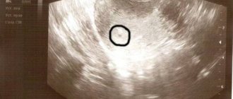
- Ultrasound. There are still a lot of rumors and speculations about the dangers of ultrasound examination during pregnancy. But experts (including the famous pediatrician Komarovsky) claim that sound waves and vibrations do not pose any danger to either the child or the mother. On the contrary, this is one of the safest and most painless hardware procedures. In the early stages of pregnancy, a regular, two-dimensional ultrasound is performed, which allows you to study the anatomy of the heart structures and listen to the heart rhythm.
- Echocardiography. This diagnostic method combines ultrasound scanning with an electrocardiogram. As a rule, it is prescribed no earlier than the 2nd trimester. The study helps determine the frequency and nature of contractions of the heart muscle.
- Doppler examination. It records the movements of red blood cells, which allows you to evaluate the speed and direction of blood flow, the volume of blood that leaves and returns to the heart.
- X-ray. This study will not establish arrhythmia, but will exclude (or confirm) heart defects and pathologies of the pericardium (heart sac). Therefore, x-ray is a concomitant, but not the main examination for suspected fetal tachycardia and for identifying its causes.
- CHT (cardiotocography). This is a relatively young technique (it appeared in the 60s and is still being improved). With its help, fetal heart rate and uterine tone are recorded. This is done using ultrasonic and strain gauge sensors. As a result, the cardiotocogram represents 2 graphs, where the tachogram reflects changes in heart rate, and the hysterogram reflects uterine contractions.
A modern cardiac monitor is a device that takes into account the motor activity of the embryo, sleep period and other important details when drawing up charts.
Fetal heartbeat during pregnancy: basic normal indicators
Heart failure can cause heart palpitations during pregnancy, and these cases are not uncommon. Frequent contraction of the heart muscle, up to approximately 100-120 beats per minute at rest, is also called tachycardia.
Pregnancy is the most important period in the life of every woman. It’s great if it proceeds without complications and there is no reason to worry. During pregnancy, all body systems experience increased stress, including the cardiovascular system.
Causes
There are many reasons for this condition, they are not fully understood and may differ for each period of pregnancy. In the early stages, the occurrence of rapid heartbeat is caused by changes in the body. The number of vessels in the uterus increases significantly, and a new (uteroplacental) blood circulation appears. This leads to increased work of the heart, the pulse rate increases.
The amount of blood circulating through the vessels during pregnancy increases by 45%. Hormone levels increase sharply. Further deterioration of the condition can be caused by anemia, excessive weight gain, thyroid dysfunction, fever, injury, stress, frequent consumption of caffeinated drinks and a number of other factors.
In the later stages, palpitations during pregnancy are inevitable, so you should not be afraid of them. It develops for natural reasons:
- the fetus enlarges;
- the load on blood vessels increases;
- the heart moves under the pressure of the growing uterus.
According to doctors, an increased heart rate for a short time during pregnancy provides the growing child with the necessary nutrients and supplies him with oxygen, because the heart works faster, the blood flow accelerates, which means that oxygen and nutrients move through the vessels faster.
When to sound the alarm?
If a woman feels normal and does not experience discomfort, there is no reason to worry. Such attacks occur from time to time in pregnant women and go away on their own. Unfortunately, attacks of rapid heartbeat are not always so harmless. The reasons for contacting a specialist are:
- increased fatigue;
- annoying headache for 2-3 hours;
- weakness and dizziness;
- sharp pain in the heart area;
- dyspnea;
- state of anxiety and fear;
- nausea and vomiting.
All these symptoms can be caused by other reasons, for example, excessive fatigue and drowsiness in the early stages are associated with an increase in the level of the hormone progesterone. If you are not sure whether this is a serious symptom, it is better to play it safe and consult your doctor.
To find out the causes of ailments, a specialist will prescribe a professional examination, ECG, EchoCG or ultrasound examination of the heart and competent and effective treatment.
To eliminate the unpleasant symptoms associated with rapid heartbeat during pregnancy, it is enough to change the woman’s lifestyle:
- adjust your daily routine;
- eat right and watch your weight;
- reduce consumption to a minimum or completely give up bad habits (smoking, alcohol);
- reduce consumption of coffee and caffeinated drinks;
- practice yoga, relaxation or any relaxation techniques;
- spend more time in the fresh air;
- protect yourself from stressful situations.
It is not recommended to self-medicate; even harmless herbal tea with a calming effect, purchased at a pharmacy, can be made from herbs that are not suitable for pregnant women. High doses of certain herbs can, on the contrary, cause heart palpitations and lead to miscarriage.
If, as a result of the examination, a pathology in the functioning of the cardiovascular system is confirmed, the doctor will prescribe drug treatment with antiarrhythmic drugs: Flecainide, Adenosine, Propranolol, Verapamil. It is very important that these drugs are used only on the recommendation of the attending physician, who will select the desired dosage and treatment regimen. Ignorant use of drugs can lead to intrauterine fetal death.
How to relieve an attack?
An attack of rapid heartbeat during pregnancy can occur suddenly, when there is no opportunity to consult a specialist. What should you do in this case? Sit down or lie down so that your head is higher than your body. Try to relax and breathe deeply. Provide fresh air and get rid of tight clothing. Use a cold compress on your face or soak your face in cold water. Drink lightly brewed tea with mint, lemon balm or chamomile.
Motherhood is a significant stage in the life of every woman. During pregnancy, many difficulties can arise. The main thing is to remain calm and take a reasonable, competent approach to solving all problems. Trust your doctor and do not hesitate to contact him if you have any concerns.
When a pregnant woman visits an antenatal clinic, the doctor determines the fetal heartbeat. This is a necessary examination that allows you to judge the state of your little life. This parameter changes, and at different periods of the child’s development, heart contractions decrease or increase.
The fetal heart rate by week usually ranges from 120 to 160 beats per minute during the prenatal period. These are echographically measurable numbers. The normal range of heart rate should fluctuate during pregnancy, and from 6 weeks increases to approximately 170 beats by the 70th day of the baby's development and decreases to 130 beats by the due date.
Fetal heart rate chart by week.
Age or size of the fetus
Ud. per minute
| Embryo 2 mm | 75 |
| Embryo 5 mm | 100 |
| Embryo 10 mm | 120 |
| Embryo 15 mm | 80–85 |
| 5 weeks | 80–103 |
| 6 weeks | 103–126 |
| 7 weeks | 126–149 |
| 8 weeks | 149–172 |
| 9 weeks | 155–195 (average value 175 bpm) |
| 12 weeks | 120–180 (average 150 beats per minute) |
| After 12 weeks | 120–160 (average 140 beats per minute) |
The above describes how the heart rate of the unborn baby changes in the first and second trimesters. During the third trimester, this figure is relatively stable. The FHR (fetal heart rate) slows down slightly in the last (third) trimester before birth, but it is still about twice the adult heart rate.
It is important to know that you can hear the fetal heart with a stethoscope from 20–24 weeks. This depends on the position of the baby, the amount of amniotic fluid and the nature of the mother's abdominal tissue. If the fetal heartbeat is normal, the chance of miscarriage is very low. A girl or boy's heart rate from 100 to 160 is considered normal. If the child’s heartbeat is not heard within 2-3 days, monitoring by ultrasound using Doppler ultrasound is necessary.
Unusually low, high or weak SBP is one of the main signs of hypoxia (lack of oxygen). Some pathological processes in the placenta or in the umbilical cord due to excessive physical activity of the mother, mental stress, genetic problems or other complications can lead to rare or frequent SBP. Hearing and counting the fetal heartbeat from 100 to 180 is considered normal if additional studies do not indicate pathology.
Causes of fetal tachycardia
Rapid heartbeat during intrauterine development occurs due to various circumstances. They can be external, caused by health problems in the mother or associated with an unhealthy lifestyle, both before pregnancy and during pregnancy.
These factors include:
- Unbalanced diet, and, as a result, a lack of vitamins and microelements;
- Taking certain medications during pregnancy;
- Smoking;
- Prolonged toxicosis, causing water and electrolyte imbalance;
- Vascular pathologies;
- Endocrine disorders (most often hyperthyroidism - increased secretion of the thyroid gland);
- Diseases of the respiratory system;
- Large blood loss;
- Chronic infections.
Tachycardia also occurs as a result of internal fetal anomalies. These include:
- Anemia (anemia, lack of red blood cells and hemoglobin);
- Chromosomal abnormalities;
- Intrauterine infections;
- Hypoxia (oxygen starvation).
Multiple pregnancy and Rh conflict between mother and fetus increases the risk of pathology.
There is also the so-called physiological tachycardia, caused by the activity of the fetus or the mother’s experiences. This is usually a temporary phenomenon that does not have any particular impact on the general condition.
What is it and why does it occur?
During pregnancy, a woman is extremely vulnerable in terms of the impact not only on her health, but also on the fetus. Any unfavorable factors matter. The causes of tachycardia in the fetus can be divided into those depending on the mother’s body and their own problems.
The influence of a pregnant woman on the fetus:
- overwork, nervous stress, stressful situations make the heart of the expectant mother beat more often, and along with it the heart of the fetus increases its work;
- altered hormonal composition in the blood, increased levels of thyroid hormones;
- the occurrence of anemia associated with a deficiency of vitamins or iron in food;
- loss of fluid during vomiting during toxicosis changes the electrolyte composition of the blood;
- taking medications, coffee, strong tea;
- Smoking contributes to nicotine intoxication.
With a long term, the enlarged uterus puts pressure on the diaphragm from below, reduces its mobility, changes the position of the heart to a more horizontal one, which contributes to tachycardia
. In addition, the expectant mother may have chronic diseases in a latent form. They begin to appear during pregnancy. Diseases that affect heart rhythm include:
- endocrine pathology (diabetes mellitus, diseases of the thyroid gland, pituitary gland);
- diseases of the blood and hematopoietic organs (anemia, leukemia);
- cardiovascular pathology (myocarditis, cardiopathy, hypertension, heart defects);
- rheumatism with damage to the heart and joints;
- activation of chronic infections (tuberculosis, viral hepatitis, brucellosis);
- frequent inflammatory diseases of the respiratory system;
- injuries with blood loss.
Direct embryonic causes are:
- abnormalities of chromosome development;
- intrauterine infection;
- anemia of the fetus due to improper formation of the placenta;
- Rh conflict with maternal blood;
- multiple pregnancy.
Tachycardia is most pronounced in the last trimester of pregnancy. This occurs due to increased gas exchange and increased oxygen demand in the fetus. Before birth, the mechanism of preparation for independent breathing is activated.
Experts always evaluate not only the health of the mother, but also her unborn child. In this case, the heart rate must be monitored. It is carried out regularly, regardless of the timing. Monitoring the child’s condition must be thorough at every examination, because problems never come on time.
How is tachycardia treated in the fetus?
Treatment depends on the nature of the pathology and the reasons that caused it. With a mild form and an occasional increase in heart rate, it is enough for the pregnant woman to follow the correct diet and diet, take vitamins (under the supervision of a doctor) and a close systematic examination of the embryo.
Any complications, external or internal, require a more serious approach. The pregnant woman is placed in a hospital to provide not only treatment, but also constant monitoring.
Self-medication for fetal tachycardia is unacceptable and criminal, as it can lead to the death of the child.
Drug therapy
Antiarrhythmic drugs are not prescribed until the 32nd week of pregnancy. Before this period, the possible negative consequences of taking them are much higher than the benefits. The exception is particularly severe cases when we are talking about a threat to the life of the mother or baby.
From the 36th week the situation changes; such medications no longer pose a particular danger. Medicines and course of treatment are selected depending on the specific diagnosis.
Severe attacks of tachycardia (over 220 beats per minute) are stopped with the help of drugs such as Sotalol or Amiadarone.
The gastric form of the disorder requires a combination of Lidocaine, Propranolol with magnesium preparations. The glucocorticosteroid Dexamethasone is prescribed in cases where myocarditis is suspected as a possible cause of palpitations.
In certain cases, medications are administered via the transplacental method.
Regime and diet
Ideally, you need to prepare your body for conception and pregnancy in advance.
Proper nutrition, proper rest, avoiding stressful situations, and so on - everyone knows how to do it, but not every woman does it. But when it comes to established tachycardia in a baby, these are not just recommendations, but a vital necessity.
You need to reconsider your diet and food intake. The following rules must be strictly adhered to:
- Eat regularly;
- Don't eat before bed;
- Increase the amount of food you eat, but reduce portions;
- Limit sweets;
- Eliminate coffee, carbonated drinks;
- Eat a lot of foods rich in magnesium and potassium (or take these vitamin complexes under the supervision of a doctor);
- The menu must contain dairy products, lean meat, fruits and vegetables, dried fruits, fish, and vegetable oils.
Slow walks in the fresh air, in a quiet, calm environment, as well as moderate physical activity are recommended. Lack of physical activity reduces blood flow, which is unacceptable when the fetal heart rate is accelerated.
If fetal tachycardia is treated at home, it is advisable to purchase a Doppler monitor. It will allow you to monitor your heart rate and take appropriate measures in case of arrhythmia attacks.
Many pregnant women are suspicious and restless, which is associated with hormonal imbalance. The expectant mother perceives any disturbances in the baby in panic, and with tachycardia this creates a vicious circle and only aggravates the condition.
What to do:
- Master breathing exercises and perform exercises if your heart rate is exceeded;
- Drink herbal teas with a calming effect (lemon balm, mint);
- Avoid conflicts, mental, physical and nervous tension;
- Swimming (if there are no contraindications).
Prognosis of tachycardia in the fetus
In 9 out of 10 cases, it is possible to restore normal heart rhythm and avoid negative consequences. A favorable outcome depends on many factors: the root cause of tachycardia, the condition of the pregnant woman, the duration of pregnancy, etc.
In case of complications, there is a possibility of fetal death, and there is also a high risk of side effects from taking drugs that stop attacks.
Tachycardia can provoke oxygen starvation, which threatens to delay the baby’s development. Possible consequences of such fetal arrhythmia include heart failure and other disorders of the cardiovascular system.
To prevent this from happening and minimize the danger, future parents are obliged to take all preventive measures.
Forecast
Further prognosis of tachycardia depends on the specific situation, that is, what form of the disease is important, at what stage of pregnancy it occurs, as well as the course of pregnancy (were there any complications).
As a rule, fetal tachycardia has a positive prognosis. Of course, in complex pathological conditions, timely therapy and monitoring of medications contained in the umbilical cord are very important.
The treatment regimen and correct dosage are also very important, since an overdose or side effects of the drug can lead to fetal death.
Many people are interested in what to do to prevent the fetus from having tachycardia. To do this, a pregnant woman must lead a healthy lifestyle, not be exposed to psycho-emotional stress, and carefully monitor her health. An important factor is timely diagnosis; for this you need to regularly undergo scheduled examinations with an obstetrician-gynecologist. And follow all doctor's recommendations.
Prevention of fetal tachycardia
Following the rules of nutrition and regimen is the first responsibility of the expectant mother. You definitely need to undergo routine examinations, take vitamin complexes (with the approval of a doctor), and create harmonious working and rest conditions for yourself.
Competent pregnancy planning is what experts call for. Both parents must first ensure that their child is born full and healthy. To do this, they need to take a closer look at their health and relationships.
This is not only about a healthy lifestyle and the absence of bad habits, but about responsibility for the family, creating a calm and comfortable atmosphere, respect and support. Those who understand this have a much better chance of having healthy offspring and avoiding tachycardia and other problems.