Bradycardia is a type of arrhythmia in which the human heart rate becomes abnormally slow. This condition is dangerous for pregnant women and the fetus they are carrying, because the saturation of the brain with oxygen depends on the heart rate.
For those planning to have children, it is useful to learn about the types, causes and diagnosis of bradycardia during pregnancy.
Reasons for development
Safe bradycardia that does not affect the health of the woman and fetus is called physiological. It is typical for those who live actively and play sports. Additionally, your heart rate naturally slows down in cold weather and during sleep.
In a pregnant woman
A decrease in heart rate to 60 beats per minute or lower, which threatens the health of the pregnant woman and the unborn baby, is called pathological bradycardia. This condition requires treatment. It develops with unstable blood pressure . The following cardiac ailments can also cause bradycardia:
- atherosclerosis;
- coronary heart disease;
- myocarditis;
- cardiosclerosis;
- myocardial dystrophy.
The pulse of the expectant mother may slow down due to a disturbance in the composition of the blood. Bradycardia in pregnant women is caused by gastrointestinal pathologies, various injuries, as well as the following diseases:
- hypothyroidism;
- tumors;
- infections;
- poisoning;
- renal failure.
In the fetus
Some medications cause a slowdown in the fetal heart rate. Rhesus conflict also leads to bradycardia - incompatibility of the blood of the woman and the fetus according to the Rh factor. In addition, heart rhythm disturbances in the fetus occur for the following reasons:
- due to a toxic environment;
- maternal anemia;
- mental stress in a pregnant woman;
- developmental defects;
- early aging of the placenta;
- failure in the accumulation of amniotic fluid.
A decrease in the fetal heart rate can occur if a pregnant woman smokes or drinks.
Causes, symptoms and treatment of bradycardia during pregnancy
A decrease in heart rate is dangerous in itself, and bradycardia during pregnancy can lead to a number of dangerous consequences for the mother and her child. It may be associated with heart pathologies, or it may develop against the background of physical activity. Despite the fact that tachycardia, compared to this disease, is much more common during pregnancy, bradycardia does not become less dangerous. How it differs and how to treat it, we will tell you below.
If at first there is only weakness and dizziness, then later you will feel severe hypoxia and a faint state. Due to the lack of proper nutrition of the brain, Adams-Morgagni-Stokes syndrome develops. In the absence of any measures, health will only worsen, fainting, clouding of consciousness will occur more often, and breathing and heartbeat may even stop. Involuntary urination occurs both at night and during the day.
How is it diagnosed?
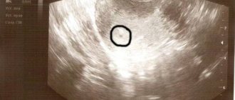
Bradycardia during pregnancy is detected in the fetus during a routine CTG ultrasound examination. It is impossible to see bradycardia by external signs without instrumental diagnostics.
At any week of pregnancy after 8, the fetal heart rate can be clearly determined. Severe sinus bradycardia can reach 70 beats per minute or even less, which is already considered a critical condition. Diagnostic signs of bradycardia at 28-30 weeks:
- the movement of the fetus stops, it freezes, sometimes convulsions are observed;
- attacks of apnea up to cardiac arrest;
- bluish skin tone.
The appearance of bradycardia is already considered a full-fledged diagnosis, therefore the doctor does not wait for other signs of the disease, but prescribes special treatment for the woman. Unfortunately, today there is a high mortality rate when a deviation appears at 20-30 weeks of gestation.
Bradycardia during pregnancy is detected in the fetus during a routine CTG ultrasound examination. It is impossible to see bradycardia by external signs without instrumental diagnostics.
Types and symptoms
There are two types of pathology based on the location of the source of the disorder:
- sinus bradycardia;
- bradycardia resulting from sinoarterial or atrioventricular heart block.
If a woman before pregnancy regularly exercised, worked using physical labor, or suffered from sinus bradycardia, then this phenomenon does not pose a danger to her body and the child. But if the root cause of the decrease in heart rate is heart block, then such serious disorders should be monitored by the attending physician. Symptoms of bradycardia during pregnancy include:
- difficulty breathing;
- increased sweating;
- decreased brain activity and memory function;
- short-term visual impairment;
- feeling of tightness in the chest area;
- constant fatigue;
- dizziness;
- loss of consciousness.
This syndrome is characterized by dilated pupils in humans.
Morgagni-Adams-Stokes syndrome is characterized by severe bradycardia and requires immediate medical attention. Its symptoms include:
- respiratory dysfunction;
- panic attack;
- dizziness;
- sudden loss of consciousness;
- blue lips, fingertips;
- swelling of blood vessels in the neck;
- dilated pupils;
- convulsions;
- involuntary urination.
A prolonged decrease in heart rate below 40 beats per minute is fatal.
There are two types of pathology based on the location of the source of the disorder:
Causes
Both at the beginning of pregnancy and in the last trimesters, a woman’s body begins to experience enormous stress, including on the cardiovascular system. Against this background, heart rate can either increase or decrease, and this does not always indicate the presence of any pathology. Some medications and chemical components can cause a problem. It also develops against the background of heart pathologies of an inflammatory nature or affecting metabolic processes. These are myocardial dystrophy, ischemia, myocarditis. Less dangerous causes of bradycardia include:
- problems in the gastrointestinal tract;
- stressful situations, especially if the nervous system is weakened;
- respiratory tract pathologies;
- poor diet, bad habits;
- endocrine diseases;
- dysfunction of metabolic processes.
Sinus bradycardia, even of a pathological nature, is not considered an indication for medical abortion. Moreover, it rarely causes significant deterioration of the condition.
Important! Planned visits to the doctor cannot be ignored, as he monitors the course of the disease. Only with complete control is it possible to prevent danger to the child and avoid dangerous pathologies.
Pregnancy in patients with bradycardia
The condition of patients with an already diagnosed disease may worsen after they become pregnant. In addition, a decrease in a woman's heart rate will affect the fetus. A slow heart rate in the potential mother leads to chronic embryonic hypoxia , in which developing tissues lack oxygen. At the same time, the unborn child lags behind in development, his brain is formed incorrectly, which in the worst cases threatens with mental retardation.
To avoid undesirable consequences for themselves and the baby, women with bradycardia need to plan their pregnancy taking into account the recommendations of a cardiologist. During pregnancy, patients should be observed by this specialist.
Symptoms
It is quite difficult to establish the presence of the disease, since the woman does not show symptoms. Often, the expectant mother feels well, and a slight deterioration in her health is attributed to fatigue or stress.
Signs of the disease are clearly manifested in a newborn child. Most often, the pathology is characterized by a sudden stop in breathing. You can also slow down your heart rate while walking.
Bradycardia in the fetus can be detected through ultrasound examination using the following signs:
- Deterioration of the heart muscle.
- Slowing down of fetal movement and stopping of movement may occur.
- Rare breathing or periodic stopping.
- Presence of convulsive seizures.
- Stopping the heartbeat or critically reducing the number of heartbeats.
Sinus bradycardia is considered the most dangerous heart rate disorder. The number of beats can be reduced to 70. In certain cases, this may indicate the presence of serious disturbances in the performance of the heart muscle. In this case, the following signs are observed:
- Freezing of movements.
- Cramps.
- Paleness of the skin or cyanosis.
- Heart failure.
This condition requires immediate assistance from specialists, as the risk of frozen pregnancy and fetal death increases significantly.
Fetal pulse is normal and abnormal
The embryo's heart begins to beat on the 21st day of development. At 3-5 weeks it contracts 75-85 times per minute. Over time, the pulse quickens, and after the formation of the nervous system it gradually decreases. Normally, the indicators change as follows:
- At 5-6 weeks, the heart contracts 80-100 times every minute.
- At 6-7 weeks, the pulse is close to 100-120 beats per minute.
- At weeks 7-9, the heart rate reaches a peak of 140-190 beats every minute.
- At 10-12 weeks, the heart rate drops to 160-180 beats per minute.
- After 3 months of pregnancy, the fetal pulse does not exceed 140-160 beats every minute.
- By the 9th month, the heart beats 130-140 times per minute.
Fetal pathology is diagnosed if in the 2nd trimester the pulse is less than 110-120 beats per minute.
Norms at different stages of pregnancy
The normal embryonic heart rate is 110-170 beats/min. Indicators differ at different periods of gestation depending on its development. The number of measurements taken increases if it is slow. An excess is called tachycardia, a rare heartbeat in the fetus is called bradycardia.
Did you know! At the beginning of bearing a child, the rhythm rate is relative. Doctors are faced with the task of making sure that there is no frozen pregnancy.
The numerical indicator may fluctuate within the normal range depending on the size of the embryo at the same period in different women. For example, if its length is within 5 mm, then the optimal value is 100 beats/min. With large sizes, the heart rate is 120-130 beats.
In the last trimester, to diagnose pathology or the level of fetal development, it is possible to determine the position of the myocardium and the characteristics of the pulse (weak or rhythmic heartbeat).
Heartbeat correspondence table by week:
| Gestational age in weeks | Beats per minute |
| 5 weeks | 81-103 beats/min |
| 6 weeks | 104-126 beats/min |
| 7weeks | 127-149 beats/min |
| 8weeks | 149-172 beats/min |
| 9weeks | 172-175 beats/min |
| 10weeks | 170 beats/min |
| 11weeks | 165 beats/min |
| 12weeks | 162 beats/min |
| 13weeks | 159 beats/min |
| 14weeks | 157 beats/min |
| 22-34 weeks | 140-160 beats/min |
| 38-40 week | 130-140 beats/min |
As can be seen from the data presented, in the first trimester there is an increase in numerical indicators. After the formation of the nervous system, on the contrary, the numbers decrease.
Interesting fact! The value of the parameters is influenced by the physiological characteristics of the woman in labor, her emotional state, and the genetic factor.
The main indicator of the norm is the lower limit – 85, the upper – 200 beats/min.
Fetal bradycardia in early and late stages
If at 6-8 weeks of development and later the embryo’s pulse does not exceed 85 beats per minute, it is not bradycardia of the fetal heart that is suspected, but rather arrest or developmental defects. In other cases, a decrease in fetal heart rate that occurs in the early stages is not considered a pathology.
Bradycardia is diagnosed only after 20 weeks of gestation if the unborn baby’s heart beats less than 120 times per minute.
Types of embryonic disease
There are 3 types of embryonic bradycardia: basal, decelerant and sinus . Typically, these conditions develop due to fetal hypoxia, which occurs for the following reasons:
- due to compression of the head;
- due to maternal anemia;
- due to low blood pressure in the mother.
Basal bradycardia is diagnosed if the fetal heart beats less than 120 times per minute. If you treat the malfunction in time and eliminate its cause, you will be able to avoid harm to the woman and the fetus.
If the decelerant nature is violated, the heart rate of the embryo does not exceed 72 beats per minute. This condition requires hospital treatment with bed rest.
With the sinus form of the disease, the fetal pulse drops to 70-90 beats per minute. In this case, the woman needs urgent hospitalization with intensive care and observation until childbirth, because a failure threatens the fading of the pregnancy.
We talked about the causes of bradycardia in children of different ages, as well as whether it is dangerous and what to do in this case, in a separate article on our website.
Treatment
Bradycardia is not an independent disease, but only a symptom of problems in the cardiovascular system. The treatment regimen will depend on the identified cause of the disease, its severity and gestational age. With proper treatment of the underlying disease, heart rate is restored to normal (60-80 beats per minute). Physiological bradycardia does not require treatment.
If the heart rate decreases due to hypothyroidism, hormonal therapy is prescribed for the entire period of pregnancy. The dosage of hormonal drugs is selected individually for each woman. Hypothyroidism can cause miscarriage, delayed fetal development and other serious complications.
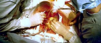
Surgical treatment is indicated for severe bradycardia due to organic heart pathology. During the operation, a pacemaker is installed, which sets the heart the desired rhythm and frequency. Surgical treatment during pregnancy is possible only for health reasons. In other cases, the operation is performed as planned after the birth of the child.
How to detect a slow heart?
Bradycardia of the mother and fetus manifests itself through symptoms of oxygen starvation. In this case, the pregnant woman faces the following ailments:
- with weakness;
- with dizziness;
- with headache;
- with noise in the ears;
- with shortness of breath;
- with low blood pressure;
- with chest pain.
Cognitive failures, expressed in memory and attention impairments, are possible.
A slow pulse is also indicated by a pre-fainting state. An anomaly that exclusively affects the fetus does not affect maternal well-being . It can only be detected using modern diagnostic methods.
What will auscultation show?
After 18-20 weeks, each examination by an obstetrician-gynecologist includes auscultation - listening to the embryo's heart using a special stethoscope, a hollow tube made of wood or metal. To hear the heartbeat of the unborn child, the doctor proceeds as follows:
- Places one end of the stethoscope on the pregnant woman’s belly and puts her ear to the other end of the device.
- Finds the point at which the fetal heartbeat is clearly audible.
- It records the minute during which it counts fetal heartbeats.
The disadvantage of auscultation is its inaccuracy : diagnosis can be hampered by many factors, including maternal obesity and increased fetal motor activity during examination.
Ultrasound of the heart
Instrumental methods, such as ultrasound diagnostics, allow for more accurate research. In this case, a special sensor is used, which is applied to the pregnant woman’s abdomen or inserted transvaginally. It transmits data to the screen, which is decrypted by a specialist.
The following abnormalities indicate a slow pulse during ultrasound of the fetal heart:
- slow movements of the embryo;
- convulsions;
- stopping the movement of the embryo.
Rare breathing or heartbeat, as well as their periodic stopping, also indicate bradycardia. Ultrasound can be performed as early as 3-5 weeks of pregnancy.
CTG and other diagnostic methods
Cardiotocography is used for diagnosis from 32 weeks of pregnancy. The essence of CTG is recording and comparing the pulse of the unborn child with the frequency of uterine contractions. The results of the study are deciphered as follows:
- 8-10 points - the condition of the fetus is normal;
- 6-7 points - mild bradycardia occurs;
- 6 points or less - the fetus is in serious condition.
This method is one of the most accurate.
It is used for complicated pregnancies, as well as if the mother has chronic endocrine or cardiovascular diseases. Another effective way to identify the disease is phonoelectrocardiography , in which the electrical impulses of the heart are recorded and analyzed. This method is a combination of ECG with phonocardiography - listening to heart murmurs.
When is an ECG used?
An electrocardiogram is resorted to if the pregnancy is proceeding with disturbances or there is a threat of any anomalies. From the embryo side, the following indications are distinguished:
- developmental delay;
- previously diagnosed cardiac pathologies;
- suspicion of developmental pathologies.
An ECG is performed on pregnant women who are over 38 years of age or have previously given birth to children with developmental defects. Also from the mother’s side there are the following indications:
- hypothyroidism;
- hyperthyroidism;
- diabetes;
- cardiovascular diseases;
- severe infections.
Bradycardia is indicated by the appearance of a P wave on the cardiogram, as well as a significant TP and PQ interval. The most accurate data is obtained by performing an ECG at 18-24 weeks of pregnancy.
Diagnostics
Since bradycardia is quite easily diagnosed by a doctor by calculating the heart rate and taking into account the patient’s complaints, it is important to determine the root cause of the disease. If the instability of the heart is associated with cardiovascular diseases, the patient is registered with a cardiologist and further studies are carried out. These include:
The expectant mother may be prescribed an ultrasound scan of the thyroid gland.
- ECG. Determines the stability of heart contractions.
- HM ECG. Gives a complete analysis of the work of the heart muscle during the day.
- X-rays of light. Determines heart failure, since the image shows the vessels of the lungs overflowing with blood.
- Ultrasound of the thyroid gland and hormone analysis. Helps detect pathology of the endocrine system.
At-risk groups
Those who suffer from chronic diseases are prone to bradycardia. The risk group also includes women living in conditions of physical or psychological discomfort.
To reduce the likelihood of complications when carrying a child, you need to monitor the course of the underlying disease, give up bad habits and turn to physical therapy. In this case, regular visits to an obstetrician-gynecologist and other specialized specialists are required .
Bradycardia can lead to pregnancy loss or death of the woman. To prevent this from happening, expectant mothers should be careful about their own health. A timely visit to the doctor and correct diagnosis will allow you to identify the problem before it causes irreparable harm.
Prevention measures
Bradycardia in the fetus is established in the later stages, after the 20th week. To prevent the development of the disorder, the expectant mother needs to take her health seriously and follow the following preventive measures:
- Stop drinking alcohol and smoking.
- Eat properly. The diet should include not only fresh fruits and vegetables, but also nuts and dairy products.
- Get outdoors regularly.
- Maintain a work and rest schedule.
Adequate sleep is also of particular importance. A healthy person should sleep at least 8 hours. But the female body experiences serious stress during pregnancy. This is why the expectant mother should get enough sleep.
When visiting a doctor and undergoing diagnostic tests, mothers who have been diagnosed with the disease are interested in what fetal bradycardia is and what danger it poses to the baby. The prognosis depends on the degree of development, the woman’s health status, the type of disorder and the effectiveness of therapy. In some cases, bradycardia does not pose a threat to the fetus. That is why the disease requires careful monitoring by specialists.
Symptoms
It is quite difficult to establish the presence of the disease, since the woman does not show symptoms. Often, the expectant mother feels well, and a slight deterioration in her health is attributed to fatigue or stress.
Signs of the disease are clearly manifested in a newborn child. Most often, the pathology is characterized by a sudden stop in breathing. You can also slow down your heart rate while walking.
Bradycardia in the fetus can be detected through ultrasound examination using the following signs:
- Deterioration of the heart muscle.
- Slowing down of fetal movement and stopping of movement may occur.
- Rare breathing or periodic stopping.
- Presence of convulsive seizures.
- Stopping the heartbeat or critically reducing the number of heartbeats.
Sinus bradycardia is considered the most dangerous heart rate disorder. The number of beats can be reduced to 70. In certain cases, this may indicate the presence of serious disturbances in the performance of the heart muscle. In this case, the following signs are observed:
- Freezing of movements.
- Cramps.
- Paleness of the skin or cyanosis.
- Heart failure.
This condition requires immediate assistance from specialists, as the risk of frozen pregnancy and fetal death increases significantly.
How can a decrease in heart rate be diagnosed, and at what term?
Diagnosis is carried out in the prenatal diagnostic room. First of all, auscultation of the fetal heart is done using a special tube (plastic or wooden).
Auscultation means listening to heart sounds and measuring heart rate per minute. The first diagnosis is carried out at the 20th week of pregnancy and subsequently at each visit to the doctor. If heart rate is less than 110 beats. per minute, this indicates DP, which requires additional assessment of the biophysical profile of the fetus (FPP).
BPP is first performed at 30 weeks of pregnancy. A comparison is made of the sum of scores of some biophysical parameters of the fetus, which include assessment of respiratory movements, tone, cardiac and motor activity.
Additionally, a non-stress test is carried out - an assessment of the reactivity of the fetal heart after motor reactions according to cardiotocography data (a study using an ultrasound sensor that is placed on the stomach of a pregnant woman in the place of best listening to the fetal heartbeat; with each registration of the heartbeat, a graphic curve is recorded on paper).
On the cardiotocogram you can see decelerations (episodes of heart rate deceleration by 15 beats per minute or more, which last 15 seconds or more) and accelerations (episodes of heart rate increase by 15 beats per minute, which last 15 seconds or more).
The presence of accelerations is considered a favorable sign, since it indicates normal oxygen saturation of the fetal blood.
In addition, Doppler ultrasound of blood flow velocity in the umbilical cord arteries or Doppler ultrasound of uteroplacental blood flow is performed. An assessment of blood flow is also carried out by an obstetrician-gynecologist, the diagnostic results are assessed in a complex, after which the treatment tactics for the pregnant woman are decided.
These research methods are important indicators of the normal or pathological course of pregnancy; the table shows the main clinics where you can be examined.
| Name of institution | Address (Moscow and St. Petersburg) | Telephone | Procedure | We're standing. (rub) |
| "Capital Medical Clinic" | Moscow, st. Sretenka, 9 | 6493903 | Cardiotocography (CTG) | 1150 |
| "SM-Clinic" | Moscow, st. Klara Zetkin, 33/28 | 6494661 | Cardiotocography (CTG) | 1900 |
| "Tsar's Clinic" | Moscow, st. Profsoyuznaya, 58, bldg. 4 | 1146555 | Cardiotocography (CTG) | 1800 |
| "Deltaclinic" | Moscow, Nastavnichesky lane, 6 | 5193499 | Dopplerography of uteroplacental blood flow | 1300 |
| "MEDINNOVA" | Moscow, st. Gilyarovskogo, 50 | 1146476 | Dopplerography of uteroplacental blood flow | 1710 |
| "Crede Experto" | Moscow, Tovarishesky lane, 10, building 2 | 1146600 | Dopplerography of uteroplacental blood flow | 2200 |
Help for the fetus when bradycardia is detected:
The greatest danger posed by a slow heartbeat is the death of the fetus. If not death, then serious developmental pathologies are guaranteed. The earlier the pathology is detected, the more effective the treatment will be. Fetal bradycardia in the early stages of pregnancy is often compensated and does not affect the further development of the baby and the general condition of the mother.
The goal of diagnosis is to eliminate the identified cause completely or reduce its impact. To determine if the heart is not functioning properly, the following is used:
— Ultrasound diagnostics; — cardiotocography; - doppleroscopy; — studies of the fetal myocardium directly; - carry out blood and urine tests.
All studies are carried out at certain intervals in order to see the dynamics of the condition. The frequency and intervals between diagnostic studies are determined by the gynecologist observing the pregnancy. The minimum is two more ultrasound examinations with an interval of 3 or 7 days after the first suspicion. The diagnosis is considered confirmed if the fetal heart beats at a rate of 110 beats/min or less for 10 minutes or more.
Any treatment is prescribed exclusively by a doctor, regardless of whether it is medication or traditional medicine recipes! Bradycardia in the later stages is a good reason for delivery by cesarean section.
The entire therapeutic course includes:
— normalization of nutrition; - complete cessation of bad habits (if any); — correction of motor activity of a pregnant woman; - prescribing iron-containing drugs for anemia in women; — the safest possible therapy for a pregnant woman in the presence of infectious or internal non-communicable diseases.
After a course of treatment during the initial diagnosis, the following is prescribed:
- for fetal bradycardia in the early stages: constant follow-up monitoring of the heartbeat using an ultrasound vaginal control sensor; - for fetal bradycardia in late stages: monitoring heart function using CTG, transabdominal ultrasound or auscultation (listening to the heartbeat).
The main drugs that are always administered to a pregnant woman for fetal bradycardia are:
• carboxylase and sodium bicarbonate drip; • solutions of ascorbic acid and glucose intravenously; • calcium gluconate intravenously.
Prevention measures
Bradycardia in the fetus is established in the later stages, after the 20th week. To prevent the development of the disorder, the expectant mother needs to take her health seriously and follow the following preventive measures:
- Stop drinking alcohol and smoking.
- Eat properly. The diet should include not only fresh fruits and vegetables, but also nuts and dairy products.
- Get outdoors regularly.
- Maintain a work and rest schedule.
Adequate sleep is also of particular importance. A healthy person should sleep at least 8 hours. But the female body experiences serious stress during pregnancy. This is why the expectant mother should get enough sleep.
When visiting a doctor and undergoing diagnostic tests, mothers who have been diagnosed with the disease are interested in what fetal bradycardia is and what danger it poses to the baby. The prognosis depends on the degree of development, the woman’s health status, the type of disorder and the effectiveness of therapy. In some cases, bradycardia does not pose a threat to the fetus. That is why the disease requires careful monitoring by specialists.