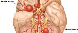
What is a cerebral aneurysm and what are the consequences of its rupture?
A brain aneurysm occurs when there are pathological changes in the vessels, and their shape changes.
They become thin and fragile, they stretch and bulge. An aneurysmal sac forms at the site of deformation, which may subsequently burst, leading to intracranial hemorrhage. The probability of death if a vessel ruptures is very high, therefore, if the diagnosis is confirmed, surgical intervention is performed urgently. In this case, clipping of the brain aneurysm is most often done, except in situations where the sac is located too deep.
When talking about what aneurysm clipping is, we mean the procedure of disconnecting the aneurysm from the general blood flow. This occurs by placing a clip on the neck of the affected vessel.
Depending on the shape of the aneurysm, double-sided clamping may be required. To gain access to the desired area, craniotomy is performed.
The patient is given general anesthesia. Trephination of the desired area of the skull is performed. A hole is cut with a cranite. The dura mater of the brain is opened. The affected area is identified and separated from other tissues. The aneurysm is disconnected from the general blood flow by applying a clip. The skull is being restored. The excised hole is secured with plates and screws.
Performing an operation requires precision and attention from the surgeon. During the procedure, various microsurgical equipment is used. If the doctor sees that the vessels are thinned, he can wrap them in surgical gauze or muscle particles. This will reduce the risk of rupture when pressure increases.
When choosing a treatment method for an aneurysm, a number of examinations are carried out. Mainly:
- General blood and urine analysis. Blood test for biochemistry and infectious diseases. Radiography. Cardiogram. Examination by a therapist and neurologist, sometimes by other specialists, depending on the symptoms. Magnetic resonance angiography. Indicated for aneurysms over 3 mm. CT is necessary to obtain a general picture for tumors larger than 5 mm. It can be used to detect calcifications and blood clots inside the aneurysm. Digital subtraction angiography allows you to see formations up to 3 mm.
Before clipping of cerebral vessels, it is necessary to prepare the body for surgery. To do this, they normalize existing diseases: they compensate for diabetes, blood pressure and other chronic diseases that occur in an acute form.
After examination by the surgeon, anesthesiologist and filling out the consent, the date of the operation is set. The day before, it is not recommended to consume food or liquid after 6 pm.
In order for the patient to quickly return to his normal lifestyle after surgery, he is shown rest and a positive attitude.
When performing a planned operation, the patient is left for several days in the intensive care unit in order to be able to provide timely medical assistance in case of complications. After the specified time has passed, the patient is transferred to the general ward.
In the postoperative period, fatigue and weakness may be a concern. Therefore, complete rest and bed rest are recommended.
An attack of headache is also a frequent accompaniment of an aneurysm clipping operation. This symptom can be eliminated with narcotic drugs, so if you have severe and frequent migraines, you should consult your doctor.
The total rehabilitation period is about two months. It is important to take into account the presence of other diseases and their severity, the condition in which the patient was at the time of clipping. If the operation was carried out as planned and the size of the formation was insignificant, then the procedure is easier to tolerate and the body recovers faster.
Deterioration of the condition after surgery is quite rare. According to statistics, this number does not exceed 10%. But when agreeing to treat an aneurysm, the patient must evaluate all the risks involved.
The consequences can be very different: ranging from minor disturbances in speech, memory, attention, constant headaches and ending with ischemic complications, pulmonary edema, and in some cases, death.
But you should not refuse the operation; the main condition for successful treatment is the choice of qualified personnel, compliance with all doctor’s recommendations and timely diagnosis of complications in the postoperative period.
In most cases, complications arise from preoperative rupture of the aneurysm or bleeding during the procedure.
- Loss of coordination of movements or decreased sensitivity of the limbs, paralysis. Dysfunction of the speech apparatus. Decreased vision. Blockage of blood vessels. Psychological disorders. The appearance of epilepsy.
In general, if all recommendations are followed and the operation is performed in a timely manner, the patient’s life expectancy is not reduced.
If treatment is refused, the aneurysm will gradually increase in size, and eventually it will rupture and hemorrhage, which often leads to death.
The main recommendations for a speedy recovery are:
- Revision of nutrition. Rationing of physical activity. Observation by a neurologist. Rejection of bad habits. Undergoing MR angiography and CT 6 months after surgery. Monitoring the condition to avoid new formations.
Cerebral aneurysm is also known as cerebral, intracranial or intracranial. It is a bulge on the vessel.
Sometimes it ruptures, causing a cerebral hemorrhage, in which blood spills into the space around the brain. These areas are called subarachnoid areas and hence this type of brain hemorrhage is called subarachnoid hemorrhage.
It is a cause of stroke and can lead to serious brain damage and death.
A cerebral aneurysm is a bulging of the worn-out wall of one of the arteries of the brain, filled with blood. Those who have it have a “time bomb” inside their skull. The wall of the artery in the place where this protrusion formed does not have a muscle layer and membrane, which causes a lack of elasticity and strength of the vessel in this place.
The disease is especially dangerous due to the fact that the thinned wall of the artery can rupture at any second, resulting in hemorrhage in the brain. An aneurysm can also compress nearby brain tissue and nerves.
Hemorrhage may occur in the meningeal spaces of the brain or in the ventricles of the brain. In any case, cerebral edema occurs and intracranial pressure increases. Blockage of the cerebrospinal fluid pathways may occur with subsequent displacement of brain structures. Blood begins to disintegrate over time, and its breakdown products cause an inflammatory reaction in the brain tissue, which leads to necrosis of these areas. This means that the functions for which these areas of the brain were responsible will be lost.
With subarachnoid hemorrhage, a complication such as cerebral vasospasm may occur. That is, the peripheral vessels of the brain sharply contract, as a result of which the blood flow in them slows down or becomes impossible, which leads to ischemia of brain tissue.
Symptoms
If a vessel in the head bursts, signs of intracerebral bleeding appear - sudden pain in the head area, intensifying after exercise, nausea, which is often accompanied by vomiting. In elderly patients, pain in the head is sometimes absent or moderate.
In cases where the hemorrhage focus is large, neurological symptoms may be supplemented by confusion. A typical symptom is focal seizures (affecting specific areas of the body) or generalized seizures (distributed throughout the body).
https://youtube.com/watch?v=P6_b6TaQCeE
Sometimes, before a vessel in the head bursts, precursor symptoms appear, which include a feeling of heat, visual dysfunction, and increasing pain in the head. More often, a stroke develops rapidly without previous signs. Typically, rupture of the vascular wall occurs during the daytime and is caused by physical or emotional stress.
If a vessel bursts in the head, the condition is accompanied by specific symptoms, which manifest themselves acutely with a tendency to increase. The clinical picture develops quickly. Confusion, stupor, and fainting are often observed. In severe cases, stunning and coma occur. Focal neurological symptoms depend on the location of the hemorrhage:
- Hemispheres. The pathology is accompanied by hemiparesis (paresis in one half of the body).
- Posterior fossa of the skull. Symptoms of damage to the structures of the brainstem and cerebellum are observed - paralysis of the eye muscles, narrowing of the pupils to a pinpoint state, stertorous breathing (noisy breathing, accompanied by inspiratory and expiratory wheezing, characteristic of obstruction, blockage of the airways).
- Ventricular system. The most life-threatening form, accompanied by meningeal (stiff neck, Kernig and Brudzinski symptoms, changes in reflexes) and brain stem (tachypnea - rapid, shallow breathing, tachycardia, stupor or stupor, increased blood pressure, increased tone of skeletal muscles) symptoms, hyperthermia (accumulation of excess heat in the body), hormetonic-type convulsions, rapid depression of consciousness.
We invite you to familiarize yourself with the Basic rules for installing an air conditioner
When blood enters the ventricular system, hydrocephalus of the occlusive type may develop, occurring in an acute form. The focus of hemorrhage localized in the frontal lobe leads to a pronounced impairment of cognitive abilities (memory, mental activity, speech).
A supratentorial (located in the cerebral hemispheres) hematoma, accompanied by cerebral edema, can lead to transtentorial (the temporal lobe is wedged through the foramen of the cerebellar tentorium and under it) herniation, which in turn causes compression of the trunk and the appearance of secondary foci of hemorrhage in the area of the midbrain and pons .
Hematomas localized in the cerebellum, when enlarged, can block the 4th ventricle, which leads to the development of hydrocephalus, which occurs in an acute form. Foci of hemorrhage located in the cerebellar area with a diameter exceeding 3 cm can provoke dislocation of the median structures. The described conditions often lead to severe depression of consciousness, the development of coma and death of the patient.
Extensive intracerebral bleeding usually (70% of cases) ends in death. In surviving patients, neurological symptoms gradually regress in parallel with the process of resorption of blood poured into the cranial cavity. Hemorrhages cause less damage to brain structures than infarctions, so neurological deficits are often quickly corrected.
If a small vessel ruptures with the subsequent formation of a small focus of hemorrhage, focal neurological symptoms usually develop without depression of consciousness. In this case, the headache is moderate. Nausea may not be a clinical sign.
The first choice methods for making a differential diagnosis are MRI and CT studies. Differentiation is made in relation to ischemic stroke and subarachnoid (intrathecal) hemorrhage. When making a diagnosis, it should be taken into account that acute neurological deficit can develop as a result of an epileptic seizure or hypoglycemia (a critical decrease in blood glucose levels). A blood test shows your glucose level. Computed tomography allows you to establish:
- Exact localization of the source of hemorrhage.
- Volume and prevalence of the pathological process.
- The presence and severity of cerebral edema.
- The presence and severity of dislocation of brain structures.
If MRI and CT examinations are not possible, a cerebrospinal fluid sample is taken as an alternative. Blood is detected in the cerebrospinal fluid if the focus of hemorrhage is in the area of the subarachnoid space or the posterior fossa of the skull. The attending physician will tell you what to do if a blood vessel in your head bursts, taking into account the results of the examination.
List of diseases for obtaining disability
September 24, 2020 503742 Unfortunately, becoming disabled today is not a rare occurrence; it can arise as a result of an accident, occupational or congenital disease.
The procedure for establishing disability in Russia is carried out by the relevant bodies, in particular the medical and social expert commission (MSEC), and carries not only medical, but also legal significance, giving a person who cannot carry out partial or full work activities the right to receive a number of social benefits and pension payments.
From a genetic perspective, there is little that can be done to prevent an aneurysm. It is necessary to warn the doctor about the presence of the disease in the family, in order to carry out timely treatment and preventive measures aimed at maximizing the protection of the walls of blood vessels from adverse effects.
It is mandatory to prevent atherosclerotic changes in the vascular walls. This fact is significantly influenced by smoking, so it should definitely be avoided.
In addition, high blood pressure should be mentioned, which can be associated in particular with a poor lifestyle and a lot of stress or insufficient exercise.
Obesity and high levels of lipids (fats) in the blood plasma are another potential “creator” of atherosclerosis. As a rule, preventing the deposition of fat in blood vessels requires general lifestyle changes, including diet and inclusion of a sufficient amount of exercise in daily life.
Diagnosis of the disease
Instrumental diagnostic measures are the main ways to determine the location of an aneurysm and the stage of its development. The following procedures are used:
- Ultrasound of arteries;
- ECHO of the heart;
- angiography and ventriculography (injection of a contrast agent into the vessels);
- CT;
- X-ray.
X-ray diagnostics
X-ray is the most common diagnostic method, allowing visualization of the abdominal and chest organs. The boundaries in the image allow you to track the density of the tissues. With an aneurysm, swelling or dilatation of blood vessels is visible.
Contrast radiation (injection of a special substance into a vessel) allows you to most accurately determine the border of the vessel, the shape and size of the formation. Aortography is rarely used due to the high risk of aneurysm damage and rupture.
Thanks to the ability to measure the speed of blood flow with an ultrasound device, it is possible to recognize areas with an aneurysm where the passage of blood is complicated. Safety, high speed, accuracy - the advantages of this type of examination make it most popular for identifying an aneurysm.
MRI is the formation of the most accurate image using an electromagnetic field. The volume, shape and even thickness of the walls of the formation are monitored. This makes it possible to make a prognosis and develop effective treatment.
With an ECG, it is possible to exclude ischemia (the symptoms are similar), identify areas of accumulation of atherosclerotic plaques, and track specific tissue properties.
With an aneurysm, blood and urine tests do not make it possible to recognize the disease, since the indicators remain unchanged. However, they are prescribed necessarily in the preoperative period and during the recovery stage. With x help, it is possible to assess the general condition of the body and its ability to recover.
Does a cerebral aneurysm give disability with such a diagnosis?
Medical and social examination and disability in case of cerebral vascular aneurysms Cerebral vascular aneurysms are clinically manifested in young and middle-aged people and often lead to severe disability. When assessing the ability to work, the type of aneurysm (arterial, arteriovenous), its location, the nature and degree of cerebral vascular aneurysm are taken into account. and focal disorders, the presence and frequency of epileptic seizures, mental disorders, the state and compensatory capabilities of cerebral hemodynamics, the course of the disease, the effectiveness of surgical intervention.
In each specific case, when assessing work ability, social factors are taken into account, in particular the profession and working conditions of the person being examined. The asymptomatic course of the disease and the difficulties of clinical diagnosis are the reason that in most patients, cerebral aneurysm is recognized after the first subarachnoid hemorrhage.
Indications and contraindications for surgery
The decision to perform the operation is made by the attending physician after the patient consents to the procedure. The main indications for clipping are:
- aneurysm reaching 7 mm or more; genetic predisposition to rupture of the aneurysmal sac.
For diseases of the circulatory system. For decompensation of diabetes mellitus. In the presence of acute inflammatory and infectious processes. If bronchial asthma is severe. With exacerbation of chronic diseases.
Clipping is not performed when the aneurysm is located deep enough.
How to properly prepare for the procedure
When choosing a treatment method for an aneurysm, a number of examinations are carried out. Mainly:
- General blood and urine analysis.
- Blood test for biochemistry and infectious diseases.
- Radiography.
- Cardiogram.
- Examination by a therapist and neurologist, sometimes by other specialists, depending on the symptoms.
- Magnetic resonance angiography. Indicated for aneurysms over 3 mm.
- CT is necessary to obtain a general picture for tumors larger than 5 mm. It can be used to detect calcifications and blood clots inside the aneurysm.
- Digital subtraction angiography allows you to see formations up to 3 mm.
Before clipping of cerebral vessels, it is necessary to prepare the body for surgery. To do this, they normalize existing diseases: they compensate for diabetes, blood pressure and other chronic diseases that occur in an acute form.
After examination by the surgeon, anesthesiologist and filling out the consent, the date of the operation is set. The day before, it is not recommended to consume food or liquid after 6 pm.
Price policy
The question of how much the operation costs can be answered in different ways. During planned hospitalization, the patient has the right to free treatment. To do this, when contacting the Ministry of Health, you must fill out paperwork and provide the relevant documents.
In this case, it may take several weeks or months to review the application and allocate funds from the budget.
If you don’t have time, you can contact the clinic privately, then the cost of the procedure will be from 80 to 180 thousand rubles. This depends on the complexity of the operation, the prestige of the clinic and the qualifications of the doctors, as well as the price of the materials that will be used in the clipping process.
Disability due to cerebral atherosclerosis
› The main cause of acquired dementia and various mental disorders in adulthood is cerebral atherosclerosis.
The danger of cerebrosclerosis is that the disease is asymptomatic and, as a rule, ends in disability.
How to recognize an insidious enemy, is it possible to get rid of him and who is at risk of disability? The main suppliers of blood to the brain are the paired vertebral and carotid arteries. Then they are crushed into small vessels and a capillary network one and a half thousand kilometers long!
Cerebrosclerosis is a disorder of vascular conduction that leads to stroke or aneurysm (dilation of the vessel walls). The consequences of these diseases are the most serious.
Disability registration procedure
Among the causes of mortality, cerebrosclerosis is one of the first places in the world. The danger of the disease is that the patient earns disability not so much because of his sclerosis, but because of its complications, which are not long in coming in the absence of adequate treatment and prevention.
Second degree atherosclerosis significantly limits the ability to work. If the work requires concentration and psychomotor functions, the patient is not able to perform it as before.
Often, cerebrosclerosis provokes extrapyramidal tremor of the hands, then precise work with small parts is not possible. Based on such restrictions, medical and social examination assigns a third disability group. You can ask your doctor or health care facility representatives for a referral for examination.
Disability is prescribed when diagnosing vascular stenosis, when the slightest overexertion can provoke a stroke and other serious consequences. The likelihood of complications depends on the immune system, genetic predisposition and the presence of risk factors. Without constant medical monitoring and prevention, the disease will only progress.
With atherosclerosis of cerebral vessels, disability is given:
- For transient (micro-strokes) and persistent cerebral cerebrovascular accidents;
- Acute coronary blood flow disorders;
- Obliterating atherosclerosis;
- Aortic stenosis and aneurysm.
Each of these pathologies is difficult to treat, and therefore leads to disability in advanced stages. After a stroke, the patient becomes disabled due to paralysis, paresis, decreased activity of the limbs, and reduced mental capabilities.
We suggest you read: Is it possible to apply for temporary disability after surgery?
A disabled person from a medical point of view is a patient with persistent dysfunctions of the body that limit life activity and require social protection. The presence of individual signs of cerebral atherosclerosis does not provide grounds for obtaining a disability group.
Invalidation is carried out after a medical and social examination. Health indicators are assessed in a comprehensive manner, analyzing all aspects: clinical-functional, social-living, professional-labor and psychological. In this case, generally accepted classification and criteria are used.
Refers the patient to a medical institution after examination, treatment and rehabilitation course, if necessary. Bodies of social and pension protection of the population also have powers if the applicant has the necessary medical documents and signs of limited viability.
If the medical institution refuses to issue a referral, you can obtain a certificate with which the patient will undergo an examination independently after submitting an application and documents, as well as papers describing the social and labor status of the person.
The examination can be done in absentia, in a hospital hospital or at home.
In the latter option, health indicators are confirmed by the conclusion of the health care facility. If necessary, an additional examination program is proposed to identify the degree of limitation of vitality and rehabilitation possibilities.
The commission makes a decision by counting a majority of votes, and in the presence of all members it is communicated to the examinee. The applicant receives a certificate of disability and a note on the certificate of temporary incapacity for work. A personal rehabilitation program is offered.
The refusal can also be made in writing. 2 months before the end of the disability period, a re-examination is carried out. For disabled people of the first group, the term is 2 years, the second and third - one year.
According to medical statistics, the mortality rate from stroke in the Russian Federation is second only to cardiac pathology. With a pronounced narrowing of the lumen of the cerebral artery, 15% of patients die during the first month.
Half experience cerebral hemorrhage, 40% have subarachnoid bleeding, 10% of victims lose their former ability to work, and 20% become disabled requiring outside care. All categories require lifelong expensive treatment.
Visit a cardiologist regularly, control your blood pressure, avoid stress, overeating and overwork. Pathologies of cerebral vessels are life-threatening, so atherosclerosis can only be defeated with the help of a doctor. Don't get carried away with self-medication!
Recommendations from neurologist Dr. M.M. Sperling on improving the quality of life in cerebral atherosclerosis - in this video
Statistical indicators
In approximately 0.2-3.0% of cases, a cerebral aneurysm ruptures and bleeds. For example, in the United States there are 30,000 such patients annually, of whom 10-15% die before reaching the hospital, and more than 50% die despite appropriate treatment within the first 30 days.
Even of those who survive, more than half suffer permanent neurological disability. You can get a cerebral aneurysm at any age, but most often at the age of 35-60 years (more often in women).
Causes
First of all, it is necessary to mention the genetic predisposition, which results in various defects on the vascular wall - thinning or lack of elasticity.
Therefore, it is good to know in advance whether anyone in the family has suffered from vascular diseases, which may have a genetic origin.
Often an aneurysm affects people who have certain genetic disorders and circulatory disorders.
Other causes include atherosclerosis, head injuries, and various infections of the nervous system.
Risk factors include smoking, high blood lipids (hyperlipidemia), high blood pressure and drug use.
Many studies have identified several factors that significantly increase the risk of aneurysms.
- Hereditary factor - with a deficiency of type III collagen, thinning of the muscle layer of the arteries occurs. Aneurysms are especially often formed in this case in the area of bifurcations (bifurcations) of arteries and in places where the artery is highly tortuosity. This is also accompanied by other pathologies, for example, coarctation of the aorta, hypoplasia of the renal arteries History of arterial injuries Hyalinosis of the vascular walls Smoking Drug use High blood pressure Arterial embolism - transfer with the bloodstream of small “pieces” of malignant tumors or a conglomerate of fungal or bacterial microorganisms Effect of radioactive radiation any duration Atherosclerosis of cerebral vessels
- Cerebral aneurysm is a disease of adults 30-60 years old Women are susceptible to aneurysm more often than men The risk of its development is high due to hereditary disposition In the USA, for example, 27,000 patients rupture every year
Causes of rupture of blood vessels in the head
Intracranial hemorrhage most often occurs due to the rupture of a small vessel that has undergone atherosclerotic changes, with blood entering directly into the brain tissue. The main reason that provokes the pathological process when blood vessels in the head burst is long-term arterial hypertension (80-85% of cases).
Other causes of occurrence are associated with congenital and acquired vascular pathologies - atherosclerosis, aneurysms, arteriovenous and other types of vascular malformations. A history of arterial hypertension may be combined with damage to elements of the vascular system or blood pathologies (dyscrasia). If a vessel bursts in a young patient, the reasons may be related to cocaine addiction. Other causes and provoking factors:
- Aneurysm of the mycotic type, provoked by infectious diseases of the central nervous system or injuries in the cranial area.
- Cerebral infarction of hemorrhagic type.
- Excessive therapy with anticoagulants - drugs that prevent blood clotting.
- Dissection (dissection) of intracranial arteries.
- Disturbances in the functioning of the hemostasis system (maintains the fluid state and other properties of the blood).
- Tumor processes localized in brain tissue.
- Deficiency of vitamins and nutrients.
- Intoxication is chronic and acute.
- Inflammatory processes that affect blood vessels in the brain.
- Excess body weight.
- A diet high in saturated fat.
- Taking sympathomimetic (stimulating the sympathetic nervous system) drugs.
- Smoking, alcohol abuse.
In old age, burst vessels can be a consequence of cerebral amyloid angiopathy. The disease is accompanied by the deposition of protein compounds that accumulate and form fibrils in the walls of the cerebral arteries.
The pathogenesis of hemorrhagic stroke is based on the mechanism of angiodystonic disorders, which affect the dynamics of regional blood circulation in brain tissue. When asked whether a vessel in the head can burst and for what reason, the doctor will primarily name a chronic increase in blood pressure and hypertensive crises.
Hypertensive crises are conditions requiring emergency medical care, characterized by an excessive increase in blood pressure. Crises provoke the development of paralysis and spasms of the walls of arteries and arterioles that supply the brain. Disorganization of the walls of blood vessels is more often associated with metabolic disorders and disorders of neurohumoral regulation.
Due to pathological changes, the permeability of the artery wall increases, which easily allows plasma cells and red blood cells to pass through. This condition is called diapedesis and often precedes wall rupture. In the mechanism of development of the focus of hemorrhage, disruption of blood clotting processes plays an important role.
Clinical picture
Minor blood leaks or sudden expansion of the walls of the aneurysm can manifest themselves as so-called warning headaches, which, however, are difficult to distinguish from “ordinary” headaches that arise for other reasons.
With major bleeding, a very severe headache is typical. It often appears after some stress.
Pain should always be a signal for proper examination. It can be accompanied by a disorder of consciousness, movement, and in the worst case, the patient dies.
An unruptured cerebral aneurysm may not manifest itself at all, but in about 40% of patients, any of the following symptoms or a complex of them are sometimes present:
- impaired peripheral vision (to the sides, above and below); difficulty thinking or performing certain activities; speech problems; problems related to perception (touch, heat, cold, pain); sudden changes in behavior; impaired balance and coordination of movements; significant decrease in the ability to concentrate; impairment of short-term memory; fatigue.
A ruptured brain aneurysm is accompanied by distinct symptoms
- headache (patients sometimes describe it as “the worst headache they have ever experienced”); vomiting or at least the urge to vomit; stiff neck or neck pain; impaired or double vision; weakening or numbness in the limbs (sometimes one half of the body); pain above and behind the eyes; dilated pupils; sensitivity to light; Impaired skin sensitivity (to touch).
Is the operation dangerous, what should you prepare for after clipping?
Deterioration of the condition after surgery is quite rare. According to statistics, this number does not exceed 10%. But when agreeing to treat an aneurysm, the patient must evaluate all the risks involved.
The consequences can be very different: ranging from minor disturbances in speech, memory, attention, constant headaches and ending with ischemic complications, pulmonary edema, and in some cases, death.
But you should not refuse the operation; the main condition for successful treatment is the choice of qualified personnel, compliance with all doctor’s recommendations and timely diagnosis of complications in the postoperative period.
In most cases, complications arise from preoperative rupture of the aneurysm or bleeding during the procedure.
- Loss of coordination of movements or decreased sensitivity of the limbs, paralysis.
- Dysfunction of the speech apparatus.
- Decreased vision.
- Blockage of blood vessels.
- Psychological disorders.
- The appearance of epilepsy.
Goals and methods of therapy
CT scan of the brain (computed tomography) is the basis for diagnosing cerebral hemorrhage - in the presence of subarachnoid hemorrhage, it is necessary to look for a cerebral aneurysm. CT is a technique in which the patient passes through a large, round device; During this process, X-rays pass through the thin layers of the brain very quickly and the result is processed by a computer.
Lumbar puncture. If the CT results are normal, but the patient’s condition suggests a ruptured aneurysm, a spinal (lumbar) puncture is performed - a puncture of the spinal canal (an incision in the back to the spine, carried out under local anesthesia, sitting or lying on the side). When collecting bloody fluid (CSF), bleeding from a ruptured cerebral aneurysm is determined.
Cerebral angiography. To determine the exact position, size and shape of the aneurysm, cerebral angiography is performed - imaging of the cerebral arteries using injections of a contrast agent. Classic angiography is performed by inserting a thin catheter through the groin area under local anesthesia;
The goal of treating a cerebral aneurysm is to prevent recurrent bleeding, which determines a high percentage of the patient’s threat of death or other serious consequences in the event of a rupture.
Microsurgical method - clipping of a cerebral aneurysm (from the English Clip - clamp). It involves clamping the blood vessels while maintaining their patency. Clipping is performed by microsurgical operation with opening of the cranial cavity (craniotomy). The endovascular method is performed under X-rays using a catheter.
During this therapy, special metal coils are inserted into the bulge, on which a blood clot forms, causing the bulge to close. In recent years, the introduction of reinforcing stents in this way has become increasingly common to further expand the capabilities of this treatment method. Therapy is carried out by inserting a catheter through the groin into the femoral artery, and then through the vascular system to the corresponding vessels of the brain.
Ideally, a decision is needed within 3 days of the onset of bleeding due to the high risk of recurrent bleeding from an already ruptured aneurysm; after 72 hours, due to the breakdown of hemoglobin and the release of toxic substances, cerebral vascular spasms occur, so the indication for surgery at this time is set on a strictly individual basis.
Deferred decision is performed quite rarely - when diagnosis is late or when the patient's condition is so severe that there is a risk of serious complications with immediate therapeutic intervention. The procedures in this case are the same as in the acute phase, but are used after the spasms of cerebral vessels have been eliminated.
The course of each therapy method is determined by the specific location and shape of the bulge, the patient’s condition, his age and other factors. Each of them has its own advantages and disadvantages.
The endovascular method is more convenient from the point of view that the patient does not open the cranial cavity surgically. But, along with this, some complications are possible during the operation, for example, rupture of the aneurysm during the intervention.
The disadvantage also lies in the absence of a 100 percent guarantee that the coils inserted by the catheter will not move and the aneurysm will not begin to fill again.
In contrast, clipping with opening of the skull is considered an operation in which the patient is cured completely, however, open brain surgery is more difficult for the patient. A team of specialists, consisting of neurosurgeons and radiologists, decides which treatment method to use.
Rarely, an aneurysm can be detected by random examination before it ruptures. But usually diagnostic methods are applied after its rupture.
- Angiography is an x-ray method using contrast agents. A drug is injected intravenously, which will allow you to see in the pictures all the vessels of the brain, the places of their narrowing, tortuosity, and determine where exactly the aneurysm is located. Computed tomography (CT) is a non-invasive research method that allows you to determine in which part of the brain the hemorrhage occurred and the volume of damaged tissue. CT angiography is a combination of the two previous methods. Computed tomography with preliminary administration of a contrast agent. Magnetic resonance imaging (MRI) - allows you to more accurately visualize blood vessels. MRI can accurately determine the location and size of the aneurysm. Examination of cerebrospinal fluid - a spinal puncture and collection of cerebrospinal fluid are performed under local anesthesia. In a ventricular rupture hemorrhage or subarachnoid hemorrhage, there will be blood in the cerebrospinal fluid.
In the absence of symptoms of an aneurysm, frequent diagnosis is not justified; only if there are 2 or more immediate relatives with this disease, regular screening is recommended, and regular diagnosis is indicated for patients who have already had a ruptured aneurysm, since the risk of developing a new aneurysm is 1-2 percent per year.
If you have an unruptured aneurysm, you may not need treatment right away. Most often, only condition monitoring and regular examination of the patient are required. When deciding on the treatment of a cerebral aneurysm, the risks of surgery are compared with the risk of its rupture. This takes into account the size and type of aneurysm, the patient’s age, its location, the patient’s health status and heredity.
Even the most experienced specialist will not be able to predict whether an aneurysm will ever rupture or not by diagnosing an aneurysm. The consequences of its rupture are very serious, almost fatal, but the decision to perform surgical intervention should be approached very individually, since surgical treatment of an aneurysm (operation) also poses a considerable risk for the patient.
Based on numerous studies, scientists come to the conclusion that if the size of the aneurysm is less than 10 mm, the probability of its rupture is not high, and in this case the operation carries a greater risk. According to various expert estimates, the occurrence of complications after surgery is 4-15 percent, and death is 0-7 percent.
1. Aneurysm clipping with craniotomy. A complex neurosurgical operation, its goal is to exclude the aneurysm from the general blood flow. At the same time, the blood flow in the vessel on which it is located is not disrupted. In this case, the skull is opened in the desired projection, and a vessel with an aneurysm is located. A clip is placed on her neck. A special microscope and microsurgical technique are used.
2. Endovascular operations. In this case, access is made through the femoral artery. A catheter is inserted into it, at the end of it there is a balloon or coil, which is passed to the vessel with the aneurysm under CT control. A balloon or coil is installed in this place, which allows you to turn off the damaged vessel from the bloodstream.
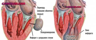
General information about aneurysm
An aneurysm is a protrusion on a vessel whose structure is damaged. Most often this is a consequence of the destruction of muscle tissue. The structure of the heart resembles blood vessels, so an aneurysm can form in it, most often after a heart attack. It can also form in the abdominal aorta.
Causes
The disease can have congenital and acquired etiology. Among the first group of causes are Marfan syndrome and congenital elastin deficiency. All abnormalities in the body in which the structure of connective tissue is disrupted can lead to the formation of an aneurysm.
- arterial injury;
- ischemia;
- postoperative changes;
- lesions of viral, bacterial, fungal origin;
- autoimmune changes;
- damage to vascular walls by atherosclerotic plaques.
Aneurysm can be classified:
- Taking into account the degree of the course - into acute and chronic forms.
- Depending on the dislocation - into local and peripheral types.
- Taking into account the shape - fusiform and saccular protrusion.
On a note!
A false aneurysm is a compaction formed by a tight fit of surrounding tissue.
Symptoms
The initial stages of the disease are characterized by an asymptomatic course. At a later stage, compression occurs, characteristic of a tumor or stroke. Neurological symptoms manifest themselves when aneurysms form in the brain. Vascular insufficiency in the form of shortness of breath, increased sweating, fluid accumulation, manifests itself with cardiac dislocation. Also against this background, a decrease in heart rate develops.
Complications
A complicated disease manifests itself as stratification of the vascular walls. Because of this, new passages are formed in the bloodstream system, which may remain empty. The second consequence is the accumulation of blood clots, which is the cause of thromboembolism. Hemorrhage is the most unfavorable outcome for the patient. Depending on the location of the formation, the symptoms of this condition change:
- When a formation in the brain ruptures, the condition is similar to a stroke, a non-traumatic outpouring.
- Rupture of the aorta is fraught with death against the backdrop of global blood loss.
- Due to damage to the cardiac aneurysm, hemotamponade is formed, which causes cardiac arrest.
Attention!
Rupture of aneurysms is the most serious consequence of the development of the disease, which very often becomes the cause of death. You should respond to this disease in a timely manner and keep the patient’s condition under control.