What does electrocardiography show?
Not only patients with complaints, but also healthy people are prescribed an ECG. Why is this procedure done and what can it show? Using this research method you can determine:
- Regularity and heart rate.
- Chronic and acute myocardial damage.
- Disturbances in the metabolism of potassium, magnesium and calcium.
- The cause of pain in the heart area is whether it is caused by the work of the heart or, for example, by a pinched nerve.
- General condition and thickness of the myocardial walls (which may be normal or increased).
- The condition of an electrical pacemaker implanted in the heart.
Elena Golukhova: “A sick heart does not forgive carelessness”
- Is your heart rate and rhythm normal?
- are there any changes indicating that the heart is experiencing oxygen starvation, that is, its blood supply is insufficient;
- whether hypertrophy (thickening) of certain parts of the heart is observed.
In what cases is an ECG performed?
Indications for this study will be a number of conditions. An electrocardiogram is prescribed if the patient experiences discomfort in the chest area.
It should also be done in case of rapid heartbeat and tachycardia (arrhythmia).
After deciphering the results of the cardiogram, treatment will be prescribed or adjusted if necessary.
Any doctor can give you a referral for an ECG. This examination is done not only if there is a suspicion of cardiac dysfunction, but also during preventive examinations, during pregnancy, before any operation, when completing documentation for a sanatorium, when issuing a certificate allowing you to engage in sports.
How many times a year should I be examined? Your doctor may recommend having your heart examined annually with an electrocardiograph if you are over 40 years old.
At a more mature age, the procedure is done once a quarter.
Disadvantages of ECG:
- The study does not detect every pathology. With its help, it will not be possible to detect a short-term disturbance if it does not occur precisely when the patient is having a cardiogram;
- An ECG cannot detect heart murmurs, heart defects, or tumors;
- The evaluation of ECG results must be carried out taking into account the results of other studies, because different pathological processes on the graph may give a similar picture.
How to do an ECG correctly: preparation and conduct of the procedure
For those who do not know how to do an ECG correctly, do not worry: conducting electrocardiography does not require special training. However, some nuances still exist. It is advisable to refrain from eating heavy food 2 hours before the procedure.
Also, do not be nervous, play sports, drink energy cocktails or alcohol, as well as strong coffee or tea. Before the examination, women do not need to apply lotion or cream to the body; they should remove any jewelry from the wrists and chest area: bracelets, rings, chains, etc.
The chest electrodes have a special suction cup that is attached to the body due to the vacuum created. The specialist taking the readings knows very well how to do an ECG correctly, so it is unlikely that he will be able to confuse the wires connecting the suction cups to the device.
Before starting work, the device must be warmed up (3-5 minutes is enough). After this, the position of the recorder pen is adjusted, giving a calibration signal by turning on a special button.
There are no contraindications for conducting an ECG - the study can be performed even on infants.
In this case, the procedure for collecting data from a child is similar to that carried out by adults. Only the result will be different - for example, babies have a higher heart rate.
Some children are afraid of all people in white coats, so they can be very nervous before the procedure. Before it starts, parents should relieve stress in their children - give them a favorite toy, show a funny picture or photo (you can do it on your phone). An older child can be told about the study in advance and shown in a playful way how to do an ECG correctly.
The examination procedure may be difficult for those with complex chest injuries, high obesity, or excessive chest hair - in this case, the electrodes will not adhere tightly to the skin and the examination result will be distorted. The presence of a pacemaker will also lead to incorrect results.
Transesophageal examination cannot be performed in the presence of tumors or other diseases of the esophagus. ECG with stress is contraindicated in acute infectious diseases, chronic heart failure, coronary heart disease, complex rhythm disturbances, and in the acute period of myocardial infarction. Also, you should not do this if there is an exacerbation of diseases of other body systems - urinary, respiratory, digestive.
Although cardiography is a routine research method, it also has indications. To determine the cause of pain or discomfort in the chest area, the patient consults a therapist or cardiologist. The doctor initially collects anamnesis, examines, measures blood pressure and pulse, auscultates the heart, and then sends it for examination to find out what the cardiogram shows.
Indications for ECG:
- chest pain (suspicion of angina or myocardial infarction);
- dyspnea;
- discomfort in the heart after viral or bacterial infections;
- pathological heartbeat, interruptions in the functioning of the heart muscle.
An ECG is mandatory in the following cases:
- when hospitalized in an inpatient department of any profile;
- before surgical interventions;
- during preventive examinations of adults;
- for schoolchildren when choosing a group of physical education classes.
An electrocardiogram of the heart is used both for the primary diagnosis of pathological conditions and for monitoring the dynamics of the disease. When prescribing drugs, the doctor relies on both the patient’s subjective sensations and ECG data, which reflect actual changes in the cardiovascular system.
A classic ECG helps to see how the myocardium and conduction system of the heart behave at the current moment. In many cases (preventive examinations, normal pregnancy), a regular cardiogram is sufficient. But situations arise when the patient complains of pain or shortness of breath only during stress or physical activity, or at a certain time of day, and the film does not show characteristic changes in rhythm or pathological waves. In such episodes, additional types of cardiography are used.
With angina pectoris, it is not always possible to record changes on the ECG, so you have to use a stress ECG or treadmill test. This method involves performing physical exercises (treadmill or bicycle ergometer) while recording a cardiogram.
Indications for performing a stress test:
- diagnosis of angina pectoris and determination of its functional class;
- monitoring the effectiveness of treatment of coronary artery disease and angina pectoris.
In addition, there are a number of contraindications to this procedure:
- acute period of myocardial infarction;
- unstable angina;
- arrhythmia, severe blockades;
- heart failure in the stage of decompensation.
Another specialized type of ECG is Holter (24-hour heart monitor). To perform this procedure, electrodes and the recorder itself are attached to the patient’s body, which is small in size and measures electrical potentials around the clock. Read more about this type of cardiography in the article “Method of Holter ECG monitoring”.
Any doctor can give a referral for this procedure, however, most often this is the domain of cardiologists. But when you are referred for an ECG, which doctor performs this procedure, each hospital may answer you differently. This is mainly done by functional diagnostic doctors, but often this procedure is also performed by nurses.
1. The subject lies down on the couch.2. The electrode attachment points are degreased with ethanol.3. Then a conductive gel is applied to them (this is sometimes replaced with wet gauze wipes).4. Electrodes are attached to his chest, hands and ankles, fixed with a suction cup.5. Wires from the electrodes extend to the device itself, which receives and processes cardiac impulses.6.
If there are serious deviations in the diagram, the attending cardiologist should immediately get involved in assessing the results.
DETAILS: What ointment to use for wound healing. The use of antibiotic ointments in the treatment of purulent wounds
In order for the ECG procedure to be successful, it is important to follow some rules:
- During the procedure, the patient should breathe evenly and not worry. For this purpose, it is desirable that the subject lie on the couch for at least five minutes.
- The last meal before the procedure should be no later than two hours.
- The room where the electrocardiographic examination is performed must be sufficiently warm. Otherwise, physiological tremors caused by cold can distort the pattern of cardiac activity, which will be reflected in incorrect cardiogram data.
- Patients with severe shortness of breath during an ECG are recommended not to lie down, as usual, but to sit, since it is in this position that all cardiac arrhythmias are most clearly recorded.
In addition to how this procedure is done, many people have a question: how long does an ECG take? Let's answer: no more than a couple of minutes.
Despite the fact that this procedure does not bring any discomfort, it is still worth knowing one more thing about the ECG: how often can this examination be done?
People over 40 years old are recommended to have an ECG once a year.
How often to do an ECG for older people is decided by their attending physician, but it is recommended once a quarter.
Rules for successfully completing an ECG
Exercise studies are prohibited for people with the following conditions:
- Acute infectious diseases;
- Tachycardia at rest and other types of arrhythmias;
- Unstable angina;
- Diseases of the musculoskeletal system;
- Heart attacks and strokes;
- Thrombophlebitis and thrombosis;
- Other acute cardiac diseases.
Before undergoing a stress ECG, you should consult with a cardiologist and provide him with the most complete information about chronic and current diseases. This will help the doctor correctly dose the load during the procedure.
The main requirement for the patient is to remain calm while taking cardiac cardiography. Even breathing is another important factor. To do this, it will be better if the person lies down on the couch for about 5 minutes, and only then the ECG will be prepared and taken.
The last meal is no later than 2 hours before your appointment with the cardiologist.
The room in which the ECG is performed must be warm, otherwise body tremors may distort the results.
If the patient has severe shortness of breath, it is better to sit during the ECG, but not lie down. Only in this case are various arrhythmias best recorded.
Secondary questions:
- How long does it take to do an ECG? No more than 5 minutes.
- How often should the examination be performed? For people over 40 years old, it is advisable to do a heart cardiogram once a year. For older patients – once a quarter. If a person has not yet reached 40 years of age - at the request of a doctor.
How many times a year can you undergo a computed tomography scan of the lungs and other organs?
No special preparation is required for an ECG. However, it is better to arrive a little early to catch your breath and calm down. The doctor will ask you to undress and lie down on the couch. Electrodes are placed on the arms, legs and chest, which are connected to an electrocardiograph device. During the procedure, you need to lie still and try to completely relax.
The study takes from three to ten minutes, the results are usually ready within 24 hours.
Chest pain: what is the cause and what to do?
On the recommendation of a doctor, an ECG is performed with drug tests, for example, nitroglycerin. In this case, during the examination you need to take the drug in the dosage that the cardiologist will select.
How often can a CT scan be done without harm to the body? This question is asked by many patients, since computed tomography is considered one of the high-tech diagnostic methods that helps doctors make an accurate diagnosis. The study uses special X-ray equipment, so the person receives a certain dose of radiation. Computed tomography was discovered in 1972. The creators of this method received the Nobel Prize.
Computed tomography is a modern non-invasive diagnostic method, during which the patient’s body is scanned using X-rays, and an image of the organs being examined is displayed on the computer.
This is an absolutely painless procedure that allows you to obtain information about:
- tumors;
- injury;
- inflammation;
- abscesses;
- developmental anomalies.
Mechanism of functional research
The technique for recording an electrocardiogram is universal and involves placing four electrode plates on the hands and ankles, as well as placing six electrode suction cups on the chest. These electrodes are connected by wires to the recording unit.
Electrodes are applied according to a specific algorithm. Those that are applied to the limbs are marked with colors for convenience:
- red,
- yellow,
- green,
- black.
To make it easier to memorize this algorithm, medical university students are asked to memorize the humorous phrase “every woman is eviler than the devil,” in which the first letter of each word indicates a specific color of the electrode.
You should start applying the plates with your right hand and move clockwise. To improve conductivity and increase signal accuracy, wet cloth wipes are placed under the electrode plates.
For the same purpose, the skin of the chest is degreased with an alcohol solution, and the “chest electrodes” are lubricated with a special contact gel. Their location also corresponds to the established algorithm: at a certain distance from each other, starting from the center of the chest to the area of the left armpit.
The resulting diagram of the heart includes teeth of different lengths and heights. The procedure algorithm involves research over four cardiac cycles. In a healthy state, the teeth obtained in each cycle should not differ from each other by more than 10 percent.
Execution technique
Carrying out cardiography does not require particularly complex skills, so middle and junior medical personnel know how to make a heart cardiogram. A device for such manipulation is a cardiograph. It can be stationary and permanently located in a specially equipped room, which each clinic has, or mobile - for convenient ECG recording at the patient’s bedside.
When performing an ECG, the patient lies on his back. The points where the electrodes are applied are freed from clothing and moistened with an isotonic sodium chloride solution to improve conductivity. Electrodes in the form of plates are attached to the limbs: red - on the right hand, yellow - on the left, green - on the left leg and black on the right.
Six electrodes in the form of suction cups are installed on the chest. They are called chest leads (V1-V6), and electrodes from the limbs are considered basic (I, II, III) and amplified (aVL, aVR, aVF). Each of the leads is responsible for a specific area in the heart. If pathological processes along the posterior wall of the heart muscle are suspected, additional chest leads (V7-V9) are used.
It is important that before a scheduled electrocardiogram the patient does not drink alcohol or coffee. When removing it, it is undesirable to move or talk, as this leads to distortion of the examination results.
The cardiogram is recorded as a graph on special paper or in electronic form. It is important to record at least four cardiac cycles to obtain objective data on the condition of the heart. The film is signed indicating the full name, gender (male, female), date of the study, and age of the patient, since an adult and a child have different values of normal parameters. After this, the recording is transferred to the doctor, who interprets the ECG in detail.
The recording of the potential difference between two points in the electrical field of the heart by an electrocardiograph is called a lead.
When recording a cardiac cardiogram, standard leads are recorded from two limbs by alternately connecting paired electrodes. Three standard positions form a triangular figure (Einthoven triangle).
A cardiac cardiogram is recorded when the patient is at rest. In some cases, the specialist records the ECG during inspiration, asking the patient to take a deep breath.
When analyzing ECG results, the cardiologist must have the necessary knowledge and skills to interpret the graphic image.
Electrocardiography is prescribed not only for existing or suspected heart disease. Your doctor may recommend an ECG as a preventative measure, as well as during a medical examination and annual physical examination.
If there are no suspicions of abnormalities, a cardiac cardiogram is done upon receipt of a medical book for employment. Children are given an ECG upon admission to kindergarten, and according to the new rules, they are required to provide it to the head of the sports section and the children involved in it. In addition, ECGs are often performed on pregnant women before giving birth. Patients with diabetes mellitus must be examined without fail, even in the absence of indications.
The referral for the study is given by the attending physician or cardiologist. Indications for an urgent procedure include pain in the heart, fainting, dizziness, hypertension, swelling of the legs, and weakness in the joints.
Preparing for electrocardiography
The main components of any cardiograph are a galvanometer, a lead switch, an amplification system and a recording device. Weak electrical impulses arising in the heart of the subject are perceived by electrodes, amplified and then detected by a galvanometer. Data on changes in the electromagnetic field enters the recording device, and the recorders leave graphs of the work of various parts of the heart on a uniformly moving paper tape. There are teeth of different sizes, depending on the strength of the signals this department sends.
But it is important not only how an ECG is done, but also the correct interpretation of the recordings. The norms for the duration and height of each tooth were established experimentally, and any deviation from these norms indicates a certain problem. Knowing how an ECG of the heart is done, being able to decipher it, as well as the ECG norm in adults, a specialist is able to make an accurate diagnosis.
A cardiograph is a device that records the bioelectric impulses of the heart. The examination result is displayed on thermal paper or display in the form of a graphic curve.
This graph is called an “electrocardiogram”. Modern models of cardiographs save the results in electronic form for subsequent computer processing.
An ECG is an informative, relatively inexpensive and accessible test that is done when you need to obtain information about the work of the heart muscle.
What can't you see?
Although the ECG is the most prescribed examination in cardiology, in some cases it is not informative. Thus, it is impossible to judge from a standard electrocardiogram:
- how often heart rhythm disturbances occur, under what conditions they appear;
- in what places are the arteries supplying the heart narrowed;
- what is the volume of the heart cavities, are there blood clots in them;
- with what force the heart pumps blood into the vessels.
For all these purposes, other diagnostic methods are used, although the doctor may ask for an ECG before giving a referral.
Is ECG harmful?
The natural question of whether ECG is harmful can be answered based on the advantages of this diagnostic method:
- reliability of information
- safety and comfort of the session
- efficiency (10 min)
- no health or pregnancy restrictions
As you already understand, it is impossible to harm the health of an ECG, since this method is based on reading heart rhythm indicators and does not produce any radiation or effects on the body. Moreover, people whose work involves constant physical activity undergo electrocardiography almost every day, which once again confirms its absolute harmlessness.
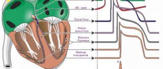
How does electrical wiring work in the heart?
So, an electrocardiogram, according to its name, records electrical processes occurring in the heart. Let's figure out what and how is happening there. In the depths of the heart muscle there are special groups of cells that make up the so-called conduction system of the heart. For simplicity, you can imagine it in the form of electrical wiring embedded in the wall, although in reality everything is a little more complicated.
DETAILS: How many days can you take Zodak
The “power source” of a healthy heart is the sinus node, which is located in the right atrium. For those who are familiar with electricians, it can be compared to a capacitor. The sinus node accumulates a charge and then emits electrical impulses at a certain frequency that cause the heart to contract. Consequently, if “the battery is good,” then the first line of the cardiogram conclusion will say: sinus rhythm.
Sinus rhythm is the normal physiological rhythm of the heart.
The heart has four chambers - two atria and two ventricles. The atria contract first, then the ventricles. In order for this to happen in exactly this sequence, it is necessary that the electrical impulse first excites the atria and then switches to the ventricles. This switching occurs in the so-called atrioventricular node. More often it is called in Latin the atrioventricular node (atrium - atrium, ventriculum - ventricle), and even more often - simply the AV node.
Two “wires” emerge from the AV node, which, according to the author’s surname, are called the bundle branches. Through the right bundle branch, the electrical signal is mainly conducted to the right ventricle, and through the left bundle branch, of course, to the left ventricle. Since the left ventricle is the largest chamber of the heart, and it needs a lot of electrical supply, the left leg is also divided into anterior and posterior branches.
How often should an electrocardiogram be done?
An ECG during early pregnancy can be shown more than once so as not to miss a dangerous condition. Here are the cases in which doctors refer for additional examination:
- for complications: severe toxicosis, gestosis, low or polyhydramnios, high/low blood pressure, pressure surges;
- with rapid heartbeat, pain in the heart area, in the left side of the chest and regular pain in the area of the left shoulder blade (be sure to tell your doctor about these symptoms!);
- with frequent dizziness, darkening of the eyes;
- in the presence of hectic work and other factors in the life of a pregnant woman that affect her nervous system.
If there are no complications, the procedure is performed three times. The first time is as early as possible, ideally at 5-6 weeks. If the deadline is missed - during registration. The second is carried out during general screening at the 12th week. When applying for sick leave for maternity leave, the doctor may order an ECG for the third time.
Decoding the results
Decoding the electrocardiogram of the heart is an important and crucial stage in making a diagnosis and prescribing treatment. For correct interpretation, it is necessary to understand the essence of the teeth and lines on the graph.
The ECG printout contains three important elements:
- tooth – concavity or convexity of a line. Encrypted with Latin letters P, Q, R, S, T;
- interval includes segments and teeth;
- segment – the distance between two teeth.
When describing a cardiogram, the duration of the intervals, the height of the teeth, the position and shape of the segments are taken into account. Important factors are the film recording speed with which the electrocardiograph works (usually 25 or 50 mmsec) and artifacts (patient movement during the procedure, isoline drift):
- Wave P – displays processes in the atrium, normally positive, up to 2.5 mm high and lasting 0.1 s.
- Q wave – shows impulses in the interventricular septum, duration – 0.03 s.
- The R wave is the highest, displaying the impulses of the ventricles themselves.
- The S wave is negative and shallow, indicating the completion of the impulse in the ventricles.
- The T wave reflects the repolarization of the ventricles.
The next important indicator of a normal ECG is sinus heart rate. Criteria: the P wave is in front of all QRS, equal to PQ (0.12-0.2 s) in all leads and the heartbeat is 60-80 beats/min.
Next, the electrical axis of the heart (EOS) is determined, which characterizes the conductive and fiber organization of the organ. It can be vertical (70-90 degrees), horizontal (0-30) and normal (30-60).
Who is doing
A doctor of any specialty has at least a minimal understanding of how to read a cardiogram of the heart and be able to recognize the signs of serious conditions. Most often, cardiograms are deciphered by therapists or cardiologists, because they prescribe this study. Paramedics and EMTs read the films to quickly make decisions about medical support or admission to a cardiac hospital.
Modern cardiographs at the end of the recording provide a preliminary result of the study indicating the size of the intervals and waves, heart rate, position of the electrical axis of the heart and signs of such pathologies: blockade, arrhythmia, hypertrophy of the myocardial walls. This makes the doctor’s job easier in counting and measuring segments, but sometimes the program misinterprets the results. The doctor rechecks the pathological signs on the ECG and makes the correct conclusion.
In some cases, the conclusion of an electrocardiogram of the heart does not completely resolve the diagnostic issue. The doctor may ask to see previous films and findings from other examinations. When making a diagnosis, data from the medical history, course of the disease, and medication intake are taken into account.
Many patients want to know how to independently decipher a cardiac cardiogram, because they often want to quickly find out the result of the study in order to reassure themselves. But it is better to entrust this task to a doctor, having received competent advice, although some ECG data are easy to interpret even for beginners. The procedure is easier to do if the recording is of high quality and there are no artifacts on the film.
To understand how to read a heart cardiogram, you need to know about the parameters of rhythm and heart rate. To determine the number of contractions, count the number of large squares on the film between the two nearest R waves. At a speed of 50 mms, divide 600 by the number of squares, and at 25 mms, divide 300 by the number of squares.
Afterwards the EOS value is indicated. As mentioned earlier, the position of the axis can be normal, horizontal or vertical. Normal: vertical in thin people, horizontal in hypersthenics (stocky, with a wide chest). Deviation of EOS is interpreted as hypertrophy of the myocardial walls, blockade of conduction pathways or other pathologies.
Normal cardiogram of a healthy heart and what it looks like
In a healthy adult, a normal cardiogram (cardiogram of a healthy heart) is considered to be a curve with sinus rhythm.
The heart rate (HR) is 60-80 beats per minute, the EOS (electrical axis of the heart) is in the standard position.
PQ interval (period of the excitation wave passing through the atria and atrioventricular node to the ventricular myocardium) - 0.12-0.18 seconds. (up to 0.2).
No changes in rhythm or tone (arrhythmia, bradycardia, tachycardia) were detected.
Increased heart rate is possible in pregnant women or overly emotional individuals.
In elderly patients, on the contrary, there is a slowing of the heart rate or morphological pathologies of the myocardium.
Only a specialist with a medical education can correctly decipher the cardiogram and describe the obtained ECG parameters.
Electrocardiography can accurately diagnose various diseases of the cardiovascular system - ischemia, abnormalities in the development of conduction pathways, cardiac aneurysm, extrasystole, angina pectoris and many others.
The most serious diagnosis with electrocardiography is myocardial infarction.
It is here that you can first detect areas of damaged or dead tissue, determine the specific location (in which wall of the heart) and the depth of the lesion. An ECG easily distinguishes the acute phase of a heart attack from old scars and aneurysms.
In case of a heart attack, the ECG procedure is performed more than once.
The first time this happens is during the first contact with the patient - at home, in an ambulance or in a hospital waiting room. If there are no changes in the graphic image, but if symptoms are present, the procedure is repeated after 6 hours - by this time the symptoms usually appear in full force.
After this, diagnostics are carried out daily, and during recovery - once every few days. Thus, over the entire period the patient is examined at least 10 times.
The patient must always remember that taking care of his health should only be entrusted to a specialist. This fully applies to the electrocardiography procedure. You should not neglect the doctor’s prescription and you should not try to decipher the ECG yourself, even if you are sure that the result will be a normal cardiogram.
A cardiogram of a healthy heart, like an ECG with abnormalities, can only be read correctly by a doctor.
Only a person with a medical education is capable of assessing the risk of a critical condition obtained from the examination, clinical symptoms and the results of the study. Otherwise, there is a possibility of underestimating the ECG, which can lead to fatal consequences.
Electrocardiography is an inexpensive, accessible and quite informative research method that was invented more than a century ago. Despite such a long period of time, this invention has not lost its relevance and, moreover, continues to be improved to this day, which indicates its relevance.
Using this method, you can not only detect heart rhythm disturbances, but also assess the condition of the myocardium. Our article will tell you in detail about the features of the study and how often an ECG can be done.
Clinical examination: who should not have an ECG
The golden rule of medicine is that any research must be justified. Our colleagues abroad strictly adhere to it. Even if studies are performed on healthy people, they must be done for certain indications and in certain risk groups. Research that is done just like that, just in case, on the principle of “what if something happens,” often not only does not provide useful information, but often even misleads and confuses.
This fully applies to the ECG. As we have already said, an ECG is just a recording of electrical signals that are produced in the heart, and which doctors have agreed to interpret in a certain way.
Any doctor learns to interpret a cardiogram throughout his life. There are a lot of standard warrants. The more experienced the doctor, the more variants of the norm he knows. In our clinic, a long time ago, its late director, Professor V.I. Makolkin forbade functional diagnostic doctors from “deciphering” the ECG. Every doctor should learn to read an ECG independently, with the help of senior colleagues if necessary.
Thus, after several years of work, even a young doctor already had a huge baggage of viewed ECGs, and not just looked at, but “linked” directly to the patient. And this is an important condition for analyzing the cardiogram. Often, when a doctor “deciphers” a cardiogram without seeing the patient, he can give a conclusion that is completely untrue.
So, there is no need to take a cardiogram for young healthy people just in case. Young people have a large number of individual characteristics that do not require treatment. This may be pacemaker migration, sinus arrhythmia, high signal voltage, rare extrasystoles. ECG in children often differs from the standard to which we are accustomed. It would be good if this child met a competent doctor who said that nothing needs to be done.
DETAILS: How to clean the blood vessels of the brain using folk remedies
One day a mother came to an appointment with her 18-year-old daughter. In their hands they held a thick volume with cardiograms, echocardiograms, extracts, and prescriptions. The more I leafed through these documents, the more convinced I became that the girl did not have a single serious illness. All these years she was treated for completely non-existent diseases and absolutely harmless changes on the ECG.
So, in the absence of symptoms, a healthy person does not simply need to have a cardiogram. The likelihood of seeing some minor deviation from the norm, which will be incorrectly interpreted, is much higher than accidentally identifying some serious pathology. It is much more important that the doctor measure your blood pressure, listen to you, and do routine tests. But if he heard something there, if the pressure is elevated, then he needs to react and do a cardiogram.
Where to get tested
Any doctor in the hospital can give a referral for examination. In most cases, this is a scheduled inspection at the request of the employer or educational institution (school). But you can also undergo the procedure at your own request.
The doctor who performs the procedure is a cardiologist. This is his direct specialization, but data can be collected by functional diagnostics workers, as well as nurses. The main thing is that the conclusion is carried out by a specialized doctor (cardiologist).
First of all, determine for yourself on what basis you want to make a cardiogram. If it is free, then you should go to the hospital where you live and get a referral from your doctor. The procedure may be unpaid if you register with a prescription for treatment.
If you want to get thoroughly checked and get results quickly, it is better to go to a paid clinic. The cost of services usually does not exceed $10. Find a private hospital in your city, call, check the cost, and you will be given a time and day to come for a cardiogram.