Is it possible to do an ecg if a child is coughing?
You don’t have to buy a new toy for your child—make it together with your child from scrap materials.
A little glitter and colored yarn, a little imagination - and the new toy is ready.
A few colored cocktail straws, glue and a shoebox lid are all you need.
You can play it for real while having fun with your family or friends.
Hello. 2 days ago the holter went away, there was a strong cough at night. I woke up 2 times during the night. Can it affect Holter readings?
Doctors' answers
Theoretically, at the time of coughing, it is of course possible that some changes in cardiovascular activity may occur. You did not indicate what kind of daily monitoring was carried out - ECG or blood pressure. Therefore, I will write down possible changes in both cases: With HM, blood pressure with an intense hacking cough, with straining - during an attack, an increase in blood pressure is possible, but it (the increase) should be short-term, and when deciphering the monitoring result, the doctor (especially if you indicated that the cough was bothering you) ) will not consider these readings as reliable, especially if during registration the blood pressure readings were normal. If an HM ECG was performed, then also during a coughing attack, a change in the heart rhythm is possible; tachycardia and arrhythmia may appear. If any disturbances in rhythm and conduction appear during coughing, in general this is even diagnostically important, since many rhythm disturbances do not appear at rest, but can occur during stress or excitement. And if this happens, the doctor will be able to prescribe adequate antiarrhythmic therapy in a timely manner. As for your testimony specifically, I cannot answer, since I do not know the conclusion and cannot assess the reliability of these changes. Much depends on the purpose for which the CM was carried out.
Consultation is available 24 hours a day
We need to know your opinion. Leave a review about our service
CREATE NEW MESSAGE.
But you are an unauthorized user.
If you register, you will be able to further track responses to your messages and continue the dialogue on interesting topics with other users and consultants. In addition, registration will allow you to conduct private correspondence with consultants and other users of the site.
Left ventricular hypertrophy on the ECG: recommendations from a cardiologist
The left ventricle is the part of the heart that, when contracting, pumps blood into the aorta. This is the main chamber of the heart, providing blood flow throughout the body. Left ventricular hypertrophy is an increase in mass, thickening of its wall. Often, at the same time, an expansion of the cavity of the left ventricle occurs - its dilatation. Hypertrophy is both an anatomical and electrocardiographic term. Anatomical hypertrophy of the left ventricle is manifested on the electrocardiogram (ECG) by a number of signs. A functional diagnostics doctor or cardiologist takes into account the number and severity of such signs. There are several diagnostic criteria that more or less correctly identify hypertrophy (from 60 to 90% probability). Therefore, not all people with signs of left ventricular hypertrophy on the ECG actually have it. Not all patients with anatomical hypertrophy show it on the ECG. Moreover, the same ECG can be described differently by different doctors if they use different diagnostic criteria in their work.
What diseases does this happen in?
- Left ventricular hypertrophy occurs in young people who are constantly involved in sports. Their heart muscle works intensely during training and naturally increases its mass and volume;
- occurs in diseases associated with difficulty in the exit of blood from the left ventricle into the aorta and with an increase in vascular resistance in the body;
- this ECG sign may be the first symptom of severe heart defects - aortic stenosis and aortic insufficiency. These diseases cause deformation of the valve separating the left ventricle and the aorta. The heart works under a heavy load, but the myocardium copes with it for a long time. A sick person does not feel any discomfort for a long time;
- Left ventricular hypertrophy occurs in a serious disease – hypertrophic cardiomyopathy. This disease is manifested by severe thickening of the walls of the heart. Thickened walls “block” the exit from the left ventricle, and the heart works under load. The disease does not appear immediately; shortness of breath and swelling gradually appear. In advanced cases, this disease may be an indication for heart transplantation.
- This is one of the manifestations of heart damage due to arterial hypertension. It can also develop with a moderate but constant increase in pressure. It is precisely to stop the progression of left ventricular hypertrophy that recommendations are aimed at constantly taking medications for hypertension, even with normal pressure.
- may appear in older people with severe atherosclerosis of the heart valves. This narrows the opening from the left ventricle into the aorta.
What could this lead to?
If a person has signs of left ventricular hypertrophy on an ECG, but it is not confirmed by echocardiography (ultrasound of the heart), there is no reason to worry. This ECG feature is probably due to increased body weight or a hypersthenic constitution. The ECG phenomenon of left ventricular hypertrophy itself is not dangerous.
If hypertrophy on the ECG is accompanied by a real increase in muscle mass, in the future this can cause heart failure (shortness of breath, edema) and severe heart rhythm disturbances (ventricular extrasystole, ventricular tachycardia). Athletes should not forget about this when creating a training regimen.
Changes in the myocardium on the ECG - what does this mean for making a diagnosis?
Description of the procedure
Have you been struggling with HYPERTENSION for many years without success?
Head of the Institute: “You will be amazed at how easy it is to cure hypertension by taking it every day.
An electrocardiogram (ECG) is one of the most informative, simple and accessible cardiological studies. It analyzes the characteristics of the electrical charge that causes the heart muscle to contract.
Dynamic recording of charge characteristics is carried out in several areas of the muscle. The electrocardiograph reads information from electrodes placed on the ankles, wrists and chest skin in the area where the heart is projected, and converts them into graphs.
OUR READERS RECOMMEND!
Our readers successfully use ReCardio to treat hypertension. Seeing how popular this product is, we decided to bring it to your attention.
Normal and deviations - possible reasons
Normally, the electrical activity of the myocardial areas, which is recorded by the ECG, should be uniform. This means that intracellular biochemical exchange in heart cells occurs without pathologies and allows the heart muscle to produce mechanical energy for contractions.
If the balance in the internal environment of the body is disturbed for various reasons, the following characteristics are recorded on the ECG:
- diffuse changes in the myocardium;
- focal changes in the myocardium.
The reasons for such changes in the myocardium on the ECG can be either harmless conditions that do not threaten the life and health of the subject, or serious degenerative pathologies requiring emergency medical care.
- rheumatism as a consequence of scarlet fever, tonsillitis, chronic tonsillitis;
- complications of typhus, scarlet fever;
- consequences of viral diseases: influenza, rubella, measles;
- autoimmune diseases: rheumatoid arthritis, systemic lupus erythematosus.
One of the reasons for changes in muscle tissue may be cardiodystrophy - a metabolic disorder in heart cells without damage to the coronary arteries. Lack of cell nutrition leads to changes in their normal functioning and impaired contractility.
Causes of cardiac dystrophy:
- Ingress of toxic metabolic products into the blood due to severe impairment of kidney and liver function;
- Endocrine diseases: hyperthyroidism, diabetes mellitus, adrenal tumor, and, as a result, excess hormones or metabolic disorders;
- Constant psycho-emotional stress, stress, chronic fatigue, starvation, unbalanced diet with nutritional deficiencies;
- In children, a combination of increased stress with a sedentary lifestyle, vegetative-vascular dystonia;
- Lack of hemoglobin (anemia) and its consequences - oxygen starvation of myocardial cells;
- Severe infectious diseases in acute and chronic form: influenza, tuberculosis, malaria;
- Dehydration of the body;
- Avitaminosis;
- Alcohol intoxication, occupational hazards.
Determination by cardiogram
With diffuse lesions of the heart, deviations from the normal pattern are observed in all leads. They look like numerous areas with impaired conduction of electrical impulses.
This is expressed on the cardiogram as a decrease in T waves, which are responsible for ventricular repolarization. With focal lesions, such deviations are recorded in one or two leads. These deviations are expressed on the graph as negative T waves in the leads.
If focal changes are represented, for example, by scars remaining in the connective tissue after a heart attack, they appear on the cardiogram as electrically inert areas.
Diagnostics
Decoding the electrocardiogram data takes 5-15 minutes. Its data can reveal:
- Size and depth of ischemic lesion;
- Localization of myocardial infarction, how long ago it occurred in the patient;
- Electrolyte metabolism disorders;
- Enlarged heart cavities;
- Thickening of the walls of the heart muscle;
- Intracardiac conduction disorders;
- Heart rhythm disturbances;
- Toxic damage to the myocardium.
Features of diagnosis for various myocardial pathologies:
- myocarditis – the cardiogram data clearly shows a decrease in the waves in all leads, a violation of the heart rhythm, the result of a general blood test shows the presence of an inflammatory process in the body;
- myocardial dystrophy - ECG indicators are identical to the data obtained for myocarditis; this diagnosis can only be differentiated using laboratory test data (blood biochemistry);
- myocardial ischemia - ECG data show changes in the amplitude, polarity and shape of the T wave in those leads that are associated with the ischemic zone;
- acute myocardial infarction – horizontal displacement of the ST segment upward from the isoline, trough-shaped displacement of this segment;
- necrosis of the heart muscle - irreversible death of myocardial cells is reflected on the ECG graph as a pathological Q wave;
- transmural necrosis - this is an irreversible damage to the entire thickness of the heart muscle wall, expressed in the cardiogram data as the disappearance of the R wave and the acquisition of the QS type by the ventricular complex.
When making a diagnosis, you should additionally pay attention to the symptoms of concomitant diseases. This may include pain in the heart with myocardial ischemia, swelling of the legs and arms with cardiosclerotic changes, signs of heart failure as a result of a heart attack suffered on the legs, hand tremors, sudden weight loss and exophthalmos with hyperthyroidism, weakness and dizziness with anemia.
The combination of such symptoms with diffuse changes detected on the ECG requires an in-depth examination.
What diseases do they accompany?
Pathological changes in the myocardium detected on the ECG may be accompanied by impaired blood supply to the heart muscle, reprolarization processes, inflammatory processes and other metabolic changes.
A patient with diffuse changes may exhibit the following symptoms:
- dyspnea,
- chest pain,
- increased fatigue,
- cyanosis (blanching) of the skin,
- rapid heartbeat (tachycardia).
Diseases accompanied by changes in the heart muscle:
- Myocardial dystrophy is a violation of biochemical metabolic processes occurring in the heart;
- Allergic, toxic, infectious myocarditis - inflammation of the myocardium of various etiologies;
- Myocardiosclerosis – replacement of cardiac muscle cells with connective tissue as a consequence of inflammation or metabolic diseases;
- Disturbances of water-salt metabolism;
- Hypertrophy of parts of the heart muscle.
Additional examinations are needed to differentiate them.
Additional diagnostic tests
These cardiograms, despite their information content, cannot be the basis for making an accurate diagnosis. In order to fully assess the degree of changes in the myocardium, the cardiologist prescribes additional diagnostic measures:
Shower for hypertension: how to take it correctly with high blood pressure
Have you been trying to cure HYPERTENSION for many years?
Head of the Institute of Treatment: “You will be amazed at how easy it is to cure hypertension by taking it every day...
Read more "
Today it is difficult to find a completely healthy person. Even doctors have such a term as “conditionally healthy person.”
Every person wants to feel energized in their body in the morning and wake up in a great mood. But not everyone succeeds.
When it comes to a healthy lifestyle and hardening, there are not many people who want to do this.
However, in our time there is one proven way to stay healthy and be treated for hypertension - to take a contrast shower.
Benefits of shower
Not everyone can decide to take a contrast shower. It really doesn't feel pleasant when taken. It brings discomfort and tension. But everyone knows that a contrast shower helps make a person more alert.
It can improve blood circulation in the body and thereby get rid of blood pressure. Also, a contrast shower will strengthen the immune system, improve the condition of the skin, and help you lose weight even with high blood pressure. But you need to take a contrast shower correctly.
Some people find many reasons not to have this procedure. But before you refuse it, you need to know what benefits and harm it can cause to a person and his body.
A contrast shower can train the body. It will strengthen blood vessels, which is useful for high blood pressure and hypertension, restore cells and capillaries, train muscles and help the body feel more energetic. A shower will also strengthen your immune system.
If you regularly take a contrast shower correctly, this will also help get rid of excess weight and hypertension. The body will intensively burn calories during the procedure, as well as improve blood circulation, which is very useful for high blood pressure.
To maximize the effect of such a shower, you need to massage with jets. They need to be used in areas where fat deposits are observed, as well as in the chest for hypertension. You should take such a contrast shower every day after sleep.
The majority of people do not like to take a contrast shower due to the fact that during it you can experience unpleasant sensations. This is especially difficult for the fair sex to decide on, since their skin is much more sensitive.
But the discomfort will not last long. After 4-5 times everything will return to normal, and a contrast shower in the morning will become a habit. In order for the body to adapt faster, it is recommended to start doing such procedures in the summer.
How to shower?
on
To learn how to shower properly, you need to follow the simple rules that will be given below:
- Initially, you need to calm down and tune in to the procedure.
- Use a hard towel, preferably terry, for drying after a shower.
- You need to rub yourself vigorously after a shower. This will help improve blood circulation and stabilize blood pressure.
It should be remembered that you need to start dousing correctly with warm water. Next, it needs to be made hotter. You need to add water gradually. You need to stand under the hottest water possible for about 1-2 minutes, and then suddenly turn on the cold water.
Hypertension will go away, and the pressure will be 120 over 80 if you include it in the diet...
Hypertension will disappear forever! Here's the secret...
You should stay under the cold jets for 30-40 seconds. The time can be gradually increased. This also applies to lowering the temperature. The water does not need to be made ice cold initially. The water temperature should gradually decrease.
It is necessary to accustom the body to take a contrast shower for 30-40 days. At one time, you can sharply change the water temperature no more than 3-4 times at the initial stage. Finish your shower with cold water only, and then immediately rub yourself vigorously. The skin should turn pink when rubbed.
Nuances
- It is not recommended to put your head under a contrast shower, as a pressure drop may occur or a cold may appear.
- During the procedure, you need to step from foot to foot so that your feet receive the same amount of energy.
- You can take such a shower several times a day, but this must be done daily.
- If you have a cold, you should not stop the procedure.
After regularly taking a contrast shower, you will notice that your immune system will be strengthened. The condition of muscles and blood vessels will improve. The skin will look younger and fresher. It will also become more elastic.
It should be remembered that the rejuvenating and healing effect of this procedure will occur only if it is taken regularly for a sufficient amount of time.
Due to the fact that contrast water affects the cardiovascular system, blood circulation in the body improves, which leads to a more abundant supply of oxygen to the blood and organs.
Harm of a contrast shower
If the procedure is performed incorrectly, it can cause harm, like any other therapeutic effect on the body. A contrast shower can be stressful for many, especially for those who are often sick.
For this reason, many people begin to douse themselves not with cold water, but with warm water (19-20 degrees). As a result, the body does not receive the required hardening and such people develop colds or inflammation.
The whole point here is that warm water cannot turn on the body’s defense mechanisms, which begin to work only in an extreme situation. For this reason, the chance of catching a cold increases significantly.
You need to gradually accustom your body to cold water. As soon as the body gets used to water with a temperature of 19-20 degrees, it should immediately begin to accustom it to cold water.
At the same time, a quick dousing with ice water will not allow the body to cool down too much, but will help to “awaken” the body’s defense mechanisms. All systems will begin to work several times more actively.
You also need to take a contrast shower correctly, namely:
- Warm water.
- Hot water.
- Cold water (30 s).
- Hot water (30-40 s).
- Cold water (more than 60 s).
- Hot water (up to 60 s).
At the same time, you should not tolerate water of different temperatures, as this will not give a good result. Everything should be normal.
Contraindications
Like all other medical procedures, contrast showers also have their contraindications. It should not be taken for thrombophlebitis, heart and blood diseases.
If a person has high blood pressure, blood circulation in the brain is impaired and vascular spasms are observed, then experts do not recommend taking a contrast shower. But you can try this procedure after consulting a doctor.
For any chronic diseases of the body, it is recommended to consult a doctor before starting a contrast shower - this must be remembered. Also, in case of inflammatory processes in the body, you should stop taking a contrast shower.
You should take a contrast shower in the morning after exercise. It can also be taken throughout the day. In this case, it is necessary to calculate the time of admission so that after the procedure at least half an hour passes before going outside. Also, after taking a contrast shower, you should rub yourself well with a towel.
Knowing these points, you can make the right decision for yourself and start taking a contrast shower. If there are any contraindications or doubts, it is recommended to consult a doctor for advice. In conclusion, we suggest that you familiarize yourself with the video in this article on the topic of hardening for hypertensive patients.
on
Important points about how an ECG is done for patients of different ages and genders
Electrocardiography is the recording of bioelectric potentials that occur during contraction of the heart muscle. This method is accessible, does not require special preparation, and is safe for the patient. At the same time, the information received by the doctor can help in making a diagnosis of coronary artery disease, arrhythmia, and conduction disorders.
Read in this article
Note to parents
If your child starts coughing, you shouldn’t immediately be afraid that he has coronavirus. In addition to COVID-19, there are many different diseases that cause coughing. If the child has not had contact with people with coronavirus infection, at home or in the classroom, and no one is sick in the kindergarten, then where does the coronavirus come from? You need to see a doctor and identify the cause. Most likely, 98% of the time it won’t be coronavirus. Moreover, coronavirus infection does not begin immediately with a cough. In addition to it, there must be other symptoms.
Operating principle of an electrocardiograph
An ECG recording device consists of electrodes that are attached to the patient's body, a galvanometer, an amplifier, a recorder and a lead switch. The impulses that are formed in the heart muscle must first be amplified, then they are perceived by the galvanometer. It converts electrical waves into mechanical vibrations.
The recorder records, using thermal paper recorders, a typical graphical curve called an electrocardiogram.
Using an ECG study, you can judge the condition of the heart muscle by the following indicators:
- impulse conductivity;
- rhythm of heart contractions;
- enlargement of one or more parts of the heart;
- blood supply to the myocardium;
- areas of necrosis (infarction) their size, depth and duration of occurrence.
How to properly prepare for an ECG, what not to do
Electrocardiography does not require lengthy preparation, which is one of the advantages of this method. It is removed for emergency reasons in any condition of the patient. But if a planned study is prescribed, then before conducting it it is recommended:
- Do not consume food or caffeine-containing drinks at least 3 hours before the procedure.
- You need to have a good rest before the examination.
- Eliminate physical and emotional stress.
- Take a shower, after which do not use the cream.
Clothing is selected in such a way that it is easy to attach electrodes to the skin of the ankles, wrists and chest.
On the day of the study, drinking alcoholic beverages and smoking is strictly prohibited; you must avoid sports and a hearty breakfast. The best drink to drink is plain drinking water, weak tea or fruit juice.
How to do an ECG
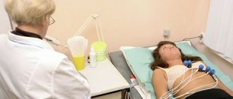
To take an electrocardiogram, the patient is placed on a couch and a healthcare professional places electrodes on the legs, wrists and chest. If there is difficulty breathing in a horizontal position, the procedure is performed while sitting.
Rules for the procedure
To ensure good contact between the skin and the electrode, the attachment site is degreased with ethyl alcohol and a special conductive gel is applied. After this, readings are taken using an ECG diagnostic device.
The whole procedure takes about 10 - 15 minutes.
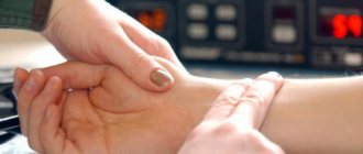
In order to get a reliable result, you need to be in a calm, relaxed state, and not hold your breath. Muscle tremors from excitement or cold can lead to data distortion.
The generally accepted leads are 3 standard, 3 reinforced and 6 chest. Each lead will record at least 4 cardiac cycles. After this, the device is turned off, the electrodes are removed, and the functional diagnostics doctor is given a signed tape, which he must decipher.
For information on the ECG recording method, watch this video:
Are there any special features during pregnancy?
In the body of a pregnant woman, the load on the heart muscle changes, since it must provide blood supply to the fetus located in the uterus. The electrocardiogram may show abnormalities that are not an indication of heart disease.
Therefore, starting from 3 - 4 months, when deciphering the testimony, an amendment is made for the presence of the gestation process.
When preparing and conducting the procedure itself, standard research techniques are used.
How to do an ECG for women
For women, the rules for installing electrodes are the same as for men. They should be located in the heart area, directly on the skin, so before performing an ECG, you must completely remove all clothing from the chest, including your bra. Please note that tights or stockings will interfere with attaching the sensors to the lower leg.
Disadvantages of ECG examinations
Despite its high diagnostic value, a regular ECG cannot detect changes in heart function outside the time of its taking. Therefore, along with the traditional technique, the patient may be prescribed additional Holter monitoring and exercise testing during the day.
Using this method, it is impossible to recognize heart murmurs, therefore, if structural defects of the valves or septa are suspected, echocardiography, phonocardiography or ultrasound of the heart should be performed.
If it is planned to install a stent or shunt for myocardial ischemia, then coronary angiography is required to determine the localization of the narrowing of the coronary arteries. Tumor processes are diagnosed by X-ray or MRI examination.
We recommend reading about acute coronary syndrome. You will learn about the causes of the development of pathology, pathogenesis, symptoms, diagnosis and treatment. And here is more information about ECG for myocardial ischemia.
Interpretation of ECG indicators
On the tape, the curve obtained after taking the cardiogram has 5 teeth. They occur with sequential contraction of the atria and ventricles. The following designations are accepted:
- The P wave is an indicator of the work of the right (first half) and left atrium.
- P Q – interval of impulse passage to the ventricle along the Hiss bundle.
- QRST - complex occurs during contraction of the ventricles, while the highest R wave reflects the excitation of the ventricular myocardium, and Q and S are the partitions between them, T - occurs during the period of myocardial recovery after systole.
Teeth and intervals
Normal in adults
A doctor can fully evaluate the electrocardiogram, since making a diagnosis requires knowing the symptoms of the disease and data from other research methods (blood tests, ultrasound, echocardiography). The general characteristics that a specialist evaluates in a healthy person are as follows:
- The rhythm of contractions is from 60 to 80 per minute.
- The size of the intervals should not exceed normal values, or be shorter than average values.
- Electrical axis - normally R exceeds S in all leads except aVR, V1 - V2, sometimes V3.
- The ventricular complex is no more than 120 ms.
- T is positive and longer than the QRS complex.
ECG (normal)
During pregnancy
As the uterus grows, it raises the dome of the diaphragmatic septum and after 24 - 24 weeks the apex of the heart moves to the left. This is reflected in the electrocardiogram by an increase in the amplitude of R in the first lead, and S and Q in the third lead, the ventricular complex decreases along with the ST segment. Changes in conduction through the heart muscle are also associated with the influence of hormones produced by the placenta.
Characteristic features:
- Shift of the heart axis to the left.
- T biphasic and negative in the right chest leads.
- The ventricular complex is wider than normal.
- Rapid rhythm, single extraordinary contractions.
Respiratory arrhythmia in pregnant women
Is it possible to do an ecg if you have a cough?
Electrocardiography is the recording of bioelectric potentials that occur during contraction of the heart muscle. This method is accessible, does not require special preparation, and is safe for the patient. At the same time, the information received by the doctor can help in making a diagnosis of coronary artery disease, arrhythmia, and conduction disorders.
What is sinus rhythm and ECG interpretation
Anton Rodionov, cardiologist, candidate of medical sciences, associate professor of the department of faculty therapy No. 1 of the Sechenov First Moscow State Medical University
Who can decipher an ECG and when a cardiogram does more harm than good, cardiologist Anton Rodionov said in his new book.
Deviations that the device can detect
By taking and interpreting an electrocardiogram, you can identify signs of the following diseases:
- angina pectoris and heart attack;
- type of arrhythmia, location of the pacemaker;
- blockade due to decreased conductivity;
- myocardial hypertrophy and its localization;
- signs of myocarditis and pericarditis;
- pulmonary embolism;
- symptoms of pulmonary hypertension;
- disorders of the electrolyte composition of the blood.
3rd degree AV block
Current patient questions
The ECG method is traditional and has been used for a long time in medical practice. But patients often have concerns about its use. The most common questions:
- How often can an ECG be done? The body is not affected by either an electric or magnetic field. Therefore, there are no restrictions on the frequency of its implementation. You can take a cardiogram every day, but this is most often used in a hospital setting.
- Is ECG harmful to the body? The procedure is completely safe, as it does not require injections, radiation or traumatic intervention. It can be prescribed without fear to children and pregnant women.
- Is it possible to do an ECG for coughs and colds? Colds are not a contraindication, but at the time of coughing on the ECG there will be a distortion in the shape of the waves and intervals, and there may also be signs of respiratory arrhythmia. Therefore, this type of study is not routinely prescribed.
Thus, an ECG is a time-tested, accessible type of diagnosis, which is used both for preventive examination during clinical examination and for making a diagnosis in the presence of complaints about cardiac dysfunction. Such research is safe and informative.
Recognizing myocardial infarction on an ECG can be difficult due to the fact that different stages have different signs and variations of waveforms. For example, the acute and acute stage may not be noticeable in the first hours. Localization also has its own characteristics: the infarction on the ECG is transmural, q, anterior, posterior, transferred, large-focal, lateral, different.
The heart needs to be examined under different circumstances, including at 1 year of age. The ECG norm in children differs from that in adults. How is an ECG done for children, deciphering the indicators? How to prepare? How often can you do it and what to do if the child is afraid?
The location of the heart is determined by various parameters. An important role is played by the electrical axis of the heart, which can be normal, sometimes there are deviations to the left and right. Vertical and horizontal position, as well as displacement, do not always indicate pathology, especially in a child. How to determine on an ECG?
Holter ECG monitoring, which is important for the patient, can be daily or even biennial. The decoding will show deviations in the functioning of the heart, and the device is worn without interruption. Monitoring is safe even for children.
Sometimes it is not possible to accurately listen to noises. In this case, cardiac phonocardiography comes to the rescue. The method allows you to listen to even the fetus without harm to mother and child. But initially auscultation will be required.
Heart examinations are rarely performed using a cardiovisor. The screening system works like a camera - it takes a picture of the work of the myocardium. The test results allow you to assess the condition of the heart and its rhythm.
In general, a cardiointervalography examination is carried out to study the functioning of the autonomic nervous system. The procedure is safe and painless, so it can also be performed on children. What is the device?
It's quite unusual to undergo cardiac mapping. This examination is also called dispersive, color. Cardiac complex for non-invasive mapping can be performed on a large number of people.
The rather unusual method of vectorcardiography is not used very often. The concept means transferring the work of the heart to a plane. The doctor evaluates special loops.
Reviews about Electrocardiography
I have had an electrocardiogram done more than once for both myself and my daughter, mainly during medical examinations. Everything was always fine. She came, undressed to the waist, lay down on the couch, the doctor installed all the necessary suction cups, 5 minutes and you can get dressed, take the result and go to the therapist.
But this situation happened to my son. At two years old, the pediatrician heard a heart murmur in the child and sent him for an ECG. We did the procedure, but the result was not very good. Not the worst, of course, but it made me worry. With a thoughtful and mysterious expression on her face, the pediatrician sent us for an ultrasound of the heart. I thought of all sorts of things while I was sitting in line with my child, and I was terribly worried. It's good that there was a very experienced doctor there. Ultrasound showed an additional chord in the left ventricle - the cause of the murmur. A completely harmless phenomenon and very common. But the bad cardiogram did not explain it. Then the ultrasound doctor drew attention to the child’s runny nose and explained that the clogged nose caused the distorted result.
To reassure ourselves, we did an electrocardiogram after recovery and everything was fine.
Electrocardiography is prescribed to all patients who complain of heart problems, as well as those who get a job or undergo a professional medical examination. This procedure is quite simple, but, unfortunately, it does not always give a complete answer about the condition of the heart. There are cases when this method cannot detect such a dangerous condition as myocardial infarction. If the doctor has doubts, and the patient has symptoms, then in this case the diagnosis should be carried out using a blood test. In recent years, this procedure can be done by the patient himself at home. To do this, some medical institutions, for a fee, offer to rent a small device that can take all the necessary readings of the heart and send a signal to the hospital for decoding using a regular telephone. This is very convenient if the patient has concerns about his condition.
Colds
06/09/2019 admin Comments No comments
One of the blog readers asked how to avoid a cold turning into a prolonged cough.
, which ends almost any ARVI in her children. And then I remembered that I had my own method of prevention on this topic in my senior years at medical school. However, by the time I graduated from high school, I rarely caught a cold and did not have time to accumulate enough statistics, so today I invite everyone to check this method of preventing cough during acute respiratory viral infections (ARI).
Its meaning is in the preventive use at the very beginning of an acute respiratory infection of one of the drugs that improves sputum discharge.
Where did the method come from?
I studied in Minsk (first MGMI, then BSMU). Moving to the capital, difficult studies in the first three years, daily use of public transport, and dormitory conditions created conditions for the active transmission of various cold viruses, so I developed ARVI more often than at school.
I also noticed that all my colds were of the same type.
:
- the first 2-3 days my throat hurt,
- then the throat subsided, but for 1-2 days a stream flowed from the nose,
- then the nasal secretion became thicker, less abundant, and within a week everything went away,
- In parallel with the decrease in nasal discharge, a cough often appeared. Sometimes small, and sometimes strong, deep, for 2-3 weeks.
To treat cough I used:
- always - drugs that improve sputum separation (usually bromhexine
due to its low price), - occasionally - some antibiotics due to suspicion of atypical mycoplasma pneumonia ( macrolides, doxycycline
), but there was no significant effect from them. According to my current understanding, there were not sufficient grounds for their use at that time.
Antibiotics are indicated
in cases:
- tonsillitis
(
streptococcal, anaerobic
). To differentiate between viral and streptococcal tonsillitis, it is recommended to do the Streptatest express test; - acute otitis media
; - sinusitis
- inflammation of the paranasal sinuses (in the presence of clinical and radiological changes in the sinuses 10-14 days after the onset of ARVI, because in the first days of acute rhinitis, sinusitis is present in almost 100% of patients); - pneumonia
, including atypical, - in the presence of purulent
complications (
purulent sinusitis, purulent lymphadenitis, paratonsillar abscess, descending laryngotracheitis
).
According to the recommendations of the US Academy of Pediatrics, mucopurulent runny nose
It is also not an indication for antibiotics if it lasts less than
10-14 days
.
Sometimes my cough lasted up to 2-3 weeks, although I never smoked, and non-smokers' cough always goes away faster. And then I came to the conclusion that it is necessary to take a drug to thin the sputum not when the cough has already begun, but when the cold is just starting
and my throat hurts.
Theoretical background
To avoid the transformation of a cold into a long cough, I came up with the idea of starting to take any expectorant or mucolytic drug in advance, literally from the 1-2 day of ARVI
.
After all, it’s better to take 5-7 days for prevention
than to then take 2-3 weeks for treatment, right?
a theoretical basis for early use of a sputum thinner
:
- an increased amount of sputum contributes to its rapid removal from the bronchi and trachea through coughing (coughing), which means mechanical removal of infectious agents
. The ciliated epithelium of the respiratory tract, with the help of the movement of cilia, constantly removes mucus along with foreign particles and microorganisms that have entered the respiratory tract; - It has long been known that inhaling dry air promotes infection
.
The mucus secreted by the epithelium contains local immunity factors - secretory immunoglobulins class A (sIgA), lysozyme, interferon, lactoferrin, proteases
, etc.
When phlegm, mucus, and mucus dry out, they turn into a breeding ground for bacteria and do not perform a protective function. additionally humidify dry air
during the heating season in winter (you can read about the importance of humidified air for children in the blog of pediatrician Komarovsky, and I described the simplest free air humidifier in the form of a wet rag on a battery here). Dry air is more dangerous for children than for adults.
Is there any harm possible?
Can there be harm from early use of sputum thinners? Children are not in danger of stagnation of sputum - they cough it up. There is some danger only for a small category of people who cannot cough on their own ( very young or weakened children, patients with weakness of the respiratory muscles
etc.) - there needs to be caution in dosages and sometimes
postural drainage
(
giving the body special positions in which sputum, under the influence of gravity, independently flows into the main bronchi and trachea, from where it is removed by coughing
).
Is it possible to do an ecg for colds and coughs?
Electrocardiography is the recording of bioelectric potentials that occur during contraction of the heart muscle. This method is accessible, does not require special preparation, and is safe for the patient. At the same time, the information received by the doctor can help in making a diagnosis of coronary artery disease, arrhythmia, and conduction disorders.
Where did the method come from?
I studied in Minsk (first MGMI, then BSMU). Moving to the capital, difficult studies in the first three years, daily use of public transport, and dormitory conditions created conditions for the active transmission of various cold viruses, so I developed ARVI more often than at school.
I also noticed that all my colds were of the same type.
:
- the first 2-3 days my throat hurt,
- then the throat subsided, but for 1-2 days a stream flowed from the nose,
- then the nasal secretion became thicker, less abundant, and within a week everything went away,
- In parallel with the decrease in nasal discharge, a cough often appeared. Sometimes small, and sometimes strong, deep, for 2-3 weeks.
To treat cough I used:
- always - drugs that improve sputum separation (usually bromhexine
due to its low price), - occasionally - some antibiotics due to suspicion of atypical mycoplasma pneumonia ( macrolides, doxycycline
), but there was no significant effect from them. According to my current understanding, there were not sufficient grounds for their use at that time.
Antibiotics are currently NOT INDICATED in cases of uncomplicated ARVI: rhinitis, nasopharyngitis, viral tonsillitis, conjunctivitis, bronchitis, tracheitis, laryngitis
.
Antibiotics are indicated
in cases:
- tonsillitis
(
streptococcal, anaerobic
). To differentiate between viral and streptococcal tonsillitis, it is recommended to do the Streptatest express test; - acute otitis media
; - sinusitis
- inflammation of the paranasal sinuses (in the presence of clinical and radiological changes in the sinuses 10-14 days after the onset of ARVI, because in the first days of acute rhinitis, sinusitis is present in almost 100% of patients); - pneumonia
, including atypical, - in the presence of purulent
complications (
purulent sinusitis, purulent lymphadenitis, paratonsillar abscess, descending laryngotracheitis
).
According to the recommendations of the US Academy of Pediatrics, mucopurulent runny nose
It is also not an indication for antibiotics if it lasts less than
10-14 days
.
Sometimes my cough lasted up to 2-3 weeks, although I never smoked, and non-smokers' cough always goes away faster. And then I came to the conclusion that it is necessary to take a drug to thin the sputum not when the cough has already begun, but when the cold is just starting
and my throat hurts.
Theoretical background
To avoid the transformation of a cold into a long cough, I came up with the idea of starting to take any expectorant or mucolytic drug in advance, literally from the 1-2 day of ARVI
.
After all, it’s better to take 5-7 days for prevention
than to then take 2-3 weeks for treatment, right?
a theoretical basis for early use of a sputum thinner
:
- an increased amount of sputum contributes to its rapid removal from the bronchi and trachea through coughing (coughing), which means mechanical removal of infectious agents
. The ciliated epithelium of the respiratory tract, with the help of the movement of cilia, constantly removes mucus along with foreign particles and microorganisms that have entered the respiratory tract; - It has long been known that inhaling dry air promotes infection
.
The mucus secreted by the epithelium contains local immunity factors - secretory immunoglobulins class A (sIgA), lysozyme, interferon, lactoferrin, proteases
, etc.
When phlegm, mucus, and mucus dry out, they turn into a breeding ground for bacteria and do not perform a protective function. additionally humidify dry air
during the heating season in winter (you can read about the importance of humidified air for children in the blog of pediatrician Komarovsky, and I described the simplest free air humidifier in the form of a wet rag on a battery here). Dry air is more dangerous for children than for adults.
Is there any harm possible?
Can there be harm from early use of sputum thinners? Children are not in danger of stagnation of sputum - they cough it up.
There is some danger only for a small category of people who cannot cough on their own ( very young or weakened children, patients with weakness of the respiratory muscles
and etc.
) - there needs to be caution in dosages and sometimes postural drainage
(
giving the body special positions in which sputum, under the influence of gravity, independently flows into the main bronchi and trachea, from where it is removed by coughing
).
Of the side effects (I took bromhexine
) I only had
a cough
caused by increased secretion of sputum, but there was no cough as such. After stopping taking the mucolytic, the cough went away within a couple of days.
Why you need to drink enough fluids
Mucus thinners are more effective
if the patient receives enough fluid. Therefore, a patient with acute respiratory viral infection should not feel thirsty, but he should not be forced to drink too much. It is better to drink warm.
Useful
:
- anti-inflammatory tea with chamomile, thyme, sage;
- blueberry, cranberry jelly;
- table mineral water - no more than 1 glass per day for an adult and always without gas, which irritates the mucous membranes of the pharynx and stomach;
- fruit drinks, compotes.
Do not give excess vitamin C
. The daily requirement for it ranges from 30 mg in newborns to 50-60 mg in adults. Studies have shown that excessive doses of vitamin C (above 0.5-1 g) are not only useless for colds, but even harmful.
Which drug to choose
There are now many drugs that improve sputum separation
:
- means that stimulate expectoration
-
thermopsis, marshmallow, licorice
. Their effect is short-lived, so they must be taken every 2-3 hours. Increasing a single dose may cause nausea and even vomiting; - mucolytic
(or secretolytic) drugs -
bromhexine, ambroxol, acetylcysteine, carbocysteine
.
There are herbal preparations: Bronchipret
(contains thyme and ivy extracts),
Herbion primrose syrup
(primrose and thyme extracts), etc.
A detailed list of cough medications (which improve sputum discharge) can be found on the website medi.ru.
What to choose? To prevent cough, you can use any mucolytic drug in an average (usual dose) for 5-7 days
, which according to the instructions is allowed in children of this age and there are no other contraindications:
- Bromhexine
- usually available from 2-3 years of age. - Ambroxol
- can be prescribed at any age, but since 2010 in some countries (France, Italy) it is allowed only from 2 years of age due to the suspicion of severe complications in the respiratory tract in young children (when taken for longer than 4-5 days, the instructions recommend the doctor's consultation). - Acetylcysteine
- from 1 year depending on the dosage form. - Carbocisteine
- usually from 2 years. - Bronchipret
- from 3 months (syrup) and from 6 years (drops). Contains ethyl alcohol necessary for herb extraction. The usual course of treatment is 10–14 days. - Herbion primrose syrup
- from 2 years. The permitted duration of the course according to the instructions is 2–3 weeks.
I will note only 2 points:
- Bromhexine
is very cheap, and in the body it turns into
Ambroxol
. Ambroxol is more expensive, but begins to act within a couple of hours, while Bromhexine needs 1-2 days. - According to research, Bronchipret is more effective than ambroxol
or
acetylcysteine
.
conclusions
- In case of ARVI, to prevent complications (cough), you can try, from the 1st-2nd day of a cold, for 5-7 days
, to take a drug that improves sputum separation, in an average dose and taking into account the permitted age for this specified form. - The patient should receive enough fluids.
- Side effects may include coughing
associated with increased sputum secretion. This is fine. But there should not be a deep paroxysmal cough. Soon after stopping the mucolytic, the cough disappears on its own.
Source: https://orz.lesovir-c.com/mozhno-li-delat-jekg-pri-prostude-i-kashle/