Content
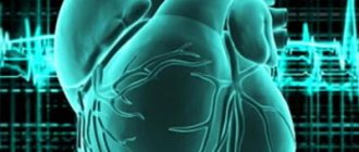
Classification of atrial fibrillation, epidemiology, prognosis, pathophysiological aspects and mechanisms
Non-drug treatments for atrial fibrillation
Operation Maze in the classic “incision-suture” version
Modified Maze operation using different ablation techniques (Maze IV)
Technique of surgery using monopolar ablation
Surgery technique using bipolar ablation
Minimally invasive techniques for surgical treatment of atrial fibrillation
Indications for surgical treatment of atrial fibrillation
Features of the postoperative period
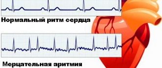
ECG signs of atrial fibrillation: replacement of normal P waves with fast oscillations, or fibrillation waves (f-waves), of various sizes and shapes, associated with abnormal frequent contractions of the ventricles with undisturbed AV conduction. ECG signs of atrial flutter: the presence of “saw teeth” (F-waves), reflecting the excitation of the atria at regular intervals. F-waves are well expressed in leads II, III, aVF, V1, there is no isoelectric line between the teeth.
Classification of atrial fibrillation
Paroxysmal form - an episode lasts less than 7 days (inclusive), recovers on its own (usually in the first 24-48 hours). If an episode of atrial fibrillation is stopped with drug therapy or electrical cardioversion until it spontaneously recovers (within 7 days), then in this case the name of the arrhythmia does not change (it continues to be called paroxysmal). Persistent form - lasts more than 7 days, does not recover on its own, but there are indications and the possibility of cardioversion. Persistent atrial fibrillation can be either the first manifestation of arrhythmia or the logical conclusion of repeated attacks of paroxysmal atrial fibrillation. Permanent form – long-term, when cardioversion is contraindicated, was not performed, or was unsuccessful. The term “isolated” atrial fibrillation applies to atrial fibrillation that occurs in young and middle-aged people (<60 years of age) without clinical and echocardiographic signs of cardiopulmonary disease and without arterial hypertension. Over time, if cardiovascular disease develops, such patients move into the general category of patients with atrial fibrillation. The term “idiopathic” atrial fibrillation implies that there is no clear cause for atrial fibrillation, and the age of the patient does not matter.
Epidemiology
Atrial fibrillation is the most common type of tachyarrhythmia encountered in clinical practice, accounting for approximately 1/3 of hospitalizations for arrhythmia. The prevalence of atrial fibrillation reaches 2% in the general population and 6% in people over 60 years of age. In patients with mitral disease admitted for surgical treatment, atrial fibrillation occurs in 60-80%. Patients with coronary artery disease suffer from atrial fibrillation in 6-10% of cases. Age-adjusted prevalence is higher in men. Recently, there has been a significant increase in the incidence of atrial fibrillation, with the number of patients expected to double over the next 20 years.
Forecast
The incidence of ischemic stroke in patients with atrial fibrillation of non-rheumatic etiology is on average 5% per year, which is 5-7 times higher than in people without atrial fibrillation. Every sixth stroke occurs in a patient with atrial fibrillation. Patients with rheumatic heart disease and atrial fibrillation have a 17-fold increased risk of stroke compared with age-matched controls (Framingham Heart Study). Mortality among patients with atrial fibrillation is approximately 2 times higher than in patients with sinus rhythm and is related to the severity of the underlying disease. In patients with prolonged tachysystole, tachycardiomyopathy develops, accompanied by dilation of the heart cavities, decreased ejection fraction, and the appearance of regurgitation on the atrioventricular valves, which leads to an increase in heart failure. The absence of atrial contribution against the background of irregular ventricular contractions leads to a decrease in cardiac output by 20-30%, which is especially pronounced when diastolic filling of the left ventricle is impaired in the case of mitral stenosis, diastolic dysfunction of the left ventricle. The presence of atrial fibrillation after cardiac surgery is associated with increased mortality, thromboembolic complications, disability and a decrease in the quality of life of patients. Surgical elimination of valvular pathology, even with the use of electrical cardioversion and antiarrhythmic drugs in the postoperative period, can lead to stable restoration of sinus rhythm in this category of patients only in 6-10%. Despite the fact that the cardiologist has a significant number of antiarrhythmic drugs in his arsenal, pharmacological treatment of atrial fibrillation still poses a significant problem. This is due to the lack of effectiveness of drug therapy, high relapse rates and serious side effects of drugs, including the development of fatal heart rhythm disturbances and sudden death. Atrial fibrillation poses a major financial challenge to the healthcare system, as the cost of treatment for patients suffering from atrial fibrillation is 35-40% higher than for patients in the same age group.
Pathophysiological aspects and mechanisms of atrial fibrillation
Atrial fibrillation is a complex arrhythmia, the pathogenesis of which is not fully understood. Of great importance in the occurrence and maintenance of atrial fibrillation is attached to ectopic focal and trigger activity, the mechanism of multiple repeated circles of excitation (re-entry) and the autonomic nervous system (ganglionic plexuses). Most often, ectopic foci are located at the mouths of the pulmonary veins (up to 80-90%), less often in the crista terminalis, Marshall ligament, the mouth of the coronary sinus, and the interatrial septum. Fibrosis, inflammation, ischemia and hypertrophy can serve as a substrate for changes in the electrophysiological properties of the atrial myocardium. In the latter case, the term “critical” mass of fibrillating atrial myocardium was even introduced. All these reasons lead to an increase in the dispersion of refractory periods in different zones of the atria and anisotropy of excitation conduction, which contributes to the implementation of the re-entry mechanism. As J. Cox has proven, re-entry circles most often function around anatomical obstacles to the homogeneous conduction of excitation through the atria (the openings of the vena cava and pulmonary veins, coronary sinus and atrioventricular valves). The longer atrial fibrillation exists, the more pronounced is the electrical remodeling of the atria (shortening of the refractory period), which reflects the postulate “atrial fibrillation generates atrial fibrillation.” A schematic representation of the mechanism of development of atrial fibrillation is presented in the figure.
Complications
Acute renal failure
- Bleeding from a punctured vessel occurs most often in the first postoperative period; there are not many causes of bleeding:
- bleeding disorder,
- improper application of a postoperative pressure bandage,
- incorrect behavior of the patient after surgery, it is necessary to unquestioningly follow the surgeon’s recommendations;
- Impaired kidney function - since the contrast is excreted through the kidneys and it is quite toxic, therefore, acute renal failure may occur against the background of underlying kidney disease;
- Thromboembolic complications - due to the need to discontinue medications that reduce blood clotting (warfarin) before surgery, blood clots may develop in the vessels, which can break off and cause various thromboembolic complications;
- Heart rhythm disturbances - the development of new types of arrhythmias is possible, and there are a huge number of reasons for this;
- This is not all, but only the main possible complications of the procedure; you can find out more from your surgeon;
- If any complications develop, the rehabilitation period after radiotherapy is extended.
Non-drug treatments for atrial fibrillation
Prophylactic pacing in patients with sick sinus syndrome
- continuous atrial pacing
- preventive stimulation algorithms (continuous dynamic overdrive stimulation, trigger overdrive stimulation)
- stimulation of the interatrial septum
Catheter ablation of the AV node (modulation of AV conduction by destruction of slow α-pathways, destruction of the AV node with pacemaker implantation - biventricular stimulation is preferred)
- indicated for patients with severe symptoms and refractory to drug therapy, but does not reduce the risk of thromboembolic complications, since it does not eliminate atrial fibrillation
Endovascular catheter ablation methods
- in the left atrium (focal ablation, ablation of ganglion plexuses, isolation of pulmonary veins) with paroxysmal and persistent atrial fibrillation
- ablation of the right atrium isthmus for atrial flutter
Surgical methods
- classic operation Maze (labyrinth) in the form of “incision-suture”
- modified Maze operation using different types of ablation (mono- or bipolar radiofrequency, cryoablation, microwave, laser or high-intensity focused ultrasound)
- minimally invasive interventions (mini-Maze) from minithoracotomy (thoracoscopy) using various options for epicardial ablation.
Treatment of arrhythmia with medications
To eliminate the symptoms of arrhythmia, drug treatment is always carried out first. Medicines in the form of tablets, mixtures and injections are prescribed depending on the type of arrhythmia and the severity of the cardiac disorder. Medications used in the treatment of arrhythmias are classified as follows:
- Antiarrhythmic drugs that contain blockers and are able to normalize heart rhythms in a short time. This type of medication is taken only after a doctor’s prescription.
- Sedatives that have a moderate calming effect and can relieve nervous tension. In the case of physiological arrhythmia, this type of medication, after consultation with a doctor, can be taken as a preventative measure.
- Tranquilizers, which are prescribed if patients have individual intolerance to antiarrhythmic drugs.
- Complexes of magnesium and potassium with a pronounced cardioprotective and restorative effect. These elements eliminate electrolytic imbalance, as a result of which the work of the heart muscle is normalized naturally.
For physiological arrhythmia, medications can be completely replaced with proper rest, which includes:
- Restful sleep;
- Light physical activity;
- Walks in the open air.
It is also important to minimize the consumption of alcoholic beverages and quit smoking. When treating physiological arrhythmia, vitamin complexes and complexes with magnesium and potassium are considered necessary medications. When the first symptoms of arrhythmia appear, you can take sedatives such as Corvalol or valerian.
Features of treatment with medications
Treatment of any type of arrhythmia is aimed at simultaneously solving several problems, namely:
- Stopping an attack and normalizing heart rhythms;
- Improving metabolic processes in the heart muscle;
- Prevention of thrombosis.
To normalize heart rhythms, antiarrhythmic drugs are most often used, which are classified into 4 groups depending on their properties. These medications are indicated for various types of arrhythmias, differing in their causes and mechanisms of development. The choice of drugs depends on many factors, including the patient’s condition. In this regard, only a doctor can decide on the use of specific antiarrhythmic drugs.
To improve metabolic processes and provide high-quality nutrition to the heart muscle, ATP preparations, potassium and magnesium complexes, Riboxin, Mexicor and others are used. Ventricular tachycardia is very dangerous.
If the attack occurs suddenly, then you should immediately try to weaken it with validol or valocardine. If possible, go out into fresh air or open indoor windows to prevent loss of consciousness.
Advice! It is imperative to call an ambulance in the event of an attack of tachycardia, especially if it was previously diagnosed against the background of other heart diseases.
After hospitalization, the patient is usually prescribed sodium channel blockers under the supervision of a physician. For example, it could be novocainomide, lidocaine or quinidine. After the arrhythmia attack is relieved, a more precise diagnosis is carried out and further treatment of the disease is prescribed.
Operation Maze in the classic “incision-suture” version
First performed at the J. Cox clinic in 1987, it underwent three modifications in the hands of the author. The original surgical technique of the Maze-I and Maze-II procedures was modified due to the negative impact on the sinus node, intraatrial conduction delay (Maze-I) and the extreme complexity of the procedure (Maze-II).
Currently, the Maze-III procedure has become the technique of choice for surgical correction of atrial fibrillation. The operation is electrophysiologically based and anatomically oriented. Based on the theory of multiple circles of macro re-entry, formed around anatomical structures: the mouths of the pulmonary veins (PV) and coronary sinus (CS), the superior and inferior vena cava (SVC and IVC), the left appendages (LAA) and the right atrium (RAA) , openings of the atrioventricular valves. The operation involves isolating these formations and interrupting the conduction pathways along the mitral and tricuspid valves. Surgical incisions are made in such a way that an electrical impulse leaving any point in the atrium cannot return to the same point without crossing the suture line (labyrinth principle). This provides one route for the electrical impulse to travel from the sinus node (SN) to the AV node, with numerous dead ends along the way to ensure activation of the entire atrial myocardium simultaneously.
Thus, the Maze operation preserves the function of the sinus node and AV junction, preserves the organized synchronized electrical depolarization of the atria and ventricles, and restores atrial transport function.
Surgical technique
- Isolation of pulmonary veins as a single block
- removal of both atrium appendages
- connection of the left atrial appendage with the collector of the pulmonary veins by a suture incision
- connection of the right atrial appendage with the fibrous ring of the tricuspid valve through a suture incision
- connection of the pulmonary vein collector with the posterior semicircle of the fibrous ring of the mitral valve
- T-shaped incision of the right atrium (vertical atriotomy from the atrioventricular groove + longitudinal incision between the superior vena cava and the inferior vena cava)
- incision of the interatrial septum from the atriotomy to the coronary sinus
- atriotomy incisions are connected to each other
Modified Maze operation using different ablation techniques (Maze IV)
Despite its proven effectiveness (up to 95-97% of patients do not have atrial fibrillation in the long-term period), the classical Maze operation has not found widespread use due to the complexity of its implementation, the need for prolonged cardiac arrest and artificial circulation. As a result, in the postoperative period there is a risk of bleeding and acute heart failure. In recent years, most of the incisions used in the Maze procedure have been replaced by the use of linear ablation to create a bidirectional electrical conduction block. For this purpose, the following have been proposed: cryotherapy, radiofrequency, microwave, laser energy and high-frequency focused ultrasound.
The Maze IV operation is much simpler to perform, but its effectiveness is somewhat lower (up to 70% in the constant form and up to 90% in the paroxysmal form).
Requirements for ablation devices
- possibility of creating transmural damage for a complete conduction block (control of transmurality by temperature, impedance, etc.)
- safety in relation to surrounding organs and anatomical structures (esophagus, coronary arteries, coronary sinus)
- low profile and sufficient flexibility for optimal positioning in the cavities of the heart
- adaptation to minimally invasive approaches with the possibility of creating transmural epicardial exposure
Cryotherapy
- use of rigid reusable electrodes based on nitrous oxide (cooling to -89.5˚C) or flexible disposable electrodes based on argon (cooling to -185.7)
- high level of safety and effectiveness during endocardial exposure in conditions of a “dry” heart
- longer than other types of exposure (application up to 2-3 minutes)
- on a beating heart, epicardial cryotherapy has a low penetrating ability due to the warming effect of circulating blood
Radiofrequency ablation (RFA)
- use of alternating current energy in the range from 100 to 1,000 kHz
- mono and bipolar mode of exposure, irrigated or dry ablation
- resistive heating of tissue occurs only in the immediate vicinity of a monopolar effect (2-3 mm) and deeper tissues are heated through passive conduction; in the case of bipolar effect, the tissue is heated only between two electrodes without the risk of affecting surrounding tissue
- electrodes in the form of a “handle” with end action or multi-pole semi-rigid are used
- ablation using monopolar electrodes is more “surgeon-dependent” than bipolar, since there is no algorithm for achieving transmurality, and the effect is assessed visually
- performing ablation near the atrioventricular valves (the ismus of the right atrium and left atrium) with a bipolar electrode is difficult
- special low-profile flexible electrodes have been created for epicardial radiofrequency ablation from minimally invasive approaches (minithoracotomy, thoracoscopy)
Microwave ablation
- is based on the emission of electromagnetic waves at a frequency of 915 MHz or 2450 MHz, causing oscillation of molecular dipoles leading to dielectric heating of tissue
- exists only in a monopolar version without a transmurality algorithm, there is temperature control and recommended programmable parameters for the duration and power of exposure
- there is an option for epicardial exposure from a minimally invasive approach
Ultrasound ablation
- high-intensity focused ultrasound is used in the frequency range from 1 to 5 MHz, the effect is based on the phenomenon of ultrasonic cavitation leading to coagulative tissue necrosis
- exists in a monopolar version only for epicardial exposure
- more effective when affecting the beating heart than monopolar RFA, since it is less susceptible to cooling by circulating blood
- high degree of safety for surrounding tissues, since the impact is carried out at a certain focus (i.e. at a certain depth)
Laser ablation
- based on the photocoagulation effect, the wavelength of 980 nm laser exposure is combined in the device with visible red light to reflect the treatment area
- special flexible fiber optic conductors are used
- the ability to cause a transmural effect has been proven experimentally, but there is no mechanism for controlling transmurality
- at the stage of clinical testing
Cauterization of the heart: principle
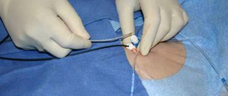
The principle of the operation comes down to the introduction of several catheters - they provide the ability to record an ECG from the inside. Such intracardiac cardiograms make it possible to determine the diagnosis with high accuracy. After identifying the point in the heart responsible for the disorder, it is eliminated using an ablation catheter, and the affected cardiac tissue is cauterized using an electrode.
Advantages and disadvantages of cauterization
This operation has many advantages :
- the ability to relieve arrhythmia that cannot be treated with medications;
- According to statistics, most patients easily tolerate the procedure;
- after surgery, a long period of time for rehabilitation is not required;
- complete absence of scars;
- there is no need to resort to general anesthesia;
- the likelihood of a medical error during the operation is reduced to zero.
However, heart cauterization has some disadvantages :
- It is not possible to process a large number of tissues in 1 manipulation. If a significant part of the heart is affected, treatment will require several manipulations;
- For some patients, a significant disadvantage will be the fact that cauterization does not involve the use of general anesthesia.
Contraindications
Even despite the fact that cauterization of the heart is one of the simplest operations, treating arrhythmia in this way has some contraindications:
- infectious diseases the patient has;
- hypertonic disease;
- intolerance to painkillers;
- hypotension;
- heart failure;
- renal failure;
- endocarditis;
- heart attacks;
- Iron-deficiency anemia.
Preparation for the procedure
As before any operation, on the eve of heart cauterization the patient will have to take some preparatory measures. So, it is important for the patient to undergo some diagnostic procedures:
- donate blood from a vein for general and biochemical analysis;
- do an electrocardiogram;
- undergo magnetic resonance imaging with contrast;
- donate blood for HIV infection.
The patient will also need:
- several days before the procedure, do not take any hormonal drugs or medications to treat arrhythmia;
- do not take food 12 hours and water 8 hours before cauterization;
- shave your armpit and groin hair, as a catheter will be inserted into one of these areas.
Carrying out the procedure
Before surgery begins, the patient is given a local anesthetic. After a certain period of time, the surgeon punctures one of the large arteries, through which a catheter is inserted directly to the heart area. Using a special electrode, the affected cardiac tissue is cauterized; the tissue temperature can reach up to 60 °C.
After complete healing of the burn, a dense scar will form, and the contractions that previously provoked the occurrence of arrhythmia will disappear. Once the operation is completed, the catheter insertion site is tightly bandaged with a sterile bandage.
The patient will have to stay in a medical facility under the supervision of specialists for 5-7 days. In the first 24 hours after surgery, an ECG is performed to assess the dynamics of the heart muscle contraction frequency. Blood pressure, body temperature and daily diuresis are also subject to control.
Rehabilitation and possible complications
The average rehabilitation time takes from 2 to 3 months; during this period, any physical activity is strictly contraindicated for the patient. During this time, doctors prescribe antiarrhythmic drugs and anticoagulants.
Reference! According to statistics, 90% of operations are successful. The likelihood of complications occurring if you follow all the recommendations of specialists is equal to zero.
During rehabilitation, the patient will have to give up:
- smoking, drinking alcoholic beverages;
- eating excessively salty foods;
- strong coffee and tea;
- eating fatty foods.
To prevent relapse of the disease, it is important for the patient to lead a correct lifestyle not only during the rehabilitation period, but throughout his life - eat right, normalize sleep and wakefulness, and eliminate any emotional experiences. Moderate physical activity is acceptable 3-4 weeks after the procedure.
Technique of surgery using monopolar ablation
The operation is performed under conditions of artificial circulation with normothermic perfusion using bicaval cannulation. It is often used when it is necessary to open the atria to intervene on the mitral and tricuspid valves. The left atrium is opened parallel to the interatrial groove, the left atrial appendage is resected with suturing of its base, or its electrical isolation is carried out with suturing from the inside. The left pulmonary veins are isolated en bloc with the adjacent wall of the left atrium and connected by an ablation line to the suture of the left atrial appendage.
Then ablation is performed in the area of the left isthmus by connecting the ablation line of the left inferior pulmonary vein to the posterior semicircle of the mitral valve. The direction of ablation should be based on the type of blood supply to the heart when evaluating coronary angiography. With a dominant circumflex branch, ablation is carried out towards the P3 segment, with a pronounced right type of blood supply - towards the P1 segment, with a balanced one - towards the P2 segment. This must be taken into account to prevent thermal damage to the envelope branch. It is recommended that when performing ablation in this area, short cardioplegia is performed for the same purpose. A short ablation line is also drawn along the coronary sinus (up to 2 cm). The right pulmonary veins are isolated en bloc by connecting the ablation line to the left atrium incision. Isolations of the left and right pulmonary veins are connected to each other in the area of the roof of the left atrium, since this zone is safer in terms of penetration of the effect on the esophagus. The right atrium is opened with a vertical incision from the atrioventricular groove towards the interatrial groove. It is not recommended to resect the right atrium due to its participation in the production of atrial natriuretic hormone, which plays a significant role in water-electrolyte homeostasis. Ablation is performed from the anterior septal commissure of the tricuspid valve to the right atrium appendage with a transition to the crista terminalis, and from the posterior septal commissure to the atriotomy incision. The longitudinal ablation between the superior vena cava and the inferior vena cava is connected to the atriotomy incision and to the ablation in the area of the interatrial septum. Ablation of the right isthmus (from the tricuspid annulus to the coronary sinus and then to the inferior vena cava cannulation site) is also recommended to prevent atrial flutter, although this is not part of the original Maze III procedure. Complications of mopopolar ablation occur rarely; the most dangerous are: damage to the esophagus, coronary arteries, and bleeding. A more frequent development of postcardiotomy syndrome was also noted.
Buy online
Classic labyrinth intervention (Maze III) is the “gold standard” for surgical treatment of patients with clinically manifested atrial fibrillation (AF) refractory to drug therapy. Despite its high efficiency, this intervention is not widely used due to technical complexity, high invasiveness and the use of artificial circulation [1].
Less traumatic interventions have been introduced as a result of increased knowledge of the pathophysiology of AF, as well as the development of ablative techniques and devices that replace the original incision-suture technique.
Trigger activity from the pulmonary veins (PVs) plays an important role in the pathophysiology of AF, especially in the paroxysmal form [2]. Recent clinical and experimental studies also indicate an important role of the autonomic nervous system in the induction and maintenance of AF [3, 4]. R. Wolf et al. [5] developed a technique in which PV isolation and radiofrequency ablation (RFA) of the ganglion plexuses (PG) of the left atrium (LA) are performed through a bilateral thoracotomy approach. Thoracoscopic surgery is less invasive and reduces perioperative complications and patient discomfort.
This study evaluates the first experience with thoracoscopic intervention in patients with various forms of AF over a 1-year follow-up period.
Material and methods
From February 2011 to August 2012, thoracoscopic intervention was performed in 30 patients with symptomatic AF refractory to drug therapy. Inclusion criteria for the study: age 18-70 years, paroxysmal AF after one or more ineffective catheter ablation, persistent or long-term persistent form of AF and LA size more than 6 cm. Exclusion criteria: previous “open” surgical interventions on the heart and lungs, duration AF for more than 10 years and LA size more than 70 cm. All patients signed voluntary informed consent for surgical intervention.
The age of the patients ranged from 37.9 to 71.8 (57.6±8.6) years. All had normal left ventricular ejection fraction and no structural heart pathology. Eight (26%) patients with paroxysmal AF and 5 (16%) patients with persistent AF had previously undergone catheter ablation. A total of 8 (26%) patients had paroxysmal AF, 15 (50%) had persistent AF, and 7 (24%) had long-term persistent AF. The clinical characteristics of the patients before surgery are presented in Table. 1.
During the intervention, the patient was in a supine position. The operation was performed under anesthesia with intubation with a double-lumen tube. Access was achieved using 3 ports on both sides of the chest. Two ports were placed in the fourth and sixth intercostal spaces in the midaxillary line. The third port was located 5 cm anterior to the midaxillary line in the third intercostal space. The endoscope was inserted through a port located in the fourth intercostal space. The remaining two ports were used for two thoracoscopic instruments.
On the right side of the chest, the pericardium was opened 2-3 cm above the phrenic nerve, then its edges were fixed with holders for optimal positioning of the ablation device. Before RFA, ultrafrequency stimulation (1000 pulses/min) was performed in the area of the PV ostia with a transpolar electrode (AtriCure, Inc., West Chester, Ohio, USA) to determine the localization of the GE. A positive vagal response was considered when asystole was at least 3 s or the ventricular rate slowed by more than 50% in the presence of AF. Radiofrequency exposure was performed in the GS area for 20 s using a transpolar electrode (AtriCure, Inc., West Chester, Ohio, USA). Microfrequency stimulation was then repeated in the same position to confirm the effectiveness of RFA. If necessary, the exposure was repeated in the presence of a persistent vagal response. The end point of this stage of the operation was the absence of a vagal response after exposure to repeated ultrafrequency stimulation.
The next stage of the operation was a “blunt” dissection of the space around the PV with sequential passage into the transverse and then oblique sinus of the pericardial cavity. The PVs were then bypassed using a dissector (AtriCure Lumitip, Inc., West Chester, Ohio, USA), followed by isolation using a bipolar clamp (AtriCure, Inc., West Chester, Ohio, USA). Isolation was performed at least 3 times in the area of the right and left PVs. The input block was confirmed using a bipolar mapping electrode in the absence of potentials at the stimulus site. Output block was confirmed by stimulation during sinus rhythm. If necessary, additional isolation of the PV was performed. The final stage was the creation of 2 ablation lines from the right upper and lower PVs, along the dome and lower wall of the left pulmonary veins, reaching the left pulmonary veins. A similar procedure was performed on the left side, with the exception of the pericardium, which was opened as low as possible under the phrenic nerve. In addition, the Marshall ligament was cut with an electrocoagulator (see Fig. 1, color plate).
Figure 1. Technique for thoracoscopic pulmonary vein isolation with radiofrequency ablation of the ganglion plexuses. a - the intervention is performed through thoracoscopic ports using a bithoracal approach (1 - 5 mm port 5 in the third intercostal space along the anterior axillary line; 2 - 10 mm port in the fourth intercostal space along the midaxillary line; 3 - 10 mm port in the sixth intercostal space along the anterior axillary line); b — with the help of atraumatic suction and grabber, the pericardial ligament is divided in the projection between the inferior vena cava (IVC) and the lower lobe right pulmonary vein (LPV), entering the space of the oblique pericardial sinus; c — with the help of atraumatic suction and grabber, the pericardial ligament is divided over the dome of the left atrium, entering the space of the transverse pericardial sinus; d — radiofrequency isolation of the right pulmonary veins using an ablation bipolar clamp; d — radiofrequency ablation of the ganglion plexuses from the right pulmonary veins, followed by the creation of lines along the roof and posterior wall of the left atrium with a linear electrode; e — radiofrequency isolation of the left pulmonary veins using an ablation bipolar clamp.
Immediately after surgery, all patients were implanted with a subcutaneous device for long-term heart rate monitoring (CPM). At the end of the intervention, patients were transferred to the intensive care unit for observation.
Patients with a proportion of AF <0.5% (according to ICM data) were classified as “responders”, i.e. to those who responded to the intervention [11]. “Non-responders” (those who did not respond to the intervention) included patients with symptomatic and asymptomatic AF (AF >0.5% according to ICM) or any other atrial tachyarrhythmia. ICM data after hospital discharge were collected every 3 months throughout the follow-up period.
The observation period was 12 months. All patients received warfarin for 3 months after surgery; further anticoagulant therapy was based on the results of testing according to the CHA2DS2VASc scale. In addition, patients received antiarrhythmic therapy for 3 months, and its discontinuation was based on the presence of recurrent AF after a blinding period.
Statistical calculations were performed using STATA 12&1 software. Treatment effectiveness was compared using the log-rank test, which was graphically expressed by Kaplan-Meier curves.
results
During the intervention, 2 (6.7%) patients underwent median sternotomy due to severe pleural adhesions and bleeding from the right lower PV. The remaining 28 (93.3%) patients did not have any complications during surgery. The average duration of the intervention was 150.4±28.7 minutes. All patients were transferred to the general ward 10-12 hours after the intervention and discharged 4-9 (6.1±1.8) days after surgery. The results of the analysis of the early postoperative period are presented in table. 2.
Perioperative and long-term mortality was 0. The average follow-up period was 12.5±2.2 months.
At the end of the observation period, 22 (73.3%) patients did not have AF or any other atrial tachyarrhythmias (Fig. 2).
Figure 2. Overall effectiveness (absence of atrial tachyarrhythmias) after thoracoscopic intervention at long-term follow-up. All 13 patients who had previously undergone catheter RFA responded to the intervention. All of them did not need to take antiarrhythmic drugs, and 14 (47%) patients did not need to take anticoagulants with CHA2DS2VASc ≤1. During observation, none of the patients had cerebrovascular accident or transient ischemic attack, and none of the patients required pacemaker implantation.
Discussion
This study presents the first experience of thoracoscopic PV isolation with HS ablation with a follow-up period of 12 months. The effectiveness for all forms of AF according to ICM data was 73.3%. The study results demonstrated that the thoracoscopic approach was both safe and effective, although 2 patients required sternotomy due to bleeding. These complications developed during this procedure in the first 6 patients, which can be attributed to the “learning period.” There were no complications in the subsequent 24 patients. In addition, this procedure is effective even in patients with a long history of AF or previously failed catheter ablation.
The technique of PV isolation and SG ablation was previously described by R. Wolf et al [5]. Our results are comparable to those of other researchers who have used this technique [6, 7]. This operation is less effective than the classic Maze III operation [1], however, the thoracoscopic approach has several advantages. The intervention is minimally invasive and does not require the use of artificial blood circulation, and is also characterized by a shorter duration of both the operation itself and the stay of patients in the intensive care unit and hospital stay. It should be noted that all patients with a history of ineffective catheter ablation responded to thoracoscopic intervention (AF according to ICM <0.5%). This may be due to insufficient transmural myocardial damage during endocardial catheter ablation, as well as the creation of additional bipolar PV isolation with the addition of HS ablation during thoracoscopic intervention.
In 1958, R. Alessi et al. [8] showed that stimulation of the vagus nerve leads to uneven atrial contraction during the refractory period in dogs. Shortly thereafter, G. Moe et al. [9] were able to induce sustained AF when stimulating the vagus nerve without triggering activity from the PV orifices. Further experimental studies showed that the initiation of AF by trigger activity depends on stimulation of the GS [10]. These experimental data cast doubt on the classical “trigger-substrate hypothesis” and show that the autonomic nervous system plays a key role in the onset and maintenance of AF. In clinical studies by S. Pappone et al. [21] demonstrated that patients with a vagal response during RFA had a higher success rate than patients without a vagal response. In turn, V. Sherlag et al. [22] were the first to compare PV isolation with and without PV ablation. Although the follow-up period was short and the number of patients was small, these data showed that the effectiveness was much higher with additional HS ablation. Some authors performed only HS ablation for AF. Efficacy rates range from 29 to 84% with a short follow-up period (4.2–17.3 months). The effectiveness of isolated HS ablation in long-term follow-up is a subject of debate [17]. In dogs, autonomic innervation is restored within 4 weeks [13], and even reinnervation occurs in completely denervated hearts in patients who have undergone heart transplantation [14].
Our work presents the first experience of 30 thoracoscopic interventions. PV isolation is a common AF treatment for patients with paroxysmal AF. At the same time, the effectiveness of treatment for patients with long-term persistent form of AF does not exceed 40-50%, and additional effects in the LA can improve the effectiveness of treatment for patients in this category [16, 17]. Thoracoscopic intervention using the Coolrail device (AtriCure, Inc, West Chester, Ohio, USA) provides additional linear influences similar to the left atrial Maze III.
In our study, bilateral thoracoscopic RFA was performed using a bipolar electrode. A number of researchers [18–20] use a one-way approach using other energy sources. All of these types of energy are capable of reaching transmural lesions. However, with a unilateral approach, positioning of the ablation device is not completely feasible under visual control and may lead to loss of contact with the epicardium.
Limitations of the study.
Our study presents the first experience in performing thoracoscopic ablation of AF. This study was not randomized and included only 30 patients. In addition, the follow-up period was 12 months. Further multicenter randomized studies will evaluate the long-term effectiveness of the thoracoscopic method, especially in patients after ineffective primary endocardial ablation.
In conclusion, minimally invasive thoracoscopic LA ablation is a safe and effective intervention in the treatment of patients with AF and preserves sinus rhythm in 73.3% of patients during the 12-month follow-up period.
Surgery technique using bipolar ablation
Bipolar ablation has a number of advantages over the monopolar option:
- ablation occurs between two active electrodes (length 7-10 cm), one of the branches of which is located on the endocardial side, the second on the epicardial side, or both epicardial, which eliminates undesirable effects on surrounding tissues
- speed (ablation line 6-7 cm is carried out in 10-15 seconds)
- control of transmurality by impedance or temperature
It can be used both for valve corrections with opening of the atria, and without opening the latter (aortic valve replacement and CABG, or in isolation).
After opening the pericardium and connecting artificial circulation to the beating heart and assisted circulation, ablation of the mouths of the right pulmonary veins is performed, then the heart is rotated and ablation of the mouths of the left pulmonary veins and left atrial appendage is performed. To achieve complete conduction block, it is recommended to perform at least 2 ablation captures of the pulmonary veins with the adjacent wall of the left atrium.
| Bipolar ablation in the left atrium |
After cardioplegia, a standard left atriotomy is performed. Connecting ablations are performed between the sites of the right and left pulmonary veins (one for left atrium sizes up to 55 mm, two for 55 mm or more), the left atrial appendage and the posterior semicircle of the mitral valve. The left atrial appendage is sutured from the inside or resected initially. If the left atrium is large (more than 60 mm), atrioplasty is performed. In the right atrium, if there is no need to correct the tricuspid defect, ablation of the free wall and appendage of the right atrium is performed. One of the branches of the bipolar device is inserted into the lumen of the right atrium through a small incision inside a purse-string suture placed near the interatrial groove.
Minimally invasive techniques for surgical treatment of atrial fibrillation
For the surgical treatment of isolated atrial fibrillation in the absence of structural heart pathology, the following methods have been proposed:
Monopolar epicardial ablation from right minithoracotomy with video support or completely thoracoscopic
It is performed using the “box lesion” technique, which means isolating the pulmonary veins as a single block.
The ablation device is positioned on the posterior wall of the left atrium using special conductors through the transverse and oblique sinuses of the heart.
Types of devices
- non-irrigated radiofrequency ablation with internal cooling (uses a device with a built-in multi-pole electrode and vacuum connection for better positioning and contact with the epicardium.)
- microwave ablation (multipole flexible antenna)
- ultrasound ablation based on high-intensity focused ultrasound (multipole irrigated circular electrode)
It is performed using flexible bipolar clamp electrodes using a “vertical” technique (separate isolation of the right and left pulmonary veins) or a “lateral” technique reminiscent of a “box lesion”.
The operation also involves crossing the Marshall ligament with bipolar coagulation, removing the left atrial appendage or clipping it, as well as performing epicardial mapping to determine the completeness of the creation of a conduction block (high-frequency stimulation of the pulmonary veins 800 imp/min with determination of heart rate deceleration and recording of the atrial electrogram before and after ablation ).
Comparative characteristics of mono- and bipolar ablation on the beating heart from mini-approaches
| Bipolar ablation | Monopolar ablation |
| Myocardial tissue is evenly distributed between the branches and the impact occurs only between them, excluding the spread of energy to surrounding organs and tissues | The effect is produced epicardially against the background of blood circulation in the atrium (“heat sinking effect” is due to endocardial and myocardial blood circulation), which requires a higher temperature and duration of exposure |
| Minimally invasive intervention takes place under full bilateral visual control, preventing complications such as perforation or stricture of the esophagus, damage to the circumflex branch of the left coronary artery, coronary sinus | The intervention is carried out under unilateral control, and the location of the electrode in relation to the left pulmonary veins is not controlled (interposition of the ear, the presence of epicardial fat) |
| More precise preparation of the left pulmonary veins is possible, incl. Marshall ligaments, as well as ablation and clipping of the left atrial appendage (increases efficiency by 10-12% and eliminates the threat of thromboembolic complications) | Visualization of the auricle and preparation of the left pulmonary veins is impossible. Epicardial fat, as a tissue with a low water content, acts as an ieolator and limits the penetration of radiofrequency energy. Creating a continuous, homogeneous transmural penetration is problematic. |
| There is control of the transmurality of the impact by analyzing the tissue impedance between the jaws, which automatically blocks excessive overheating of the tissue and the risk of damage. | There is no control of transmurality of exposure, there is control of exposure temperature |
| Preparation of the transverse sinus is performed more safely in a retrograde manner using a special navigator, which virtually eliminates damage to the right branch of the pulmonary artery or vena cava | The preparation is antegrade, that is, “from oneself,” which does not exclude injury to the great vessels |
| It is possible to perform epicardial stimulation from the right and left pulmonary veins to identify incomplete conduction block | It is possible to stimulate only the common area or the right pulmonary veins |
| Longer, requires alternate switching off of both lungs | Faster, requires separate intubation to shut down only the right lung |
| Requires two-way access | One-way access |
Benefits of Minimally Invasive Atrial Fibrillation Surgery
- no radiation exposure, unlike endovascular techniques
- anatomical ablation, under visual control
- possibility of removing the left atrial appendage to reduce the risk of thromboembolism
- the possibility of effective influence on the ganglion plexuses.
Lifestyle and prognosis after surgery
In most cases, rhythm normalization can be achieved after the first manipulation, but some patients require repeating the procedure. Sometimes, even after ablation, you may need to continue taking medications. There are a number of changes that you can make to your lifestyle and help your heart not experience rhythm problems in the future:
- Avoid taking stimulants that can cause heart rhythm problems, such as caffeine, alcohol and nicotine;
- Regularly examine blood pressure and cholesterol levels in the blood, and keep these indicators within normal limits;
- You need to maintain physical activity. It is recommended to discuss the optimal level of physical activity with your doctor and, based on this, create an exercise program;
- Avoid stress and psycho-emotional stress;
- Eat healthy food and fight excess weight;
- Get screened regularly.
Indications for surgical treatment of atrial fibrillation
According to the recommendations of the European Society of Cardiology, the American Heart Association, and the American College of Cardiology, combined surgery to eliminate atrial fibrillation along with other surgical procedures is indicated for patients with symptomatic atrial fibrillation and all patients for whom surgical treatment for atrial fibrillation can be performed with minimal risk. The type of surgical procedure and its scope are determined by the operating cardiac surgeon. In patients with paroxysmal and persistent atrial fibrillation, ablation in the left atrium is indicated for one-stage correction of the mitral defect; in patients with a permanent form of atrial fibrillation or one-stage correction of the mitral-tricuspid defect, biatrial ablation is indicated. During operations for ischemic heart disease or aortic disease, epicardial ablation of the ostia of the pulmonary veins and left atrial appendage is performed with ligation (amputation) of the latter.
Factors that determine the effectiveness of surgical elimination of atrial fibrillation
- duration of atrial fibrillation
- left atrium size
- amplitude of f-waves in lead V1 (more than 0.1 mV)
- diastolic myocardial dysfunction
- presence of concomitant ischemic heart disease
- three-valve correction
Indications for minimally invasive surgery for atrial fibrillation in patients with isolated atrial fibrillation
- symptomatic atrial fibrillation refractory to medical therapy and the patient prefers surgery
- history of thromboembolic episodes
- contraindications or difficulties of anticoagulant therapy
- ineffectiveness of endovascular catheter techniques or the presence of contraindications to their implementation (thrombosis of the left atrial appendage)
Contraindications to minimally invasive surgery for atrial fibrillation
- left atrial appendage thrombosis (for monopolar ablation)
- presence of sick sinus syndrome
- adhesive process in the pericardial cavity (previous operations with opening of the pericardium)
- adhesions in the pleural cavities (previous pneumonia, pleurisy)
- Moderate to severe COPD (prolonged unilateral ventilation is difficult)
- left atrium size more than 55 mm