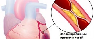
Causes of the disease
Heart rhythm and conduction disturbances can occur due to physiological characteristics and cardiac pathologies.
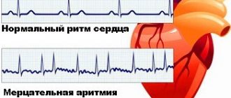
Sinus tachycardia, for example, develops during physical and emotional stress. Respiratory bradyarrhythmia is normal. Atrial fibrillation and flutter are dangerous. They usually develop against the background of diseases of the cardiovascular system. As a rule, heart rhythm and conduction disturbances occur when:
- coronary heart disease;
- vices;
- arterial hypertension;
- cardiomyopathy.
Non-cardiac diseases such as:
- acute poisoning;
- fever;
- stomach ulcer;
- hyperthyroidism;
- adrenal tumor.
Risk factors for the development of pathology include:
- smoking and alcohol abuse;
- obesity;
- concomitant diseases (mainly the endocrine system).
Decrease in heart rate
A heart rate less than 60 beats per minute is rare. But for many people, especially those leading an active lifestyle, this is not a pathology: sinus bradycardia for athletes is a normal variant that does not require therapy. The normal heart rate is different in a child and an adult, so it is important to know what sinus bradycardia is in children (detecting a heart rate of less than 70 beats per minute should force parents to consult a pediatrician). The severity of the decrease is of great importance - the slower the pulse, the less oxygen-rich blood is delivered to the vital organs, therefore, if typical symptoms appear, you should consult a specialist in time and begin treatment.
Types of conduction disorders
All pathologies are divided into two groups:
- Changes in rhythm (arrhythmias). Such pathologies can be expressed in the form of atrial fibrillation, tachycardia, bradyarrhythmia, ventricular tachycardia, etc.
- Direct blockade. Some experts classify them as a separate category of pathologies. Blockades are divided into sinoauricular, intraatrial, atrioventricular and intraventricular.
Both types of conditions are equally dangerous.
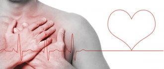
Symptoms of pathology
Clinical symptoms manifest themselves in different ways. In some patients, the pathology is diagnosed only during an ECG, while others show pronounced signs of disorders.
Pathologies are characterized by:
- slow or increased heart rate;
- feeling of lack of air;
- chest pain;
- general weakness;
- changes in blood pressure;
- dizziness;
- loss of consciousness.
Any pathology is dangerous because when it occurs, blood circulation is disrupted. As a result, organs and tissues do not receive the required amount of oxygen and nutrients. It is important to pay attention to each symptom and tell your doctor about it. This will allow him to accurately determine the disease you have and begin adequate treatment as soon as possible.
Pathology leads to the development of several complications. This:
- Collapse. This condition is characterized by a sharp drop in blood pressure, pale skin and weakness.
- Arrhythmogenic shock. Occurs when there is a sharp decrease in blood flow. The patient loses consciousness, turns pale, the heartbeat becomes rare, and the pressure drops below 60 mm Hg. Art.
- Ischemic stroke. This pathology occurs against the background of increased thrombus formation. A stroke is characterized by changes in speech, unsteadiness of gait, and paralysis of the limbs.
- Pulmonary embolism (PE). This pathology develops when a pulmonary artery is blocked by a blood clot and is manifested by shortness of breath and suffocation, blue discoloration of the face, neck and chest.
- Acute myocardial infarction. The pathology develops due to the fact that the arteries cannot provide the necessary blood flow to the heart. As a result, oxygen deficiency occurs in the tissues. An area of necrosis forms on the heart. A heart attack is manifested by acute pain in the chest.
- Asystole (cardiac arrest). This condition develops when ventricular fibrillation occurs when the vessels do not receive the required amount of blood.
All these conditions can lead to the death of the patient.
Causal factors
A rare pulse can be physiological and pathological. In the first case, this is not a disease, but a normal state of metabolic processes in the following cases:
- against the background of constant sports loads;
- in people engaged in heavy physical labor;
- during sleep.
Pathological causes of sinus bradycardia are caused by the following diseases and conditions:
- acute disturbances of blood flow in the heart (in 20% of patients with myocardial infarction in the first hours of the attack);
- chronic cardiac pathology of metabolic or inflammatory nature;
- complications of arrhythmia, ischemic heart disease and atherosclerosis;
- endocrinopathies (decreased functional activity of the thyroid gland);
- acute infectious pathology with severe course;
- poisoning;
- long-term drug therapy;
- stressful situation.
A rare rhythm in childhood is most often caused by neurocirculatory dystonia or congenital heart pathology: a child suffering from a severe form of bradycardia is less active, more often suffers from headaches and lags behind in development from his peers. Parents should contact a specialist as early as possible to maintain the baby’s health.
Diagnostics
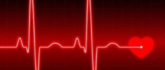
The basis of examination for suspected pathology is an ECG. During the examination, the doctor discovers the main signs of a particular condition. Extrasystoles are manifested by altered ventricular complexes, tachycardia paroxysm - by short intervals between us, etc. Do not try to interpret the ECG results yourself. Entrust the procedure to a cardiologist.
An ECG is not the only examination used to diagnose pathology.
Also performed:
- Daily monitoring of ECG and blood pressure.
- Tests with physical activity.
- Transesophageal ECG.
- Transesophageal electrophysiological study.
- MRI of the heart.
- Ultrasound of the heart, etc.
All diagnostic methods are aimed at detecting deviations from normal heart rhythm and recording partial or complete blockades. The patient may also be referred for an examination to assess the patency of various vessels.
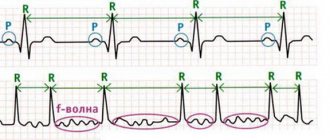
Heart block on ECG
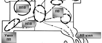
Sinus node block
Complete loss from the general rhythm of the ventricular complex.
This case occurs very rarely, but if it occurs, it can be noticed by the loss of a complete contraction. In this case, the blockade of the SA junction is not complete, since this will cause cardiac arrest.
Intraatrial block
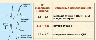
It also rarely appears. It is characterized by slow conduction of atrial impulses. On the electrocardiogram, this can be seen by a wide (more than 0.1 second) P wave, which splits and deforms.
Attention! A similar sign appears on the electrocardiogram with LA hypertrophy.
Atrioventricular block
Frederick's syndrome poses the greatest threat to the patient's life.
Obstructed rhythm from the AV junction is the most common option that occurs, which can have 3 degrees of severity.
The first degree is detected by the cardiogram by an extended PQ interval and an excess of 0.2 seconds.
Second degree AV block has two subtypes:
- Mobitz 1. Characterized by a progressive PQ interval, which is accompanied by loss of QRS. During the pause, only the P wave is recorded.
- Mobitz 2. The QRS complex regularly falls behind every second or fourth P wave.
The third degree of AV block is called complete, since the passage of impulses stops completely. The ECG displays rhythms independent of each other. The atria contract more often, because the sinus node does not stop exciting them, and third-order pacemakers respond to the contraction of the ventricles, which produce impulses with a frequency of no more than 30-40 per minute.
Here, too, there are 2 types of pathology:
- With a wide QRS complex - idioventricular block, in which contraction of the ventricles is excited by ectopic foci in the ventricles themselves.
- With a normal QRS complex - idionodal block, in which the foci of excitation are located on the lower part of the AV node.
Bundle branch block
With this type of blockade, the impulse reaches only one ventricle completely. And the second ventricle is involved in general work in an unusual way. The following electrocardiographic signs of this blockade can be distinguished:
- The ventricular QRST rhythm on the ECG widens.
- It splits.
- Opposite directions of the ends of the ventricular complex arise.
If the right leg is blocked, then:
ECG signs of right leg block
- For a long time, the QRS complex reaches 0.11 and more than 0.12 with incomplete and complete blockade, respectively.
- The QRS complex is jagged in V1-V2 and has flattened S waves in V5-V.
- The ST segment decreases and an inverted T wave appears in V1-V2, III with a dominant R wave.
- In V1-V2 there is an increase in the time of ventricular excitation.
- The EOS deviates to the right (does not always occur).
Left leg block has characteristic signs:
- The duration of the QRS complex is the same as with right bundle branch block.
- The R wave is jagged or smoothed, wide, or an M-shaped QRS complex appears in V5-V.
- The ST segment decreases in I, aVL, V5-V6 and rises in III, V1-V.
- The ventricular excitation time exceeds 0.05 seconds.
All of the above signs of rhythm disturbance on the ECG should be identified and interpreted only by a qualified specialist.
In the video you can watch a lesson on how to recognize the symptoms and signs of arrhythmia on an ECG:
Treatment
Treatment of pathologies is always carried out comprehensively. The choice in favor of one method or another is determined by:
- the patient's condition;
- his individual characteristics;
- existing concomitant diseases.
If coronary heart disease is detected, patients receive:
- blood thinners;
- nitroglycerine;
- cholesterol-lowering agents.
For hypertension, medications that lower blood pressure are prescribed.
In case of chronic deficiency, it is necessary to take diuretics and glycosides.
If a heart defect is detected, surgical correction may be performed.
Emergency care for patients involves the administration of drugs that restore heart rhythm.
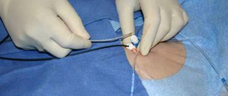
Surgical interventions
If the exact cause of the pathology is identified and the medications are ineffective, the doctor may recommend surgery. As a rule, it is aimed at installing a pacemaker. Such a device does not cure the disease, but can effectively stop attacks and reduce the risk of death.
Other operations may also be performed. Only your cardiologist will tell you about them. Surgical interventions are performed only in advanced cases when symptoms cannot be eliminated in other ways.
Our clinic in Moscow provides complex therapy for pathologies of the cardiovascular system. Experienced doctors can detect all the signs of diseases and identify factors contributing to their development and complications. Therapy is always adequate and carried out at the best price. The cost of services is indicated on the website. Our specialists will also announce prices. The exact cost of therapy will be determined by the doctor after conducting an examination and selecting appropriate tactics and methods.
Prevention
Prevention of pathologies of the cardiovascular system is aimed at:
- adherence to sleep and wakefulness;
- eliminating the risks of stressful situations;
- changing your lifestyle and giving up bad habits.
Moderate physical activity (mainly water procedures) is beneficial. To prevent arrhythmia, it is very important to follow a diet. Diet excludes salty, fatty and spicy foods, coffee and strong tea. Don't get carried away with the liquid. You need to drink 1-2 liters of water per day. It is also important to enrich the diet with vitamins and microelements.
If you have a genetic predisposition to pathologies or a history of them, it is very important to regularly visit a cardiologist and undergo a full examination. Be sure to include in the program such studies as:
- general blood analysis;
- ECG;
- ECG under load.
The examination will not take much of your time, but will help prevent the development of pathology and the occurrence of complications.