Erythrocytes or red blood cells are one of the formed elements of blood that perform numerous functions that ensure the normal functioning of the body:
- the nutritional function is to transport amino acids and lipids;
- protective - in binding toxins with the help of antibodies;
- enzymatic is responsible for the transfer of various enzymes and hormones.
Red blood cells are also involved in regulating acid-base balance and maintaining blood isotonicity.
However, the main job of red blood cells is to deliver oxygen to the tissues and carbon dioxide to the lungs. Therefore, they are often called “respiratory” cells.
Features of the structure of red blood cells
The morphology of red blood cells differs from the structure, shape and size of other cells. In order for red blood cells to successfully cope with the gas transport function of blood, nature has endowed them with the following distinctive features:
The listed features are measures of adaptation to life on land, which began to develop in amphibians and fish, and reached their maximum optimization in higher mammals and humans.
This is interesting! In humans, the total surface area of all red blood cells in the blood is about 3,820 m2, which is 2,000 times more than the surface of the body.
Red blood cell: structure, shape and functions. The structure of human red blood cells
An erythrocyte is a formed element of blood, capable of transporting oxygen to tissues and carbon dioxide to the lungs using hemoglobin.
This is a simple cell in structure, which is of great importance for the life of mammals and other animals.
The red blood cell is the most abundant cell type in the body: approximately a quarter of all cells in the body are red blood cells.
General principles of the existence of red blood cells
An erythrocyte is a cell derived from the red germ of hematopoiesis. About 2.4 million of these cells are produced per day, they enter the bloodstream and begin to perform their functions. During the experiments, it was determined that in an adult, red blood cells, the structure of which is significantly simplified compared to other cells of the body, live 100-120 days.
In all vertebrates (with rare exceptions), oxygen is transferred from the respiratory organs to the tissues through hemoglobin in erythrocytes.
There are exceptions: all representatives of the “white-blooded” fish family exist without hemoglobin, although they can synthesize it.
Since at the temperature of their habitat, oxygen dissolves well in water and blood plasma, these fish do not require more massive oxygen carriers, which are erythrocytes.
Erythrocytes of chordates
A cell such as an erythrocyte has a different structure depending on the class of chordates. For example, in fish, birds and amphibians, the morphology of these cells is similar. They differ only in size.
The shape of red blood cells, volume, size, and the absence of certain organelles distinguish mammalian cells from others found in other chordates. There is also a pattern: mammalian red blood cells do not contain unnecessary organelles and a cell nucleus.
They are much smaller, although they have a larger contact surface.
Considering the structure of frog and human erythrocytes, common features can be identified immediately. Both cells contain hemoglobin and are involved in oxygen transport.
But human cells are smaller, oval and have two concave surfaces.
The red blood cells of frogs (as well as birds, fish and amphibians, except salamanders) are spherical, they have a nucleus and cellular organelles that can be activated when necessary.
Human red blood cells, like the red blood cells of higher mammals, do not have nuclei or organelles. The size of goat red blood cells is 3-4 microns, human - 6.2-8.2 microns. In Amphiuma (tailed amphibian), the cell size is 70 microns. Obviously size is an important factor here. The human red blood cell, although smaller, has a larger surface due to two concavities.
The small size of cells and their large number have made it possible to greatly increase the ability of blood to bind oxygen, which now depends little on external conditions.
And such structural features of human red blood cells are very important, because they allow you to feel comfortable in a certain habitat.
This is a measure of adaptation to life on land, which began to develop in amphibians and fish (unfortunately, not all fish in the process of evolution had the opportunity to populate land), and reached the peak of development in higher mammals.
The structure of human red blood cells
The structure of blood cells depends on the functions assigned to them. It is described from three angles:
- Features of the external structure.
- Component composition of an erythrocyte.
- Internal morphology.
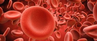
Externally, in profile, an erythrocyte looks like a biconcave disk, and in front, like a round cell. The normal diameter is 6.2-8.2 microns.
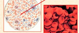
More often, blood serum contains cells with slight differences in size. With iron deficiency, the run-up decreases, and anisocytosis (many cells with different sizes and diameters) is recognized in the blood smear. With a deficiency of folic acid or vitamin B12, the red blood cell enlarges to a megaloblast. Its size is approximately 10-12 microns. The volume of a normal cell (normocyte) is 76-110 cubic meters. µm.
The structure of red blood cells in the blood is not the only feature of these cells. Their number is much more important. Small sizes made it possible to increase their number and, consequently, the contact surface area. Oxygen is more actively captured by human red blood cells than by frogs. And it is most easily released into tissues from human red blood cells.
Quantity is really important. In particular, an adult human contains 4.5-5.5 million cells per cubic millimeter. A goat has about 13 million red blood cells per milliliter, while reptiles have only 0.5-1.6 million, and fish have 0.09-0.13 million per milliliter. In a newborn child, the number of red blood cells is about 6 million per milliliter, and in an elderly child it is less than 4 million per milliliter.
Functions of red blood cells
Red blood cells - red blood cells, the number, structure, functions and developmental features of which are described in this publication, are very important for humans. They implement some very important functions:
- transport oxygen to tissues;
- transport carbon dioxide from tissues to the lungs;
- bind toxic substances (glycated hemoglobin);
- participate in immune reactions (immune to viruses and, due to reactive oxygen species, can have a detrimental effect on blood infections);
- able to tolerate certain medications;
- participate in the implementation of hemostasis.
Let us continue to consider a cell such as an erythrocyte, its structure is maximally optimized for the implementation of the above functions.
It is as light and mobile as possible, has a large contact surface for gas diffusion and chemical reactions with hemoglobin, and also quickly divides and replenishes losses in the peripheral blood.
This is a highly specialized cell, the functions of which cannot yet be replaced.
Red blood cell membrane
A cell such as an erythrocyte has a very simple structure, which does not apply to its membrane. It is 3-layer. The mass fraction of the membrane is 10% of the cell membrane. It contains 90% proteins and only 10% lipids. This makes red blood cells special cells of the body, since in almost all other membranes lipids predominate over proteins.
The volumetric shape of red blood cells can change due to the fluidity of the cytoplasmic membrane. Outside the membrane itself there is a layer of surface proteins containing a large number of carbohydrate residues.
These are glycopeptides, under which there is a bilayer of lipids, with their hydrophobic ends facing in and out of the erythrocyte.
Under the membrane, on the inner surface, there is again a layer of proteins that do not have carbohydrate residues.
Receptor complexes of erythrocytes
The function of the membrane is to ensure the deformability of the red blood cell, which is necessary for capillary passage.
At the same time, the structure of human red blood cells provides additional capabilities - cellular interaction and electrolyte current.
Proteins with carbohydrate residues are receptor molecules, thanks to which red blood cells are not “hunted” by CD8 leukocytes and macrophages of the immune system.
Red blood cells exist thanks to receptors and are not destroyed by their own immunity. And when, due to repeated pushing through the capillaries or due to mechanical damage, red blood cells lose some receptors, spleen macrophages “extract” them from the bloodstream and destroy them.
Internal structure of a red blood cell
What is an erythrocyte? Its structure is no less interesting than its functions. This cell is similar to a bag of hemoglobin, bounded by a membrane on which receptors are expressed: clusters of differentiation and various blood groups (Landsteiner, Rhesus, Duffy and others). But inside the cell is special and very different from other cells in the body.
The differences are as follows: red blood cells in women and men do not contain a nucleus, they do not have ribosomes and the endoplasmic reticulum. All these organelles were removed after the cell cytoplasm was filled with hemoglobin. Then the organelles turned out to be unnecessary, because a cell with minimal dimensions was required to push through the capillaries.
Therefore, inside it contains only hemoglobin and some auxiliary proteins. Their role has not yet been clarified. But due to the absence of the endoplasmic reticulum, ribosomes and nucleus, it has become light and compact, and most importantly, can be easily deformed along with the fluid membrane.
And these are the most important structural features of red blood cells.
Red blood cell life cycle
The main features of red blood cells are their short life.
They cannot divide and synthesize protein because the nucleus has been removed from the cell, and therefore structural damage to their cells accumulates. As a result, erythrocytes tend to age.
However, the hemoglobin that is captured by splenic macrophages during red blood cell death will always be sent to form new oxygen carriers.
The life cycle of a red blood cell begins in the bone marrow. This organ is present in the lamellar substance: in the sternum, in the wings of the ilium, in the bones of the base of the skull, and also in the cavity of the femur.
Here, from a blood stem cell, under the influence of cytokines, a precursor of myelopoiesis with a code (CFU-HEMM) is formed. After division, it will give the ancestor of hematopoiesis, designated by the code (BOE-E).
From it, a precursor of erythropoiesis is formed, which is designated by the code (CFU-E).
This same cell is called the colony-forming cell of the red blood sprout. It is sensitive to erythropoietin, a hormonal substance secreted by the kidneys. Increasing the amount of erythropoietin (according to the principle of positive feedback in functional systems) accelerates the processes of division and production of red blood cells.
Red blood cell formation
The sequence of cellular bone marrow transformations of CFU-E is as follows: from it an erythroblast is formed, and from it a pronormocyte, giving rise to a basophilic normoblast.
As the protein accumulates, it becomes a polychromatophilic normoblast and then an oxyphilic normoblast. Once the nucleus is removed, it becomes a reticulocyte.
The latter enters the blood and differentiates (matures) into a normal red blood cell.
Destruction of red blood cells
For approximately 100-125 days, the cell circulates in the blood, constantly carries oxygen and removes metabolic products from tissues. It transports carbon dioxide bound to hemoglobin and sends it back to the lungs, simultaneously filling its protein molecules with oxygen.
And as it gets damaged, it loses phosphatidylserine molecules and receptor molecules. Because of this, the red blood cell comes under the macrophage’s sights and is destroyed by it. And the heme obtained from all the digested hemoglobin is again sent for the synthesis of new red blood cells.
Source: //FB.ru/article/189174/eritrotsit-stroenie-forma-i-funktsii-stroenie-eritrotsitov-cheloveka
Formation of red blood cells
The life of an individual red blood cell is relatively short - 100-120 days, and human red bone marrow reproduces about 2.5 million of these cells every day.
Full development of red blood cells (erythropoiesis) begins in the 5th month of intrauterine development of the fetus. Until this point, and in cases of oncological lesions of the main hematopoietic organ, red blood cells are produced in the liver, spleen and thymus.
The development of red blood cells is very similar to the process of human development. The birth and “intrauterine development” of red blood cells begins in the erythron - the red germ of the hematopoiesis of the red brain. It all starts with a pluripotent blood stem cell, which, changing 4 times, turns into an “embryo” - an erythroblast, and from this moment morphological changes in structure and size can already be observed.
Erythroblast
. This is a round, large cell measuring from 20 to 25 microns with a nucleus that consists of 4 micronuclei and occupies almost 2/3 of the cell. The cytoplasm has a purple tint, which is clearly visible on a section of flat “hematopoietic” human bones. In almost all cells, so-called “ears” are visible, formed due to protrusion of the cytoplasm.
Pronormocyte.
The dimensions of a pronormocyte cell are smaller than those of an erythroblast - already 10-20 microns, this occurs due to the disappearance of the nucleoli. The purple hue begins to lighten.
Basophilic normoblast.
In almost the same cell size - 10-18 microns, the nucleus is still present. Chromantin, which gives the cell a light purple color, begins to gather into segments and the externally basophilic normoblast has a spotted color.
Polychromatophilic normoblast.
The diameter of this cell is 9-12 microns. The core begins to change destructively. A high concentration of hemoglobin is observed.
Oxyphilic normoblast.
The disappearing nucleus is displaced from the center of the cell to its periphery. The cell size continues to decrease - 7-10 microns. The cytoplasm becomes clearly pink with small remnants of chromantin (Joly bodies). Before entering the blood, normally the oxyphilic normoblast must squeeze out or dissolve its nucleus with the help of special enzymes.
Reticulocyte.
The color of the reticulocyte is no different from the mature form of the erythrocyte. The red color provides the overall effect of the yellow-greenish cytoplasm and the violet-blue reticulum. The diameter of the reticulocyte ranges from 9 to 11 microns.
Normocyte.
This is the name of a mature form of red blood cell with standard sizes, pinkish-red cytoplasm. The nucleus disappeared completely, and hemoglobin took its place. The process of increasing hemoglobin during red blood cell maturation occurs gradually, starting from the earliest forms, because it is quite toxic to the cell itself.
Another feature of red blood cells that causes a short lifespan is the absence of a nucleus does not allow them to divide and produce protein, and as a result, this leads to the accumulation of structural changes, rapid aging and death.
Degenerative forms of red blood cells
With various blood diseases and other pathologies, qualitative and quantitative changes in the normal levels of normocytes and reticulocytes in the blood, hemoglobin levels, as well as degenerative changes in their size, shape and color are possible. Below we will consider changes that affect the shape and size of red blood cells - poikilocytosis, as well as the main pathological forms of red blood cells and due to what diseases or conditions such changes occurred.
| Name | Changing shape | Pathologies |
| Spherocytes | A spherical shape of normal size with no characteristic clearing at the center. | Hemolytic disease of newborns (AB0 blood incompatibility), disseminated intravascular coagulation syndrome, specicymia, autoimmune pathologies, extensive burns, vascular and valve implants, other types of anemia. |
| Microspherocytes | Small balls from 4 to 6 microns. | Minkowski-Choffard disease (hereditary microspherocytosis). |
| Eliptocytes (ovalocytes) | Ovals or elongated shapes due to membrane abnormalities. There is no central clearing. | Hereditary ovalocytosis, thalassemia, liver cirrhosis, anemia: megablastic, iron deficiency, sickle cell. |
| Target-shaped red blood cells (codocytes) | Flat cells that resemble a target in color - pale at the edges and a bright spot of hemoglobin in the center. The cell area is flattened and increased in size due to excess cholesterol. | Thalassemia, hemoglobinopathies, iron deficiency anemia, lead poisoning, liver disease (accompanied by obstructive jaundice), removal of the spleen. |
| Echinocytes | Spikes of the same size are located at the same distance from each other. Looks like a sea urchin. | Uremia, stomach cancer, bleeding peptic ulcer complicated by bleeding, hereditary pathologies, lack of phosphates, magnesium, phosphoglycerol. |
| Acanthocytes | Spur-like protrusions of various sizes and sizes. Sometimes they resemble maple leaves. | Toxic hepatitis, cirrhosis, severe forms of spherocytosis, lipid metabolism disorders, splenectomy, with heparin therapy. |
| Sickle-shaped red blood cells (drepanocytes) | Looks like holly leaves or a sickle. Changes in the membrane occur under the influence of an increased amount of a special form of hemoglobin-s. | Sickle cell anemia, hemoglobinopathies. |
| Dental cells | Exceed the usual size and volume by 1/3. The central enlightenment is not round, but in the form of a strip. When sedimented, they become bowl-like. | Hereditary spherocytosis and stomatocytosis, tumors of various etiologies, alcoholism, liver cirrhosis, cardiovascular pathology, taking certain medications. |
| Dacryocytes | They resemble a tear (drop) or a tadpole. | Myelofibrosis, myeloid metaplasia, tumor growth with granuloma, lymphoma and fibrosis, thalassemia, complicated iron deficiency, hepatitis (toxic). |
Let's add information about sickle erythrocytes and echinocytes.
Sickle cell anemia is most common in regions where malaria is endemic. Patients with such anemia have increased hereditary resistance to malaria infection, while sickle red blood cells are also resistant to infection. It is not possible to accurately describe the symptoms of sickle disease. Since sickle-shaped red blood cells are characterized by increased fragility of the membranes, this often causes capillary blockages, leading to a wide variety of symptoms in terms of severity and nature of manifestations. However, the most typical ones are obstructive jaundice, black urine and frequent fainting.
A certain number of echinocytes are always present in human blood. Aging and destruction of red blood cells is accompanied by a decrease in ATP synthesis. It is this factor that becomes the main reason for the natural transformation of disc-shaped normocytes into cells with characteristic protrusions. Before dying, the red blood cell goes through the following stages of transformation - first 3 classes of echinocytes, and then 2 classes of spheroechinocytes.
Red blood cells end their life in the spleen and liver. Such valuable hemoglobin will break down into two components - heme and globin. The heme will in turn be divided into bilirubin and iron ions. Bilirubin is excreted from the human body, along with other toxic and non-toxic remnants of red blood cells, through the gastrointestinal tract. But iron ions, as a building material, will be sent to the bone marrow for the synthesis of new hemoglobin and the birth of new red blood cells.
– red blood cells – carry oxygen throughout the body and remove carbon dioxide. Oxygen is transported using the respiratory enzyme hemoglobin.
Changes in erythrocyte morphology
In the field of diagnostics, morphological changes in red blood cells have become a valuable subject of study. Without a detailed description of the morphological features of blood cells, it is often not possible to confidently establish blood pathology. A complete picture of the state of the blood can only be drawn upon receiving the results of this examination.
The reason for the morphological changes in blood cells lies in diseases and syndromes of the hematological type. When pathological processes occur in the body, the following characteristics of erythrocytes can be modified:
- cell diameter;
- form;
- color
In some cases, inclusions form in blood cells and nucleated cells appear.
Change in cell diameter
In the body of a healthy person, the size of red blood cells varies from 6 to 9 microns. In the presence of certain diseases, such as anemia, the diameter of the cells changes. Depending on the size of red blood cells, the content of which in the blood exceeds the permissible norms, the pathological condition may have several variations.
Types of diseases:
- Microcytosis is the appearance in the blood of a large number of cells whose diameter is below normal. They are called microcytes. The diameter of such blood cells does not exceed 6 microns, as a result of which they contain a smaller volume of hemoglobin. Typical diseases that accompany microcytosis: anemia caused by iron deficiency, hereditary spherocytosis, thalassemia.
- Macrocytosis is an increased content of macrocytes in the blood. The diameter of such cells goes beyond 9 microns. This pathology is detected in the analyzes of patients suffering from liver diseases, hypothyroidism, leukemia, deficiency of vitamin B12 and folic acid. Macrocytosis is often detected during the development of malignant tumors.
- Megalocytosis is a deviation manifested in the appearance of red blood cells in the blood, the size of which is significantly outside the normal range and exceeds 11 microns. Moreover, such cells are predominantly oval in shape and hyperchromatic; there is no clearing in their central part.
- Anisocytosis is a disease in which both macrocytes and microcytes are present in the blood plasma at the same time. This is the primary symptom of the development of anemia.
Modification of the form
A significant symptom of pathological processes in the body is poikilocytosis, which manifests itself in a change in the shape of blood cells. In this case, the appearance of red blood cells becomes different from the discoid shape. Poikilocytosis is found in severe types of anemia and is considered a more negative phenomenon than anisocytosis.
Only some of the possible forms of red blood cells are specific and indicate a specific disease. These include:
- drepanocytes - sickle-shaped erythrocytes - a clear sign of sickle cell anemia;
- microspherocytes - spherical cells without clearing in the center and smaller in diameter, found in hereditary microspherocytosis.
Attention! Other deformations in the appearance of red blood cells, called stomatocytes, dacryocytes, acanthocytes, etc., can be observed in various diseases.
Red blood cell samples with anisocytosis and poikilocytosis
Change in red blood cell color
Transformation of cells based on color comes in several variants, and the most common case is the pale color of the cell as a result of weak saturation of the erythrocyte with hemoglobin. In this case, the central part of the disk has a large uncolored area. This condition is called hypochromia of erythrocytes and is considered a characteristic indicator of iron deficiency anemia (hypochromia is often accompanied by microcytosis), thalassemia and heavy metal poisoning.
The opposite variant of pathology is hyperchromia. It is expressed in increased coloring of cells, which, in turn, is due to an increased level of saturation of red blood cells with hemoglobin. This condition is noticeably less common and develops together with macrocytosis and megalocytosis.
Hyperchromia accompanies the following ailments: malignant neoplasms of the intestines and stomach, lack of folic acid and vitamin B12, alcoholism, diphyllobothriasis.
Polychromatophilia of erythrocytes (polychromasia) is the uneven color of a cell due to its ability to perceive an alkaline or acidic dye. As a result of polychromatophilia, depending on which component predominates, the color of the red blood cell can vary from blue to grayish-pink. Normally, the presence of a small number of polychromatophilic cells is acceptable. Polychromatophilic erythrocytes significantly increase their concentration with enhanced erythropoiesis.
Red blood cells with pathological inclusions
The development of such deviations is due to pathological regeneration. The following possible variations of inclusions are distinguished:
- Cabot's rings. These are fragments of the shell of the megaloblast nucleus in the form of rings or figures of eight, painted red. Their presence is found in thalassemia, anemia resulting from vitamin B12 deficiency and folic acid deficiency. Cabot rings often accompany the course of acute erythromyelosis.
- Taurus Jolly. They also represent nuclear fragments of a megaloblast, colored violet-red. In some cases they are found in groups in one cell, but more often they are solitary. The detection of Jolly bodies in the analysis of a newborn is not considered a pathology. The presence of these cells in the blood of an adult indicates poisoning with hemolytic poisons or various types of anemia.
- Basophilic granularity is inclusions in the form of blue granules, which are an aggregated basophilic substance. This pathology appears due to thalassemia, megaloblastic anemia and lead poisoning.
- Heinz bodies are the primary sign of the onset of hemolysis. They are a single or group inclusion consisting of denatured hemoglobin. They appear as pale areas in the red blood cells. Their appearance is caused by anemia resulting from G-6-FDG deficiency, poisoning with hemolytic poisons, and hereditary forms of this disease.
Nucleated cells
Polychromatophilic, basophilic and oxyphilic normocytes are found in large numbers in various types of peripheral blood pathologies. Nucleated red blood cells in increased numbers accompany acute heart failure, posthemorrhagic anemia, tumor metastases to the bone marrow, and sometimes occur in acute leukemia.
Changes in the morphology of red blood cells are considered the most important morphological symptom, indicating the development of various types of anemia, and are of significant importance in the field of diagnosis. To obtain high-precision analyses, material is collected mainly in the morning on an empty stomach. Contacting a hematologist will allow you to determine the pathological processes occurring in the patient’s body and create a suitable treatment regimen.
Shape of red blood cells
In lower vertebrates, erythrocytes
are oval in
shape
, contain a nucleus even in adulthood, but are not capable of division. Red blood cells do not contain a nucleus and have a round shape (with the exception of camels and llamas). In humans, red blood cells have the appearance of a biconcave disc. This shape of human red blood cells increases its surface area by more than 1.5 times compared to round ones.
However, the shape of red blood cells is quite variable. In the bloodstream there are flat cells or concave on one side. Due to elasticity, they can stretch: passing through capillaries, the lumens of which are smaller than the diameter of the red blood cell, they stretch, and when they enter larger vessels, they take their usual shape. The size of red blood cells does not depend on the body weight of the animal, for example, in Proteus their diameter is 58 microns, in chicken 12 microns, in elephants 8–10 microns, in goats 4 microns, in sheep 4.3 microns, etc. The diameter of a human red blood cell is 7.5 microns, and the surface is 125 microns2. 1 mm3 of blood in men normally contains 5–5.5 million, and in women – 4.5–5.5 million red blood cells.
The structure of human red blood cells
The structure of blood cells depends on the functions assigned to them. It is described from three angles:
- Features of the external structure.
- Component composition of an erythrocyte.
- Internal morphology.
Externally, in profile, an erythrocyte looks like a biconcave disk, and in front, like a round cell. The normal diameter is 6.2-8.2 microns.
More often, blood serum contains cells with slight differences in size. With iron deficiency, the run-up decreases, and anisocytosis (many cells with different sizes and diameters) is recognized in the blood smear. With a deficiency of folic acid or vitamin B12, the red blood cell enlarges to a megaloblast. Its size is approximately 10-12 microns. The volume of a normal cell (normocyte) is 76-110 cubic meters. µm.
The structure of red blood cells in the blood is not the only feature of these cells. Their number is much more important. Small sizes made it possible to increase their number and, consequently, the contact surface area. Oxygen is more actively captured by human red blood cells than by frogs. And it is most easily released into tissues from human red blood cells.
Quantity is really important. In particular, an adult human contains 4.5-5.5 million cells per cubic millimeter. A goat has about 13 million red blood cells per milliliter, while reptiles have only 0.5-1.6 million, and fish have 0.09-0.13 million per milliliter. In a newborn child, the number of red blood cells is about 6 million per milliliter, while in an elderly child it is less than 4 million per milliliter.
Red blood cell count
There are approximately 25 trillion red blood cells in the human body with a total surface area of 3200 m2. This number of red blood cells
contains a total of about 800 g of hemoglobin. The number of red blood cells may vary depending on climatic conditions, the physical condition of the body and age. When climbing mountains, the number of red blood cells increases. The same is observed with increasing physical activity. In newborns and elderly people, the number of red blood cells is increased to 6–7 million per 1 mm3. Red blood cells are very sensitive to changes in environmental conditions. They react especially strongly to changes in osmotic pressure. In isotonic solutions they remain unchanged; when the concentration of the solution increases, they give up water and wrinkle. In a solution with a salt concentration of 0.9% they swell. With prolonged exposure to such solutions, the swollen red blood cells burst, and hemoglobin comes out, that is, hemolysis occurs. This one has a bright scarlet color and is called varnish. Hemolysis can be caused by exposure to red blood cells and other factors: chloroform, alcohol, freezing and subsequent thawing.
Red blood cell hemoglobin
Red blood cell hemoglobin is a complex protein compound that contains iron.
In the pulmonary capillaries, it easily attaches oxygen and forms an unstable compound - oxyhemoglobin. With the blood flow, red blood cells are distributed throughout the body, where in the tissues, at a weak partial pressure of oxygen, oxyhemoglobin breaks down into hemoglobin and oxygen. The latter diffuses into the cell and is consumed by oxidative processes. There, hemoglobin attaches carbon dioxide and carbohemoglobin is formed, which is transferred to the lungs and released into the external environment. The lifespan of red blood cells is about 3–4 months, on average 110 days. A complete exchange of blood in a person occurs within approximately 200 days. The red blood cell, the structure and functions of which we will consider in our article, is the most important component of blood. It is these cells that carry out gas exchange, ensuring respiration at the cellular and tissue level.
Number of red blood cells in the blood
On average, 1 liter of blood in cattle contains (5-7)•1012 red blood cells. coefficient 1012 is called “tera”, and the general form of the record is as follows: 5-7 T/l. In pigs, the blood contains 5-8 T/l, in goats up to 14 T/l. Goats have a large number of red blood cells due to the fact that they are very small in size, so the volume of all red blood cells in goats is the same as in other animals.
The content of erythrocytes in the blood of horses depends on their breed and economic use: in horses of walking breeds - 6-8 T/l, in trotters - 8-10, and in riding horses - up to 11 T/l. The greater the body's need for oxygen and nutrients, the more red blood cells are contained in the blood. In highly productive cows, the level of red blood cells corresponds to the upper limit of the norm, in low-milk cows – to the lower limit.
In newborn animals, the number of red blood cells in the blood is always greater than in adults. Thus, in calves 1-6 months of age, the erythrocyte content reaches 8-10 T/l and stabilizes at the level characteristic of adults by 5-6 years. Males have more red blood cells in their blood than females.
Red blood cell: structure and functions
The circulatory system of humans and mammals is characterized by the most perfect structure compared to other organisms. It consists of a four-chambered heart and a closed system of blood vessels through which blood continuously circulates. This tissue consists of a liquid component - plasma, and a number of cells: red blood cells, leukocytes and platelets. Each cell plays its role. The structure of a human red blood cell is determined by the functions it performs. This refers to the size, shape and number of these blood cells.
Features of the structure of red blood cells
Red blood cells have the shape of a biconcave disc. They are not able to move independently in the bloodstream, like leukocytes. They reach tissues and internal organs thanks to the work of the heart. Red blood cells are prokaryotic cells. This means that they do not contain a formal core. Otherwise they would not be able to transport oxygen and carbon dioxide. This function is performed due to the presence of a special substance inside the cells - hemoglobin, which also determines the red color of human blood.
Red blood cells / structure, formation and functions in the body
Red blood cells are red blood cells that have several different functions. They are what gives blood its red color.
How are red blood cells formed?
Red blood cells are produced in red bone marrow, which is located in the spinal column and some bones.
The entire process of birth and maturation of red blood cells includes several stages. The first stage is the formation of a megaloblast or stem cells. It consists of a nucleus and hemoglobin. Next, the megaloblast transforms into an erythroblast. The next stage is transformation into a normocyte. This cell already has the size of a normal red blood cell. For the next stage to take place, the normocyte will have to lose its nucleus. In this case, a reticulocyte appears. It is the direct precursor of a mature red blood cell. The reticulocyte enters the blood vessel, connects with the general blood flow and turns into a red blood cell within two to three hours.
This is interesting
! The total area of all red blood cells of an adult is 1.5 thousand times the area of his body.
This is interesting!
Arranged in one row, all the red blood cells of one person will form a chain 150 thousand kilometers long.
What functions do red blood cells perform?
First of all, red blood cells perform a transport function. They transport oxygen and nutrients to organs and individual tissues.
Secondly, red blood cells are involved in the respiration process. They select and transport oxygen and carbon dioxide, thus solving the issue of gas exchange throughout the body.
Red blood cells also help cleanse the body of toxins. There are protein formations on their surface. They help red blood cells pick up toxins and remove them from all body tissues.
The number of red blood cells is an indicator of the presence of the required level of hemoglobin. Iron deficiency anemia is a disease in which iron deficiency occurs in the body, resulting in impaired production of hemoglobin and red blood cells. The worst case scenario is anemia.
Red blood cells and pregnancy
During pregnancy, a woman's body undergoes many changes. A decrease in the number of red blood cells and anemia in pregnant women is no exception. This is fine. But why is this happening? A lot of water enters the blood of the expectant mother, which thins the blood. Also, for various reasons, iron levels drop, and this in turn leads to disruption of hemoglobin production. Therefore, during the period of bearing a child, it is very important to maintain hematopoiesis at a normal level, as well as monitor the level of iron in the body. The following drugs from NPPCRiZ can help with this: Bonomarlot - a bone tissue bioregulator, Vladonix - a peptide bioregulator of the thymus (responsible for immunity and bone marrow), Svetinorm - a peptide bioregulator of the liver.
- Wood ash / beneficial properties and applications for human health
- Glucose is a necessary source of energy for the human body
- Nitrogen compounds / the role of nitrogen for the human body
- In what processes of the human body is copper involved / properties of copper for health
- Betulin / beneficial properties of birch bark
- Cranberry: doctor from the swamp
- Amino acid ornithine - physical body in excellent shape
- Sodium citrate, or food additive E331 – pH under control
- Pancreatin / description of the properties of the enzyme substance
- The healing power of almonds and almond oil
The structure of hemoglobin
The structure and functions of red blood cells are largely determined by the characteristics of this particular substance. Hemoglobin contains two components. These are an iron-containing component called heme and a protein called globin. For the first time, the English biochemist Max Ferdinand Perutz managed to decipher the spatial structure of this chemical compound. For this discovery he was awarded the Nobel Prize in 1962. Hemoglobin is a member of the group of chromoproteins. These include complex proteins consisting of a simple biopolymer and a prosthetic group. For hemoglobin, this group is heme. This group also includes plant chlorophyll, which ensures the process of photosynthesis.
How does gas exchange occur?
In humans and other chordates, hemoglobin is located inside red blood cells, and in invertebrates it is dissolved directly in the blood plasma. In any case, the chemical composition of this complex protein allows the formation of unstable compounds with oxygen and carbon dioxide. Blood saturated with oxygen is called arterial. It is enriched with this gas in the lungs.
From the aorta it goes to the arteries, and then to the capillaries. These small vessels are suitable for every cell of the body. Here, red blood cells give up oxygen and add the main product of respiration - carbon dioxide. With the flow of blood, which is already venous, they return to the lungs. In these organs, gas exchange occurs in the smallest bubbles - alveoli. Here hemoglobin detaches carbon dioxide, which is removed from the body through exhalation, and the blood is again saturated with oxygen.
Such chemical reactions are due to the presence of ferrous iron in heme. As a result of combination and decomposition, oxy- and carbhemoglobin are sequentially formed. But the complex protein of erythrocytes can also form stable compounds. For example, during incomplete combustion of fuel, carbon monoxide is released, which forms carboxyhemoglobin with hemoglobin. This process leads to the death of red blood cells and poisoning of the body, which can be fatal.
The role of red blood cells in the body
The main function of the red blood cell is related to hemoglobin, which is part of it. This iron-containing protein, binding to oxygen and carbon dioxide, transports molecules of the former to tissues and organs, and CO2 is sent back to the lungs by red blood cells.
Along with the main functions - delivery of oxygen and CO2, red blood cells also have additional:
- participate in the regulation of water-salt balance;
- transport fatty acids to tissues;
- partially ensure blood clotting;
- perform protective functions - absorb toxins and transfer antibodies;
- reduce immunoreactivity and the risk of cancer;
- maintain acid-base balance at an optimal level;
- participates in the synthesis of new cells.
Red blood cells are the most important component of blood, which has respiratory, regulatory and protective functions in the body. Pathologies can be detected not only by the number of red blood cells, but also by their shape and size.
What is anemia
Shortness of breath, noticeable weakness, tinnitus, noticeable pallor of the skin and mucous membranes may indicate an insufficient amount of hemoglobin in the blood. The norm of its content varies depending on gender. In women, this figure is 120 - 140 g per 1000 ml of blood, and in men it reaches 180 g/l. The hemoglobin content in the blood of newborns is the highest. It exceeds this figure in adults, reaching 210 g/l.
Lack of hemoglobin is a serious disease called anemia or anemia. It can be caused by a lack of vitamins and iron salts in food, addiction to alcohol, the influence of radiation pollution and other negative environmental factors on the body.
A decrease in the amount of hemoglobin can also be due to natural factors. For example, in women, the cause of anemia can be the menstrual cycle or pregnancy. Subsequently, the amount of hemoglobin normalizes. A temporary decrease in this indicator is also observed among active donors who often donate blood. But an increased number of red blood cells is also quite dangerous and undesirable for the body. It leads to an increase in blood density and the formation of blood clots. An increase in this indicator is often observed in people living in high mountain areas.
It is possible to normalize hemoglobin levels by consuming foods containing iron. These include liver, tongue, cattle, rabbit, fish, black and red caviar. Products of plant origin also contain the necessary microelement, but the iron they contain is much more difficult to absorb. These include legumes, buckwheat, apples, molasses, red peppers and herbs.
Shape and size
The structure of red blood cells is characterized primarily by their shape, which is quite unusual. It really resembles a disk, concave on both sides. This shape of red blood cells is not accidental. It increases the surface of red blood cells and ensures the most effective penetration of oxygen into them. This unusual shape also helps to increase the number of these cells. Thus, normally 1 cubic mm of human blood contains about 5 million red blood cells, which also contributes to the best gas exchange.
What is RDW
Red blood cell distribution width (RDW) is the change in size (volume) of red blood cells (RBCs). Basically, this test tells you how equal or unequal your red blood cells are in size. The RDW is part of a complete blood count that also measures hemoglobin , hematocrit, and red and white blood cell counts. [, ]
LEFT – ALMOST THE SAME SIZES OF RED CELLS (RDW – NORMAL), RIGHT – SIZES OF RED CELLS VARY GREATLY (RDW – NOT NORMAL)
Low RDW values mean your red blood cells are about the same size, which is normal and desirable. But higher RDW values mean that your red blood cells are produced in different sizes. In other words, there are some problems with the production and survival of red blood cells. [, , ]
value is important , but not specific (not indicating a specific disease) value, therefore the width of the distribution of erythrocytes by volume is used in conjunction with other blood indicators.
Therefore, along with RDW, blood tests also measure such indicators as: MCV (average erythrocyte volume), MCHC (average hemoglobin concentration in an erythrocyte).
The structure of frog red blood cells
Scientists have long established that human red blood cells have structural features that ensure the most efficient gas exchange. This applies to form, quantity, and internal content. This is especially obvious when the structure of human and frog red blood cells is compared. In the latter, red blood cells are oval in shape and contain a nucleus. This significantly reduces the content of respiratory pigments. Frog red blood cells are much larger than human ones, and therefore their concentration is not so high. For comparison: if a person has more than 5 million of them per cubic mm, then in amphibians this figure reaches 0.38.
Evolution of red blood cells
The structure of human and frog erythrocytes allows us to draw conclusions about the evolutionary transformations of such structures. Respiratory pigments are also found in the simplest ciliates. In the blood of invertebrates they are contained directly in the plasma. But this significantly increases the thickness of the blood, which can lead to the formation of blood clots inside the vessels. Therefore, over time, evolutionary transformations went towards the appearance of specialized cells, the formation of their biconcave shape, the disappearance of the nucleus, a decrease in their size and an increase in concentration.
Ontogenesis of red blood cells
An erythrocyte, the structure of which has a number of characteristic features, remains viable for 120 days. Subsequently, they are destroyed in the liver and spleen. The main hematopoietic organ in humans is the red bone marrow. It continuously produces new red blood cells from stem cells. Initially they contain a nucleus, which as it matures is destroyed and replaced by hemoglobin.
Features of blood transfusion
There are often situations in a person's life that require a blood transfusion. For a long time, such operations led to the death of patients, and the real reasons for this remained a mystery. Only at the beginning of the 20th century was it established that the culprit was the erythrocyte. The structure of these cells determines human blood groups. There are four of them in total, and they are distinguished according to the AB0 system.
Each of them is distinguished by a special type of protein substances contained in red blood cells. They are called agglutinogens. People with the first blood group do not have them. From the second - they have agglutinogens A, from the third - B, from the fourth - AB. At the same time, the blood plasma contains agglutinin proteins: alpha, beta, or both at the same time. The combination of these substances determines the compatibility of blood groups. This means that the simultaneous presence of agglutinogen A and agglutinin alpha in the blood is impossible. In this case, red blood cells stick together, which can lead to the death of the body.
Functions of red blood cells
1. transfer of oxygen from the lungs to the tissues and carbon dioxide from the tissues to the lungs.
2. maintaining blood pH (hemoglobin and oxyhemoglobin constitute one of the blood buffer systems)
3. maintaining ionic homeostasis due to the exchange of ions between plasma and red blood cells.
4. participation in water and salt metabolism.
5. adsorption of toxins, including protein breakdown products, which reduces their concentration in the blood plasma and prevents their transfer to tissues
6. participation in enzymatic processes, in the transport of nutrients - glucose, amino acids.
What is Rh factor
The structure of the human red blood cell determines the performance of another function - determining the Rh factor. This sign is also necessarily taken into account during blood transfusion. In Rh-positive people, a special protein is located on the red blood cell membrane. There are a majority of such people in the world—more than 80%. Rh negative people do not have this protein.
What is the danger of mixing blood with different types of red blood cells? During the pregnancy of an Rh-negative woman, fetal proteins may enter her blood. In response to this, the mother’s body will begin to produce protective antibodies that neutralize them. During this process, the red blood cells of the Rh-positive fetus are destroyed. Modern medicine has created special drugs that prevent this conflict.
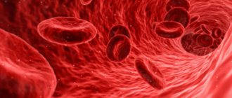
Red blood cells are red blood cells whose main function is to transport oxygen from the lungs to cells and tissues and carbon dioxide in the opposite direction. This role is possible due to its biconcave shape, small size, high concentration and presence of hemoglobin in the cell.
Blood consists of plasma (a transparent liquid of pale yellow color) and cellular, or formed, elements suspended in it - red blood cells, leukocytes and blood platelets - platelets.
Most of the blood contains red blood cells. In a woman, 1 mm sq. blood contains about 4.5 million of these blood cells, and a man has about 5 million. In general, the blood circulating in the human body contains 25 trillion red blood cells - this is an unimaginably large number!
The main function of red blood cells is to carry oxygen from the respiratory system to all cells of the body. At the same time, they also take part in the removal of carbon dioxide (a metabolic product) from tissues. These blood cells transport carbon dioxide to the lungs, where gas exchange replaces it with oxygen.
Unlike other cells in the body, red blood cells do not have a nucleus, meaning they cannot reproduce. It takes about 4 months from the moment new red blood cells appear until they die. Red blood cells have the shape of oval disks pressed in the middle, approximately 0.007-0.008 mm in size and 0.0025 mm in width. There are a lot of them - the red blood cells of one person would cover an area of 2500 square meters.
Decoding
Anisocytosis of red blood cells is determined by a hematology analyzer, which produces a histogram plotting the number of RBCs of different volumes in the sample.
Important information: What do increased segmented or band neutrophils in the child’s blood indicate?
If the analysis is carried out manually, the value of the red blood cell coefficients of variation is calculated from the histogram curve and formula. Manual calculation of parameters is rarely used, because does not provide the necessary accuracy of the result.
A positive test result is when the rate of erythrocyte anisocytosis exceeds the norm. If the result is within the normal range, the test will be negative.
The RDW designation characterizes the heterogeneity of red blood cells:
- RDW-CV in a blood test is a relative value measured as a percentage (%);
- RDW-SD is an absolute parameter measured in volume units of femtoliters (fl) or µm3.
Comparison of the scatter of RBC volumes in the sample is carried out with normal values of 80-100 fl.
Relative index is a value that reflects the distribution of red blood cells, showing by what percentage the red blood cell volumes differ from the average volume.
The value of the absolute indicator indicates how many femtoliters the volumes of red blood cells differ. The calculation is done according to the histogram graph.
The level of relative anisocytosis depends on the value of the MCV parameter (mean blood cell volume). The calculated value of the relative indicator of erythrocyte anisocytosis is obtained by multiplying 100% by the ratio of the standard coefficient to MCV.
The RDW-SD parameter in a blood test allows you to determine how many femtoliters the smallest and largest red blood cells differ from medium-sized red cells.
Normal values
The values of the distribution width of erythrocytes according to norms do not depend on gender. They are the same for an adult of any gender. With age, normal coefficient values do not change.
If heterogeneity does not exceed the norm, it means that the red blood cells are approximately the same size. The norm for absolute anisocytosis in adults is 37-47 fl.
Normal RDW levels for women and men are 11.5%-14.5%.
In women during pregnancy, the normal value of the parameter changes by trimester (%):
- first trimester - 11.7-14.9;
- second trimester - 12.3-14.7;
- third trimester - 11.4-16.6.
In children, the normal level varies. The first 6 months after birth, RDW values are higher than the adult norm. Gradually they decrease.
RDW standards for children (%):
- up to 6 months - 14.9-18.7;
- after six months - 11.6-14.8.
The result of the calculation of relative anisocytosis, which the patient receives in the analysis, is often elevated or normal. A decreased RDW-CV most likely indicates an error.
If the anisocytosis of red blood cells is slightly higher than normal, most often the deviation is random. This value becomes clinically significant for diagnosis when it exceeds 60 fl.
Important information: What are WBC leukocytes in a blood test (deciphering the norm and deviation)
A decrease in the absolute coefficient of anisocytosis has no significance for diagnosis.
Increased level
Red blood cells are heterogeneous in size and hemoglobin saturation. If anisocytosis is higher than normal, it means that there are blood cells in the bloodstream that are different in volume from the normal range.
Normally, each cell goes through a life course, during which its volume changes. Cells of different sizes are simultaneously present in the blood:
- microcytes, characterized by their small sizes, not exceeding 6 microns;
- normocytes corresponding to normal sizes, in the range of 6-8 microns;
- macrocytes, characterized by large sizes exceeding 8 microns;
- megalocytes, with a diameter > 12 microns.
Normal human RBCs fall within the normocyte range on average. An increased value of heterogeneity is possible with the appearance of microcytes and megalocytes.
The heterogeneity of erythrocytes is characterized by the degree of anisocytosis:
- The first degree corresponds to 27-50% of defective cells.
- Anisocytosis of the second degree is characterized by 55-70% of modified blood cells.
- The third degree of anisocytosis is characterized by the presence of more than 75% of such cells. Their sizes deviate from the norm.
- With the fourth degree of anisocytosis, all 100% of the cells are defective.
If the relative width of the distribution of red blood cells by volume is increased, it means that more than 50% of the cells deviate from the norm.
Relative coefficients of erythrocyte heterogeneity may increase when:
- leukocytosis with a sharp increase in the number of leukocytes;
- lack of nutrients necessary for hematopoietic processes (iron, B vitamins);
- microcytic anemia;
- myelodysplastic syndrome;
- hemolytic crisis;
- malignant tumors that metastasize to the bone marrow;
- RBC agglutination;
- poisoning with heavy metals (lead);
- Alzheimer's disease.
The anisotropy coefficient of erythrocytes increases in vascular diseases. Increased RDW in intoxication caused by alcohol abuse.
If the blood was tested after surgery, severe bleeding, heterogeneity above normal may also be noted. In such a situation, the result exceeds the acceptable standard deviations as a result of the increased production of juvenile cell species.
RDW is elevated in iron deficiency anemia, which is common in pregnant women. The RDW indicator for this disease exceeds 14%.
With vitamin B12 deficiency, macrocytes and megalocytes are found in the bloodstream.
If the anisocytosis index reaches 27%, this means that RDW in a blood test may suggest B12 deficiency anemia.
Important information: What does increased red blood cells mean in an adult?
The width of the distribution of red blood cells is increased during processes accompanied by increased destruction of red blood cells. The death of blood cells is caused by:
- increased bilirubin levels;
- an increase in the size of the liver and spleen;
- accumulation of iron ions in the body.
An increase of up to 40% is possible with thalassemia or Cooley's anemia. This disease is hereditary. Cooley's anemia is most widespread in areas affected by malaria.
Increased values correspond to changes in other erythrocyte parameters. It is necessary to take into account the MCV parameter when interpreting analyses. Simultaneous changes in parameters are observed in cases where:
- the RDW indicator is exceeded, the MCV is reduced (with iron deficiency anemia);
- above normal MCV and RWD (may mean liver pathologies, megaloblastic, hemolytic anemia, cold agglutination);
- there is an excess of the RDW norm in combination with the MCV norm (indicates the likelihood of myelofibrosis).
Reduced index
A reduced RDW in the analysis means that in the sample taken for the study, the cells have practically no difference in volume. Cases where the analysis reveals that all RBCs are the same size are most likely in error.
Red blood cells may be of equal size in anemia, leading to an increased number of defective forms in the bloodstream.
The values of erythrocyte distribution indices decrease with:
- anemia (iron deficiency, folate deficiency, B12 deficiency);
- pregnancy (with the appearance of fetal hemoglobin);
- extensive blood loss (trauma, internal bleeding, childbirth).
Hemoglobin
Hemoglobin is a red blood pigment found in red blood cells. The main function of this protein substance is the transport of oxygen and partially carbon dioxide. In addition, antigens are located on the membranes of red blood cells - blood group markers. Hemoglobin consists of two parts: a large protein molecule - globin and a non-protein structure built into it - heme, in the core of which there is an iron ion. In the lungs, iron combines with oxygen, and it is the combination of oxygen with iron that colors the blood red. The combination of hemoglobin with oxygen is unstable. When it breaks down, hemoglobin and free oxygen are again formed, which enters the tissue cells. During this process, the color of hemoglobin changes: arterial (oxygenated) blood is bright red, and “used” venous (carbon dioxide saturated) blood is dark red.