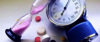
Ocular pressure is the pressure that exists between the contents of the eyeball and its membrane. You can feel it by lightly pressing your finger on the eyelid, but more often patients complain of a feeling of “fullness” in the eye orbits. It is this indicator that determines the usefulness of the functioning of the visual system.
It is an important indicator in identifying dysfunctions of the visual organs and ophthalmological diseases. Depending on the inflammatory processes occurring in the organs of vision, it may be decreased or increased. To prevent its possible deviations, it is important to know the reasons for fluctuations in intraocular pressure.
Intraocular pressure (ocular pressure or IOP)
Ocular hypertension (intraocular pressure) is one of the causes of glaucoma, cataracts and even complete loss of vision. Various factors can increase intraocular pressure (IOP) - from stress and eye fatigue to serious systemic diseases. Read more about this ophthalmological problem below.
intraocular pressure (IOP)
How dangerous is the problem?
The reasons for pressure deviations from the norm are most often associated with various diseases of the visual organ. The most dangerous is glaucoma, complete loss of vision. Therefore, it is not a harmless vision defect.
With a stable increase in pressure, changes occur in the cells and vessels of the fundus of the eye, and metabolic processes change. This poses a great danger to human health. This hides the causes of various diseases that can lead to serious complications: complete loss of vision, irreversible changes in the structure of the visual system.
The role of aqueous humor of the eye and intraocular pressure (IOP)
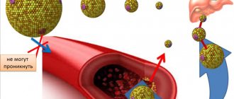
Aqueous humor fills the posterior and anterior eye chambers and is a colorless liquid with a jelly-like consistency. Its chemical composition is close to blood plasma, but with a lower protein content. Aqueous humor is released in the posterior chamber from the epithelial cells of the ciliary, or ciliary, body - a section of the choroid of the eyeball - and then enters the anterior chamber. The human eye produces an average of 3-9 ml of this liquid per day.
Functions of aqueous humor:
• the main task is to maintain intraocular pressure, the level of which depends on the ratio of the volume of moisture entering the chambers and removed;
• immune - due to the content of immunoglobulins, it helps remove infectious agents from the eye;
• nutritional - supplies the cornea, lens, anterior region of the vitreous body and the trabecular meshwork with necessary substances;
• works as a light refractive medium.
Thanks to a certain level of pressure of the internal fluid, the eye has a normal shape and size, its optical system works correctly, and the metabolism in the eyeball is maintained at the proper level.
Reference: intraocular pressure is the pressure exerted by aqueous humor and the vitreous humor on the inner walls of the eye. The normal IOP for people under 40 years of age is 12-20 mmHg. Art., at an older age this figure can reach 25 mm Hg. Art.
When deviating from the norm, the pressure inside the eye can be low (less than 12 mm Hg) or high (more than 25 mm Hg). The second variant of the disorder, otherwise called ophthalmic hypertension, or ocular hypertension, is more common.
Gymnastics for the eyes
Special exercises developed by experienced ophthalmologists help reduce IOP. To achieve a positive result, perform gymnastics at least 3 times a day.
Eye exercises:
- frequent blinking for 1 minute;
- turning the eyes clockwise and counterclockwise - perform with closed and open eyelids;
- transferring the gaze from a distant object to a near one;
- drawing geometric shapes, letters and numbers with the eyes.
Gymnastic exercises not only help a person reduce intraocular pressure, they improve the nutrition of eye tissue. They improve the functioning of the visual system, allowing oxygen and beneficial nutrients to better penetrate the tissues of the eyeball.
In addition, gymnastics prevents the development of refractive errors and accommodation spasms, which cannot be avoided as the body ages.
About ocular hypertension (intraocular pressure): what is it?
With increased intraocular pressure, its value reaches 20 mm Hg. Art. and more, while glaucomatous changes in the fundus are most often not detected - there are no degenerative disorders of the optic nerve (the number of nerve fibers is not reduced, the blood vessels are not deformed).
This is interesting! Swiss scientists have found that monitoring intraocular pressure (IOP) levels can help more accurately assess the risk of stroke.
Eye pressure tablets
In some cases, treatment can occur with the use of tablets prescribed by an ophthalmologist. With their help, you can achieve stabilization of the removal of excess eye fluid and normalize the body's metabolic processes. Most drugs are diuretics due to their high potassium content.
Before starting treatment, it is best to consult a specialist who will select combined treatment methods that will allow you to return your eye health to its previous state.
Anaprilin
Anaprilin is a synthetic drug, produced in the form of tablets of 0.01 and 0.04 g. It reduces the value of IOP, helps the heart function and actively acts as a support for normal blood and vascular pressure.
Hypothiazide
Hypothiazide has strong diuretic properties, due to which large amounts of sodium are removed from the body. The effect is achieved after one and a half hours of using the product.
Prozerin
Prozerin is produced in the form of tablets weighing 0.015 g. In addition to regulating IOP, it helps the functioning of the muscles of the bladder and uterus. Has gentle properties.
Clonidine
Clonidine is available in the form of tablets of 0.15 g. It helps to normalize and reduce blood pressure, reduces the rate of contraction of the heart muscle and stabilizes the IOP value. It is also a sedative used as a sedative.
Forms of increased intraocular pressure
According to clinical manifestations and etiology, ocular hypertension can be essential and symptomatic. Pseudohypertension of the eye is considered separately.
Essential
With this type of ocular hypertension (intraocular pressure), the tone is slightly increased. This is typical for people over 45 years of age and older people. The exact causes of the development of essential ocular hypertension have not been determined. Doctors associate them with age-related changes, in which the ratio of the intensity of production and outflow of intraocular fluid is disrupted.
An essential increase in IOP is characterized by symmetry, as well as moderate severity, which declines over time. Lesions characteristic of glaucoma are not detected.
Symptomatic
This type of increase in IOP develops against the background of pathology of the visual apparatus not associated with glaucoma, for example, keratoiridocyclitis, uveitis, iridocyclitis, etc. For such reasons, they speak of uveal ophthalmic hypertension.
The symptomatic form can also be a complication of severe systemic diseases. It may be caused by:
• inflammatory processes in the brain;
• chronic intoxication with chemicals;
• long-term use of corticosteroid drugs;
• thyrotoxicosis, etc.
This form of high IOP can also occur due to tumor tumors exerting pressure in the neck or head. Over a long period of time, symptomatic ocular hypertension often leads to secondary glaucoma.
Pseudohypertension
This is a short-term increase in pressure inside the eye as a reaction to patient stress before and during tonometry (measuring IOP) or viziometry (testing visual acuity). It is also worth mentioning the transient increase in intraocular pressure. It occurs once against the background of a hypertensive crisis or eye strain, and usually passes quickly.
Treatment
Therapy for high intraocular pressure includes instillation of special eye drops, taking vitamin preparations, physiotherapy (vacuum massage, color pulse therapy) and surgery as a radical way to get rid of the disease. This is done in one of three ways - goniotomy, trabeculectomy and laser surgery. One of the treatment measures is compliance with preventive recommendations.
Eye drops
The choice of medication is made by an ophthalmologist depending on the intraocular pressure, the patient’s age, and the presence of concomitant diseases.
With low intraocular pressure, dry mucous membranes are observed, so drops are prescribed to moisturize and eliminate discomfort (Oftolik, Visin, Korneregel, Balarpan).
In case of increased intraocular pressure, beta-blockers or antiglaucoma drugs are used to stabilize the indicator. The most prescribed medications: Timolol, Betaxolol, Pilocarpine, Latanoprost and their combinations.
For hemorrhage in the sclera, which manifests itself with increased IOP, methylethylpyridinol (Emoxipin, Emoxy-optik, Emoxibel and other analogues) is prescribed.
If the deviation in indicators is caused by an infectious-inflammatory process in the organ of vision, antibacterial and anti-inflammatory drugs are used on the recommendation of a doctor.
Physiotherapeutic treatment
The most effective for deviations of intraocular pressure from the norm are 2 types of physiotherapy: vacuum massage and color pulse therapy. They are carried out in a day or night hospital setting.
Surgical intervention
Laser surgery is a radical and relatively safe way to reduce ophthalmotonus. With the help of the beam, pathways for the outflow of aqueous humor are opened. This corrects the imbalance between the production and outflow of intraocular fluid. Stabilizes blood pressure.
Gymnastics for the eyes
Gymnastic exercises and wearing special glasses - simulators - have a beneficial effect on IOP indicators.
They are aimed at relaxing the eyes, relieving tension, improving vision and focus, and strengthening the eye muscles. They should be performed daily, especially with prolonged visual stress, after 50 years and in the presence of risk factors for the development of ophthalmotonus.
Vitamin preparations
To maintain visual acuity, reduce discomfort and signs of eye fatigue due to excessive visual stress, prevent the occurrence of eye diseases, and normalize blood circulation, multivitamin complexes are used. The following drugs and dietary supplements are available in pharmacies:
- Makulin and Makulin plus;
- Taufon tabs;
- Vitrum Vision;
- Complivit ophthalmo;
- Visionace;
- Blueberry forte with lutein;
- Lutein complex;
- Myrtilene;
- Superoptik;
- Omega-3;
- Strix.
They are used in courses twice a year, especially after operations, as prescribed by a doctor.
Other medicines
Often, IOP deviations are treated by a group of specialists depending on the provoking disease: a neurologist, endocrinologist, cardiologist, gynecologist, etc. Medicines are prescribed to eliminate the cause.
Causes of increased intraocular pressure
causes of increased intraocular pressure
Factors causing high IOP are different - from physiological to pathological. For example, short-term ocular hypertension can be caused by eye fatigue when working at a computer for a long time, or mental fatigue. At the same time, not only the pressure in the vessels of the eyeballs increases, but also the intracranial pressure. A similar situation is observed with severe stress and violent expression of emotions.
The main cause of persistent ocular hypertension is glaucoma. In most cases, it develops in the second half of life, sometimes it is congenital (the so-called hydrophthalmos of the eye, or hydrophthalmos in newborns). With glaucomatous lesions, intraocular pressure is constantly increased; crises with a sharp increase in ophthalmotonus can be observed in one eye - on the left or on the right.
Another common pathological cause of ocular hypertension is Graves' disease, or toxic diffuse goiter, in which there is excessive production of thyroid hormones. The main category of patients are women under the age of 40-45 years. With this disease, blood pressure and heart rate are increased.
Attention! Graves' disease begins unnoticed, and the symptoms of its first stage - insomnia, hand tremors, sweating, slight weight loss and mood swings - are attributed by many to overwork and stress.
Other hormonal causes of ophthalmic hypertension include Itsenko-Cushing syndrome, hypothyroidism, and pronounced menopause in women.
The development of ocular hypertension (intraocular pressure) can be a consequence of chemical or drug intoxication. For example, with long-term treatment with glucocorticosteroids (Hydrocortisone, Dexamethasone, Prednisolone, etc.), the production of prostaglandins, mediators of pain and inflammation, is inhibited. But they also improve the outflow of aqueous humor from the chambers of the eye and reduce IOP.
This is why corticosteroids, regardless of their form, can cause ocular hypertension. It develops over 2-3 years with systemic use (tablets, injections), after 5-8 weeks with local use of hormonal ointments for the eyes. There are known cases of a sharp increase in intraocular pressure 2-3 hours after intensive corticosteroid therapy.
IOP can also be increased by chemicals - furfural, tetraethyl lead, drugs with sanguinarine.
In addition to the previously mentioned inflammatory ophthalmological diseases that cause high IOP (uveitis, iritis, etc.), eye injuries can also increase it. This is due to swelling, stagnation of fluid and blood.
Also, ocular hypertension can be provoked by diseases accompanied by fluid retention in the body - heart failure, kidney disease.
Click here - all materials on the topic Pressure
All portal materials about Pressure follow the link to the photo
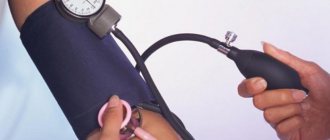
How to measure and lower eye pressure
Eye pressure is measured by the most well-known method - palpation. When palpating a closed eye, it is discovered that if the organ is normal, it is quite elastic and has a strict round shape. If the pressure is high, any touch can cause pain, and the shape will be very hard. When IOP is less than normal, the ball of the eye will dent quite strongly when touched.
In cases where eye pressure is outside the normal range, a tonometric examination is prescribed. Their working methods are quite difficult to explain. But they show the exact units of measurement of the fundus pressure condition.
To prevent eye pressure, doctors recommend first of all balancing your diet. The use of food additives – salt, sugar and other carbohydrates – is minimized. They should be replaced with nuts and dried fruits, as well as foods high in protein. It is necessary to add vitamins E and beta-carotene to the diet. On the contrary, bad habits need to be reduced. Among them, the most dangerous are: prolonged work at the computer, smoking, alcohol and eating fatty foods.
In addition, patients take medications in the form of drops and tablets for IOP. We will talk about pharmacological drugs below.
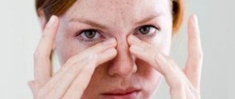
Symptoms of intraocular pressure
intraocular pressure symptoms
When intraocular pressure is slightly increased, symptoms may be completely absent, and the problem can only be determined at an appointment with an ophthalmologist.
Common complaints of patients with moderate and low ocular hypertension:
• eyes get tired and red quickly;
• headaches often occur in the temple area;
• discomfort is felt when working in front of a monitor, reading a book, or in a room with dim light;
• bothered by pain in the eyes, which is often combined with a headache.
In most cases, symptoms of ocular hypertension occur in both eyes. In glaucomocyclic crisis, only one side is always affected.
If the increase in IOP is persistent and caused by glaucoma, then the symptoms are as follows:
• visual fields are narrowed - a person sees poorly “out of the corner of the eye”;
• headaches are unilateral, migraine in nature;
• rainbow circles, blurring, and flickering of spots appear before the eyes;
• vision deteriorates noticeably, especially at dusk;
• there is pain in the eye.
Attention! An acute attack of glaucoma requires immediate medical attention! It is accompanied by severe pain in the eye, nausea, dizziness and decreased visual acuity. In this case, IOP increases to 60 mm Hg. Art. and higher.
With traumatic ocular hypertension, the patient complains of fog before the eyes, a sensation of a foreign body and pain in the eye, and less often of nausea and vomiting. These signs usually develop 3-6 hours after the injury.
The uveal form of increased pressure inside the eye (with inflammation of the tissues of the eye) is manifested by lacrimation, redness of the whites, eye irritation, and photosensitivity.
Symptoms of increased IOP
These signs need to be known in order to quickly contact a doctor and diagnose the pathology, especially if a person has close relatives with glaucoma:
- frequent headaches that cannot be relieved with medications;
- reduced clarity of vision when moving from a lighted room to a dark one;
- tearfulness and rapid eye fatigue under any visual load;
- unpleasant sensations, discomfort and severe fatigue in the evening;
- redness of the whites of the eyes due to burst capillaries;
- discomfort when moving the eyeballs to the sides and up;
- disturbing pain when looking up, manifesting itself in the evening in the temples and above the eyebrows;
- After a night's sleep during the period of awakening, vision becomes blurred; it takes time to improve its quality.
These are the main symptoms of increased IOP. If one or a number of them have been bothering you for a long time, systematically, you need to contact an ophthalmologist for a thorough examination of the organ of vision and diagnosing the cause of the concern.
Diagnosis of elevated IOP
There are several methods for determining intraocular pressure.
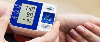
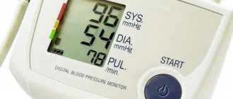
Tonometry is performed under local anesthesia, since the tonometer probe is in contact with the cornea. Proximetacaine eye drops are used for pain relief. There are many types of tonometry - electronic identification, contour dynamic, Goldman, etc. The gold standard for measuring IOP is the use of a Goldman tonometer. The resulting intraocular pressure values are measured in mm Hg. Art.
Gonioscopy is a visual examination of the anterior chamber of the eye, namely the angle between the iris and the cornea, using a goniolens and a slit lamp. After local anesthesia of the eye, the lens is applied to the surface of the eyeball. The lamp is then adjusted and the goniolens is rotated to visualize all areas of the anterior chamber angle.
By the way: in terms of intraocular pressure, gonioscopy evaluates the width of the iridocorneal angle. If it is narrowed, it impedes the outflow of fluid, increasing IOP and the risk of developing angle-closure glaucoma.
Tonography is a functional method similar to tonometry, but longer. Shows the processes of production and drainage of aqueous humor, and graphically records the pressure inside the eye.
Symptomatic ocular hypertension in Posner-Schlossmann disease can only be determined using eye biomicroscopy. This method reveals subtle swelling of the cornea and sediment of macrophages, lymphocytes and other cells on it (precipitates).
Causes of eyeball hypertension
Glaucoma concept
Glaucoma is a multifactorial pathology. There are many reasons, the one-time or single impact of which contributes to the appearance of pathology.
Causes of the disease
- Hereditary predisposition is the leading factor leading to the development of the disease. The probability of developing glaucoma in people aged 35-45 years, born from sick parents, is close to 25%.
- Systematic stress loads.
- Eye injuries.
- Surgical interventions . The most dangerous operation is to replace the lens.
- Anatomical defects of the vascular apparatus of the eyeball . In the presence of atherosclerosis or a change in the coagulation properties of blood towards hypercoagulation, the incidence of glaucoma increases several times.
- Diseases of the heart and vascular bed (hypertension, arrhythmias of any nature).
- Metabolic disorders – diabetes mellitus, thyroiditis.
Symptoms of diabetes
Possible complications
Consequences of prolonged increase in IOP:
• retinal detachment, leading to visual impairment and requiring surgical treatment;
• ulcerative lesions of the cornea or its thickening;
• posterior capsular cataract;
• ptosis, atrophy of eyelid tissue.
Left unattended, the symptomatic form of ocular hypertension is complicated by secondary glaucoma. The second consequence, also related to the first, is atrophy of the optic nerve, which leads first to partial and then to complete loss of visual function.
A little about ocular hypertension and glaucoma
Increased eye pressure not caused by glaucoma is called ocular hypertension. The exact cause of the development of this syndrome cannot be determined at the moment. However, it has been proven that the main reasons for changes in pressure fluctuations inside the eye are: nervous and physical stress, hereditary and age factors, diseases of the cardiovascular system.
The condition of ocular hypertension is conventionally divided into symptomatic and pseudohypertension. The first develops when various eye diseases are not treated in a timely manner, when high blood pressure becomes the norm. Pseudohypertension is a rare phenomenon. Caused by incorrect patient behavior during measurement or any technical errors.
The development of persistent glaucoma is accompanied by prolonged high eye pressure. Treating the disease is long and difficult. Accompanied by a narrowing of the angle of vision, a decrease in acuity, and pain in the visual system.
- Angle-closure glaucoma - compresses the tubules that provide fluid removal in the drainage system of the eye.
- Open-angle glaucoma - there is a violation of the drainage system of the eye due to deterioration in functioning. The course of the disease may not bother a person and may be detected in the later stages.
With glaucoma, IOP is constantly elevated, which negatively affects eye function and leads to irreversible processes: optic nerve atrophy, retinal detachment, and complete loss of vision.
Treatment methods for elevated IOP
Drug therapy for ocular hypertension is based on the use of antihypertensive drugs, i.e., those that reduce blood pressure. First of all, one drug from the group of beta-blockers is prescribed in the form of eye drops (Betaxolol, Timolol, etc.). If there is no effect, they resort to a combination of two such drugs or combine it with other drugs - carbonic anhydrase inhibitors (Brinzolamide, Dorzolamide, etc.) or cholinomimetics (Aceclidine, Pilocarpine, Acetylcholine, etc.).
Important! Self-medication of ocular hypertension is unacceptable. The treatment regimen should be prescribed by an ophthalmologist after a thorough examination.
Along with reducing intraocular pressure, the factor that caused its increase is eliminated, be it a disease, injury or excessive strain on the eyes. With IOP reaching 70 mm Hg. Art., the use of Glycerol and Acetazolamide is indicated. The doctor may also prescribe injections of lytic solution, urea, or Mannitol.
If medications do not help or it is necessary to remove a mechanical obstruction to the outflow of fluid from the chambers of the eye, then surgical intervention or laser iridectomy is resorted to.
Drops for eye pressure, list of effective remedies
Modern methods of pharmacology and constant scientific discoveries in the field of medicine have been able to create drops and tablets that help reduce high or increase low pressure in the fundus of the eye, the risk of developing cataracts or glaucoma. Below is a list of well-known and popular drops for eye pressure, using which you can restore healthy vision.
Azarga
Azarga eye drops are used for glaucoma and to reduce high intraocular pressure, as one of the main agents with hypertensive properties.
The drug belongs to the combination of beta-blockers, which help reduce the production of intraocular fluid.
By reducing the formation of bicarbonate ions, the effect of flow processes that affect the course of open-angle glaucoma is reduced.
Arutimol
Arutimol is a gentle remedy that has no contraindications for use. Despite the fact that the medicine prevents the development of glaucoma, it is used even in the most severe cases. The result is achieved only with prolonged use of the product. The active agent is Timolol.
Betoptik
Betoptik is a powerful means of preventing the formation of large amounts of fluid in tissues, leading to an increase in IOP. Due to the characteristics of the active ingredients, the clouding of the pupil disappears in the complete absence of tissue contraction. Prescribed for mild glaucoma. The effect of the medication is achieved after the first hour of administration.
The substance is used twice a day. Side effects include severe lacrimation, a negative reaction to light and redness of the cornea. In some cases, nervous tremors and depression may occur.
Timolol
It is used when external action of adrenergic blockers is necessary. The active ingredient is timolol, which allows you to minimize the amount of aqueous fluid in the organ. It is used to reduce eye pressure when glaucoma is actively developing and fundus pressure is greatly increased. If the drug is used as a health precaution, it is best to stop taking it, since timolol can reduce the normal blood pressure value.
Xalatan
The active ingredient of Xalatan is latanoprost, whose work is aimed at reducing fundus pressure. Xalatan is used for open-angle glaucoma. Has no side effects. The medicine begins to act after the first couple of hours and continues throughout the day. One drop is administered per eye at night.
Xalacom
The active ingredients of Xalacom are latanoprost and timolol. Due to the combination of strong active ingredients, the effect of the drops is achieved quite quickly. Within 6 hours, the increased IOP value decreases.
Okumol
It is used to reduce eye pressure, but the full effect of Okumol is achieved only after two weeks. However, due to the association of the drug with the group of beta-blockers, there are a number of restrictions on its use:
- Contraindicated for persons with pulmonary diseases;
- For heart failure;
- Pregnant and nursing mothers are prescribed the drug only in case of special consultation with the supervising physician.
Okumed
A beta blocker helps stabilize the condition of high eye pressure by reducing the outflow of fluid. Does not affect the shape and size of the pupil in any way. Okumed begins to act 20 minutes after administration. Inserted into the conjunctival sac. A decrease in eye pressure begins to occur within an hour, thanks to timolol and sodium hydrogen phosphate.
Kosopt
The active agent of Kosopta is timolol and dorzalamide, which, as in other cases, reduces the amount of intraocular fluid, which in some cases causes an increase in IOP.
Fotil
The drug Fotil is a combined beta-blocker that causes active blood formation and blood circulation in the retina. The advantages include rapid removal of the drug from the body and a decrease in fluid production, resulting in a decrease in eye pressure.
Travatan
Travatan is used in cases where serious intervention is required from medicinal methods of influencing fundus pressure. After use, the outflow of fluid is significantly reduced, which leads to improved eye health. Thanks to the use of this product, a significant improvement in IOP stabilization is achieved.
Conservative methods of treating intraocular pressure
ocular pressure (IOP)
The goal of therapy for elevated intraocular pressure is to eliminate the causes that provoked the occurrence of this condition. Treatment methods for high blood pressure may differ, depending on the advanced stage of the disease.
If high eye pressure is diagnosed, conservative therapy is prescribed at the initial stage:
· performing special gymnastic exercises;
· using eye drops to improve fluid outflow;
· reducing time spent watching TV and working with the computer;
· Wearing special protective glasses.
In addition, it is necessary to increase the time spent outdoors and give up strength and contact sports for a while.
If increased intraocular pressure manifests itself as a sign of some other disease, you need to start complex therapy.
Causes of high blood pressure
There are many factors that adversely affect the human body. Before carrying out therapy, the ophthalmologist determines the cause of IOP. Modern medicine identifies several main root causes:
- heavy mental and physical stress;
- hypertension;
- stressful situations;
- heart failure;
- kidney diseases;
- endocrine system disorders;
- hereditary factor;
- age-related changes;
- anatomical changes in the organs of vision;
- chemical poisoning.
If the reason for the change in the indicator of increased eye pressure is associated with the above ailments, then they do not treat the eyes, but rather therapy that eliminates the pathology that caused this disorder.
Use of folk remedies for treatment
If the patient prefers alternative medicine, it is recommended to use the following remedies:
Golden mustache in the form of tincture. Fill the antennae (20 pieces) with vodka (0.5 l), place them in a dark, cool place for 12 days. Take 1 teaspoon daily on an empty stomach, after straining.
Infusion of meadow clover. Pour dry grass (1 tbsp) with hot water (150 ml). We insist, strain and take every day at night for a month.
You can drink kefir every evening, adding a pinch of cinnamon to it first.
Traditional medicine recipes
There are many recipes made from natural ingredients that help relieve high eye pressure. Treatment allows you to reduce the indicators to normal and does not allow them to rise for a long time. Traditional medicine recipes are not capable of radically changing eye pressure, but it is possible to alleviate the patient’s condition by using them.
Clover decoction - brew the plant as tea, leave for a couple of hours, take half a glass at night.
Tincture of golden mustache vodka - pour seventeen crushed purple mustache rings with half a liter of vodka, leave for twelve days in a dark place. During the infusion process, shake the container with liquid every three days. Take a tablespoon in the morning thirty minutes before meals.
An equally effective remedy is ordinary kefir. Taking one glass of delicious fermented milk product daily helps normalize blood pressure, and a pinch of cinnamon added to the drink enhances the healing effect.
If you have IOP, it is recommended to engage in physical activity and move more. Gymnastics for the eyes and neck massage are useful. Often traditional methods are only auxiliary. Therefore, visiting an ophthalmologist for eye pressure is mandatory.
How to treat intraocular pressure at home
Treating Intraocular Pressure
Eye pressure can be reduced at home. To do this, it is recommended to follow these simple steps:
1. Use a high pillow to sleep.
2. To reduce visual stress, it is necessary to use sufficient lighting when working.
3. Avoid visual overload and take breaks during work to allow your eyes to rest. Clothes should have a loose collar, and the tie should not be too tight, as this will compress the blood vessels in the neck.
4. Avoid heavy lifting. A large load will negatively affect the blood vessels.
5. Quit alcohol and smoking.
6. Give up coffee and tea.
7. Avoid stress.
When performing any work, you should try to stand with your head raised.
To do this, it is recommended to follow these rules:
- when washing floors you need to use a mop;
- when weeding, you need to use a hoe with a long handle;
- when rinsing clothes by hand, the container with water should be placed on a hill;
- When putting on shoes, use special shoe spatulas.
It is useful to use dill, grapes, watermelon, birch sap, currants, and pumpkin in the treatment of high intraocular pressure.
Forecast
If the increase in ophthalmotonus is caused by fatigue and the patient quickly took the necessary measures, then the prognosis is usually positive. In the absence of any action, the disease will develop and glaucoma may appear. Patients with this diagnosis have a 75-80% chance of recovery if they go to the hospital in a timely manner. In other cases, complete blindness often occurs in one or two eyes at once.
Treatment at home for eye pressure is possible, but only under the supervision of a doctor and after a detailed examination. After completing the course of therapy, you should definitely see an ophthalmologist.
He will check the ophthalmotonus and, if necessary, recommend other methods to solve the problem.
Methods for preventing IOP
eye pressure symptoms and treatment
Violation of intraocular pressure is fraught with consequences (glaucoma and vision loss).
To prevent such conditions, it is recommended to adhere to the following rules:
1. If you have to work at the computer for a long time, you need to take regular breaks, lightly massage your eyes and perform special gymnastic exercises. You can also just sit for a while with your eyes closed and take a break from work for 2-3 minutes.
2. You need to take special vitamins for the eyes, eat blueberries, carrots, and fish. The diet should be as varied as possible. At the same time, it is recommended to exclude unhealthy foods from the diet: fatty foods, fast food, salty and spicy foods.
3. Requires daily walks outside.
4. You need to do exercises. This will help get rid of many health problems, including high intraocular pressure
5. You should regularly consult with an ophthalmologist, at least once a year. If you are prone to ophthalmic diseases, you should undergo an examination once every 6 months.
With the right approach, you can prevent problems associated with intraocular pressure in advance. You should not ignore the first symptoms of the disease; you must consult a specialist in a timely manner and begin treatment as early as possible.
Prevention
It is better to prevent any disease in time than to treat it for a long time. One of the preventive measures is, first of all, regular visits to an ophthalmologist, who will measure eye pressure.
The main ways to prevent deviations in eye pressure:
- Daily exercise for the eyes.
- Regular exercise.
- Quality rest.
- Complete nutrition.
- Taking vitamin complexes.
- It is necessary to rest your eyes and not strain your eyesight too much.
- Moderate consumption of drinks with high caffeine content.
- Complete abstinence from alcohol.
Glaucoma Hypertension Conjunctivitis Eye keratitis: photos, symptoms and treatment Optic nerve atrophy Blepharitis
Treatment of low ophthalmotonus
If there is a violation of the integrity of the membranes of the eye, then the first stage of treatment will be to achieve restoration of the tightness of the eyeball. If the human body has undergone significant loss of vitreous mass, then its substitutes (silicone or hyalon) are introduced. To correct blood pressure, the following groups of drugs are prescribed.
- Vasodilators are necessary to increase blood flow in the aqueous humor-secreting apparatus of the eye. Xanthinol nicotinate and euphilin are used.
- Drugs that increase microcirculation . Needed to restore atrophied structures and prevent degenerative changes in the future. Pentokifylline and rheopolyglucin have proven themselves to be excellent.
- Cycloplegic mydriatics - increase tension in the intraocular muscles, contributing to an increase in intraocular pressure. The only drug from this group is atropine sulfate, administered subconjunctivally.
Dynamics of pharmacological effects of cycloplegic mydriatics
If ineffective or as an addition to drug therapy, laser stimulation of the ciliary body may be prescribed. After exposure to a laser beam, the formation increases the secretion of intraocular fluid.
Acute attack of glaucoma
The disease is recurrent. Long periods of a slight increase in intraocular pressure are accompanied by attacks with sharp jumps. This episode is an emergency and requires emergency care. In the absence of timely and adequate medical support, absolute loss of vision will occur. An acute attack usually develops under the influence of causative factors:
- fatigue;
- overstrain (physical and psycho-emotional);
- long stay in a dark environment;
- intake of significant volumes of water.
Acute attack of glaucoma
The attack is accompanied by severe pain. Pain occurs not only in the area of the eyeballs, but also in the occipital areas. Vision becomes blurred, and when looking at bright light sources, white circles are visible before the eyes. During a severe attack, vomiting, nausea, and sharp pain in the abdomen or chest may occur, which allows doctors to suspect the presence of a myocardial infarction or problems with the gastrointestinal tract.
Pathogenesis of glaucoma development
The key stages of progression of this pathology in ocular hypertension can be presented as follows.
The main cause of vision loss in glaucoma
- Violation of excretion . The deviation is due to many reasons: from an increase in the muscle tone of the trabecular tubules in the presence of stress to anatomical defects.
- Pressure rise above standard values.
- Reduced blood supply to the tissues of the ocular apparatus due to compression of the supply arteries.
- Hypoxia (decreased nutrition) of the components of the optic nerve, gradually turning into ischemia (lack of nutrition).
- Ischemia in the stage of decompensation . An additional negative factor is compression of the optic nerve head by intraocular formations, the volume of which tends to increase.
- Irreversible dystrophic and destructive processes in nervous tissue.
- The occurrence of neuropathy (manifested by a complex of visual abnormalities) and complete loss of the optic nerve (leads to an absolute absence of vision).
Glaucoma is a multifactorial multifactorial disease
Taking into account the features described above, we can conclude that treatment of the disease should be aimed exclusively at achieving target pressure levels , which helps prevent the development of pathology and create conditions for the restoration of nerve cells that are in parabiosis (temporary death).