© Author: Ilya Vitalievich Soldatenkov, general practitioner, especially for SosudInfo.ru (about the authors)
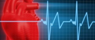
Atrial septal defect (ASD) is a congenital heart defect caused by the presence of a hole in the septum separating the atrial chambers. Through this opening, blood is discharged from left to right under a relatively small pressure gradient. Sick children do not tolerate physical activity well and their heart rhythm is disturbed. The disease manifests itself as shortness of breath, weakness, tachycardia, the presence of a “heart hump” and the lag of sick children in physical development from healthy ones. Treatment of ASD is surgical, consisting of radical or palliative operations.
Small ASDs are often asymptomatic and discovered incidentally during cardiac ultrasound. They heal on their own and do not cause severe complications. Medium and large defects contribute to the mixing of two types of blood and the appearance of the main symptoms in children - cyanosis, shortness of breath, heart pain. In such cases, treatment is only surgical.
ASDs are divided into:
- Primary and secondary,
- Combined,
- Single and multiple,
- Complete absence of a septum.
According to localization, ASDs are divided into: central, upper, lower, posterior, anterior. There are perimembranous, muscular, outflow, and inflow types of ASD.
Depending on the size of the ASD opening, there are:
- Large - early detection, severe symptoms;
- Average - found in adolescents and adults;
- Small - asymptomatic.
ASD can occur alone or be combined with other congenital heart defects: VSD, coarctation of the aorta, patent ductus arteriosus.
Causes
ASD is a hereditary disease, the severity of which depends on genetic predisposition and the influence of adverse environmental factors on the fetus. The main reason for the formation of ASD is a violation of the development of the heart in the early stages of embryogenesis. This usually occurs in the first trimester of pregnancy. Normally, the fetal heart consists of several parts, which during their development are correctly compared and adequately connected to each other. If this process is disrupted, a defect remains in the interatrial septum.
Factors contributing to the development of pathology:
- Ecology - chemical, physical and biological mutagens,
- Heredity - point changes in a gene or changes in chromosomes,
- Viral diseases contracted during pregnancy,
- Diabetes mellitus in mother
- Use of medications by a pregnant woman
- Alcoholism and drug addiction of the mother,
- Irradiation,
- Industrial hazards,
- Toxicoses of pregnancy,
- The father's age is more than 45 years, the mother's age is more than 35 years.
In most cases, ASD is combined with Down's disease, renal anomalies, and cleft lip.
Atrial septal defect in newborns
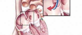
Interventricular and interatrial defects are forms of pathology. In the fetus, lung tissue normally does not yet participate in the process of enriching the blood with oxygen. For this purpose, there is an oval window through which it flows into the left side of the heart.
He will need to live with this feature until birth. Then it gradually closes, and by the age of 3 years it is completely overgrown. The septum becomes complete and does not differ from the structure of adults. In some children, depending on their developmental characteristics, the process lasts up to 5 years.
Normally, in a newborn baby, the oval window does not exceed 5 mm in size. If a larger diameter is present, the doctor should suspect ASD.
Features of hemodynamics
Intrauterine development of the fetus has many features. One of them is placental circulation. The presence of an ASD does not impair the function of the fetal heart in the prenatal period. After the baby is born, the opening closes spontaneously and blood begins to circulate through the lungs. If this does not happen, the blood is dumped from left to right, the right chambers are overloaded and hypertrophied.
With large ASDs, hemodynamics change on the 3rd day after birth. The pulmonary vessels become congested, the right chambers of the heart become congested and hypertrophied, blood pressure rises, pulmonary blood flow increases, and pulmonary hypertension develops. Congestion in the lungs and infection lead to the development of edema and pneumonia.
Then the pulmonary vessels spasm, and a transitional stage of pulmonary hypertension occurs. The child’s condition improves, he becomes active and stops getting sick. The stabilization period is the best time to perform radical surgery. If this is not done in time, the pulmonary vessels begin to become irreversibly sclerotic.
In patients, the ventricles of the heart hypertrophy compensatoryly, the walls of the arteries and arterioles thicken, they become dense and inelastic. Gradually, the pressure in the ventricles equalizes, and venous blood begins to flow from the right ventricle to the left. Newborns develop a serious condition - arterial hypoxemia with characteristic clinical signs. At the beginning of the disease, venoarterial discharge occurs transiently with straining, coughing, and physical activity, and then becomes persistent and is accompanied by shortness of breath and cyanosis at rest.
Types of ASD
Congenital heart disease in the form of atrial septal defect has several varieties. Based on the size of the hole there are:
- small hole - a defect that does not harm the functioning of the heart and is asymptomatic;
- the average hole size is more often detected in adolescents and adults;
- a major defect has obvious signs and is diagnosed immediately after birth.
Atrial septal defect has the following classification according to its nature:
- primary can reach 5 cm and occurs due to underdevelopment of the primary atrial septum;
- secondary means underdevelopment of the secondary septum and usually has a hole of 1 - 2 cm;
- the septum is completely absent, this defect is also called a three-chambered heart - 2 ventricles and one common atrium.
Depending on the location, the defect can be:
- central;
- top;
- bottom;
- rear;
- in front of him.
The intrauterine blood flow of the fetus does not affect the lungs, but goes only through the heart. The passage between the atria is necessary for the normal functioning of the unborn child. This physiological opening is called the oval window. Once the newborn baby has established circulation through the lungs, the foramen ovale closes. It is not classified as a congenital heart defect.
Isolated ASD ranks second in frequency after VSD (ventricular septal defect), but in complex defects its frequency is up to 50%. Another feature of it is that in most cases it is accompanied by other developmental defects. This could be a cleft lip, congenital kidney defects, or Down syndrome.
Symptoms
Small ASDs often do not have a specific clinical picture and do not cause health problems in children. Newborns may experience transient cyanosis when crying and fussing. Symptoms of the pathology usually appear in older childhood. Most children lead an active lifestyle for a long time, but with age they begin to worry about shortness of breath, fatigue and weakness.
Medium and large ASDs manifest themselves clinically in the first months of a child’s life. In children, the skin turns pale, the heart rate increases, cyanosis and shortness of breath occur even at rest. They eat poorly, often lift from the breast to take a breath, choke during feeding, and remain hungry and restless. A sick child lags behind his peers in physical development and has virtually no weight gain.
cyanosis in a child with a defect and fingers like “drumsticks” in an adult with ASD
Having reached 3-4 years of age, children with heart failure complain of cardialgia, frequent nosebleeds, dizziness, fainting, acrocyanosis, shortness of breath at rest, palpitations, intolerance to physical labor. Subsequently, they develop atrial rhythm disturbances. In children, the phalanges of the fingers are deformed and take on the appearance of “drumsticks”, and the nails – “watch glasses”. When conducting a diagnostic examination of patients, the following are revealed: pronounced “heart hump”, tachycardia, systolic murmur, hepatosplenomegaly, congestive wheezing in the lungs. Children with ASD often suffer from respiratory diseases: recurrent inflammation of the bronchi or lungs.
In adults, similar symptoms of the disease are more distinct and varied, which is associated with the increased load on the heart muscle and lungs over the years of illness.
Modern methods of therapy
Most often, to eliminate the pathology it is necessary to resort to surgery. However, if the hole is characterized by a small diameter, then there is still a possibility of it closing on its own. To alleviate the general condition, the baby needs to take diuretics, cardio- and inotropic drugs. Thanks to them, the body is given the necessary level of support.
You should try to protect your baby as much as possible from any excessive physical activity. Parents must do everything to ensure that he does not cry or strain too much. In such a situation, tension increases, which can negatively affect the functioning of the heart muscle. For an older child, lifting heavy objects or moving suddenly is not acceptable.
Complications
Eisenmenger syndrome is a manifestation of long-term pulmonary hypertension, accompanied by right-to-left shunting. Patients develop persistent cyanosis and polyglobulia.- Infective endocarditis is an inflammation of the inner lining and valves of the heart caused by a bacterial infection. The disease develops as a result of injury to the endocardium by a large stream of blood against the background of infectious processes existing in the body: caries, purulent dermatitis. Bacteria from the source of infection enter the systemic circulation, colonize the inside of the heart and the interatrial septum.
- Stroke often complicates the course of congenital heart disease. Microemboli come out of the veins of the lower extremities, migrate through the existing defect and clog the vessels of the brain.
- Arrhythmias: atrial fibrillation and flutter, extrasystole, atrial tachycardia.
- Pulmonary hypertension.
- Myocardial ischemia is an insufficient supply of oxygen to the heart muscle.
- Rheumatism.
- Secondary bacterial pneumonia.
- Acute heart failure develops as a result of the inability of the right ventricle to perform its pumping function. Stagnation of blood in the abdominal cavity is manifested by bloating, loss of appetite, nausea, and vomiting.
- High mortality - without treatment, according to statistics, only 50% of patients with ASD survive to 40 - 50 years of age.
How does the disease manifest itself?
In most cases, no way. Signs are detected when the phenomenon is advanced, when secondary deviations occur in the functioning of other anatomical structures of the heart.
An approximate list of symptoms in this case:
- Chest pain of varying degrees of intensity. From minor tingling to unbearable attacks, reminiscent of angina pectoris in nature. The duration of each episode is no more than 5-15 minutes. Passes without a trace. As the underlying disease progresses, relapses become more frequent.
- Arrhythmias of various types. From simple paroxysmal tachycardia, when the heart rate reaches 150-200 beats per minute to others. The ventricular form is especially life-threatening. Other possible options are extrasystole, atrial fibrillation. As the process moves forward, it becomes more and more difficult to restore the normal rhythm. Moreover, the patient stops paying attention to the manifestations, which complicates the diagnosis at the initial appointment. Everything is solved using routine methods.
- Discomfort in the chest. The symptom is directly related to the previous one. A sharp beat, heart flutters, skipping iterations, unequal time intervals between contractions. Here are the possible options.
- Shortness of breath and other disturbances of normal gas exchange. It occurs spontaneously, sometimes in a state of complete rest. In the early stages of anatomical abnormalities only with intense physical activity. It is difficult to catch the process from the first moments, perhaps through load tests.
- Cyanosis of the nasolabial triangle. Indicates tissue ischemia. Characterized by a bluish discoloration of the area around the mouth. A calling card for pathologies of the cardiovascular system in general. Nonspecific sign.
- Pallor of the skin. The dermal layer becomes like marble. This is most often noticeable in patients with weak pigmentation.
- Increased sweating, especially at night.
- Advanced cardiac pathologies are characterized by hemoptysis. This is a relatively rare sign; it is necessary to differentiate processes of cardiac origin from tuberculosis and lung cancer.
- Dizziness of unknown origin.
- Cephalgia.
- Nausea and vomiting. They are reflexive in nature. Therefore, relief does not come after episodes as with poisoning.
Signs are nonspecific and can indicate a wide range of pathologies, which are determined by dozens of conditions.
Aneurysm of the interatrial septum in adults manifests itself more clearly, especially if it is caused by congenital defects.
Diagnostics
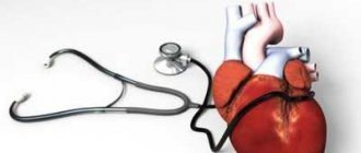
Diagnosis of ASD includes a conversation with the patient, a general visual examination, instrumental and laboratory research methods.
Upon examination, specialists detect a “heart hump” and malnutrition of the child. The heartbeat shifts down and to the left, it becomes tense and more noticeable. With the help of auscultation, a splitting of the second tone and an accent of the pulmonary component, moderate and soft systolic murmur, and weakened breathing are detected.
The results of instrumental research methods help to make a diagnosis of ASD:
- Electrocardiography reflects signs of hypertrophy of the right chambers of the heart, as well as conduction disturbances;
- Phonocardiography confirms auscultation data and allows you to record the sounds produced by the heart;
- X-ray - characteristic change in the shape and size of the heart, excess fluid in the lungs;
- Echocardiography provides detailed information about the nature of cardiac anomalies and intracardiac hemodynamics, detects ASD, establishes its location, quantity and size, determines characteristic hemodynamic disturbances, assesses the functioning of the myocardium, its condition and cardiac conductivity;
- Catheterization of the heart chambers is carried out to measure the pressure in the chambers of the heart and large vessels;
- Angiocardiography, ventriculography, venography, pulse oximetry and MRI are auxiliary methods used in case of diagnostic difficulties.
Treatment
Small ASDs can close spontaneously in infancy. If there are no symptoms of the pathology, and the size of the defect is less than 1 centimeter, surgical intervention is not performed, but is limited to dynamic monitoring of the child with annual echocardiography. In all other cases, conservative or surgical treatment is required.
If a child gets tired quickly while eating, does not gain weight well, or experiences shortness of breath while crying, which is accompanied by cyanosis of the lips and nails, you should immediately consult a doctor.
Conservative therapy
Drug therapy for the pathology is symptomatic and consists of prescribing cardiac glycosides, diuretics, ACE inhibitors, antioxidants, beta blockers, and anticoagulants to patients. With the help of medications, you can improve cardiac function and ensure normal blood supply.
- Cardiac glycosides have a selective cardiotonic effect, slow the heartbeat, increase the force of contraction, and normalize blood pressure. Drugs in this group include: “Korglikon”, “Digoxin”, “Strofanthin”.
- Diuretics remove excess fluid from the body and lower blood pressure, reduce venous return to the heart, and reduce the severity of interstitial edema and congestion. In acute forms of heart failure, intravenous administration of Lasix and Furosemide is prescribed, and in chronic cases, Indapamide and Spironolactone tablets are prescribed.
- ACE inhibitors have a hemodynamic effect associated with peripheral arterial and venous vasodilation, not accompanied by an increase in heart rate. In patients with congestive heart failure, ACE inhibitors reduce cardiac dilatation and increase cardiac output. Patients are prescribed Captopril, Enalapril, Lisinopril.
- Antioxidants have hypocholesterolemic, hypolipidemic and antisclerotic effects, strengthen the walls of blood vessels, and remove free radicals from the body. They are used to prevent heart attack and thromboembolism. The most beneficial antioxidants for the heart are vitamins A, C, E and trace elements: selenium and zinc.
- Anticoagulants reduce blood clotting and prevent the formation of blood clots. These include Warfarin, Phenilin, Heparin.
- Cardioprotectors protect the myocardium from damage, have a positive effect on hemodynamics, optimize heart function in normal and pathological conditions, and prevent the effects of damaging exogenous and endogenous factors. The most common cardioprotectors are Mildronate, Trimetazidine, Riboxin, Panangin.
Endovascular surgery
Endovascular treatment is currently very popular and is considered the safest, fastest and most painless. This is a minimally invasive and minimally traumatic method intended for the treatment of children prone to paradoxical embolism. Endovascular closure of the defect is carried out using special occluders. Large peripheral vessels are punctured, through which a special “umbrella” is delivered to the defect and opened. Over time, it becomes overgrown with tissue and completely closes the pathological hole. This intervention is carried out under fluoroscopy control.
endovascular intervention is the most modern method of defect correction
The child remains absolutely healthy after endovascular surgery. Cardiac catheterization allows you to avoid the development of severe postoperative complications and quickly recover after surgery. Such interventions guarantee an absolutely safe result in the treatment of congenital heart disease. This is an interventional method for closing an ASD. A device placed at the level of the orifice closes the abnormal communication between the two atria.
Surgery
Open surgery is performed under general anesthesia and hypothermia. This is necessary in order to reduce the body's need for oxygen. Surgeons connect the patient to an artificial heart-lung apparatus, open the chest and pleural cavity, and cut the pericardium anteriorly and parallel to the phrenic nerve. Then the heart is cut, the blood is removed using an aspirator, and the defect is directly eliminated. A hole smaller than 3 cm is simply sutured. A large defect is closed by implanting a flap of synthetic material or a piece of pericardium. After stitches and bandages are applied, the child is transferred to the intensive care unit for a day, and then to the general ward for 10 days.
Life expectancy with ASD depends on the size of the hole and the severity of heart failure. The prognosis with early diagnosis and timely treatment is relatively favorable. Small defects often close on their own by age 4. If surgery is not possible, the outcome of the disease is unfavorable.
Forecast
Patients with defects in the septum live a long time if they follow the recommendations specified by the attending physician. The prognosis for a small hole in diameter is favorable. Establishing a diagnosis makes it possible to perform an early operation and return the child to a normal rhythm of life.
After undergoing a reconstructive procedure, 1% of people only survive until they are 45 years old. Patients over 40 years of age (about 5% of the total number in this age group) who were operated on due to a heart defect die from complications associated with surgery. Regardless of the likelihood of their accession, about 25% of patients die without help.
If there is circulatory failure of stage 2 or higher, the patient is referred for a medical and social examination (MSE). Disability is established for 1 year after surgery. This procedure is performed to normalize hemodynamics. It is approved not only in the presence of surgical intervention, but also in combination with circulatory failure.
Parents should remember that timely surgery will return the child to the normal rhythm of life. Treatment of children with defects is one of the priority areas in medicine. The presence of social quotas provided by the state allows a child to undergo surgery free of charge.
Prevention
Prevention of ASD includes careful planning of pregnancy, prenatal diagnosis, and exclusion of exposure to adverse factors. To prevent the development of pathology in a child, a pregnant woman must follow the following recommendations:
- Eat a balanced and nutritious diet,
- Observe the work and rest schedule,
- Regularly attend antenatal clinics,
- Eliminate bad habits
- Avoid contact with negative environmental factors that have toxic and radioactive effects on the body,
- Do not take medications without a doctor's prescription,
- Receive immunization against rubella in a timely manner,
- Avoid contact with sick people,
- During epidemics, do not visit crowded places,
- Undergo medical and genetic counseling in preparation for pregnancy.
A child with a congenital defect and its treatment requires careful special care.
Forecasts and possible consequences
The outcome is generally favorable if the pathological defect is not complicated by accompanying processes. Mortality is minimal and amounts to 3-8% in its pure form.
If there is coronary or heart failure, a previous heart attack, or other congenital or acquired defects, mortality varies widely: 10-50% over a period of several years. You need to look at the fact, in detail. There are no generally accepted calculations.
Complications leading to fatal consequences are:
- Cardiac arrest or asystole. No pulse, no breathing. Resuscitation measures are immediate. The probability of death is maximum.
- Cardiogenic shock.
- Hypertensive crisis and subsequent stroke of hemorrhagic or ischemic type.
- Repeated or primary infarction.
- Fainting. Resulting in a fall. Often injury. Which is already dangerous.
- It is also possible to develop vascular dementia, heart failure with the prospect of other complications.
Overall survival rate is good. Treatment is associated with an even better prognosis.