Retinopathy is a pathological condition accompanied by damage to the retinal vessels of the retina. The peculiarity of retinopathy is that the disease is not accompanied by signs of an inflammatory process . It can act as an independent disease or as a complication of hypertension, diabetes mellitus, or previous injuries (in this case, retinopathy is considered background). Depending on the severity of the underlying pathology, the symptoms of retinopathy may be more or less pronounced. More details about what background retinopathy and retinal vascular changes are will be discussed in this article.
Background retinopathy and retinal vascular changes
Hypertensive retinopathy
Hypertensive retinopathy is characterized by spasms of the fundus arterioles, which provokes elastofibrosis and hyalinosis of the walls of these vessels. The severity of signs of retinopathy is determined by the severity and duration of arterial hypertension.
In its development, this type of background retinopathy goes through four stages:
- The first stage is hypertensive angiopathy, which is characterized by reversible dysfunction of the retina, arterioles and veins.
- The second stage is hypertensive angiosclerosis with an organic nature of vascular damage and compaction of the vascular walls with sclerotic plaques, which significantly reduces their transparency.
- The third stage is hypertensive retinopathy, when lesions form in the retinal tissue, including areas of hemorrhages and plasmorrhages, the release of protein fluid, fatty inclusions, and areas of oxygen starvation. Partial hemophthalmos often develops. Subjective signs noted by patients include: deterioration in visual acuity, formation of blind spots in the visual field. Treatment of arterial hypertension leads to regression of such symptoms.
- The fourth stage is hypertensive neuroretinopathy. It manifests itself as associated swelling of the optic nerve head, local retinal detachment, and the release of exudate. As a rule, such signs accompany malignant types of hypertension and pathologies accompanied by renal failure. This stage of background retinopathy requires emergency treatment, otherwise complete loss of vision will occur.
Diagnostic measures for hypertensive retinopathy include: examination by an ophthalmologist and cardiologist; performing ophthalmoscopy and fluorescein angiography. During the examination of the fundus, changes in the size of the retinal vessels, their obliteration, displacement of the veins deeper into the layers of the retina, and pressure of the hardened artery on the area of the intersection of the vessels (the so-called Salus-Gunn syndrome) are revealed.
To treat hypertensive retinopathy, laser coagulation of the retina and oxygen barotherapy are used. Treatment of the underlying disease - arterial hypertension, with the prescription of certain drugs and anticoagulants, and vitamin therapy is mandatory.
Among the complications of advanced stages of hypertensive retinopathy are hemophthalmos and blockage of the retinal veins by blood clots. Therefore, the prognosis of this background retinopathy is very serious: a total decrease in vision is possible, sometimes to complete blindness. The occurrence of hypertensive retinopathy in pregnant women is often the basis for unscheduled termination of pregnancy for medical reasons.
What it is?
Background retinopathy is an ophthalmological disease that develops due to pathological changes in the vascular walls. As a result of deteriorated blood flow to the retina and inappropriate treatment , more dangerous ophthalmological diseases develop, causing partial or complete disappearance of visual function. The nature of complications depends entirely on the severity of the condition and the type of disease occurring.
The occurrence of background retinopathy occurs with the progression of the following diseases:
- Arterial hypertension.
- Diabetes mellitus.
- Atherosclerosis.
- Blood diseases.
- Autoimmune disorders.
Pathological changes in the retinal vessels can be observed with mechanical or radiation damage to the eye, and can also act as a complication after blockage of the central artery in the retinal area.
Background retinopathy is diagnosed in equal numbers in females and males, regardless of nationality and age.
Atherosclerotic retinopathy
The main cause of this pathology is systemic vascular atherosclerosis. The phases of changes in the retina are similar to hypertensive background retinopathy. A distinctive feature is the last stage of the disease, when capillary hemorrhages form, crystals of frozen exudate are deposited along the vessels, and pallor of the surface of the optic nerve head is observed.
Ophthalmoscopy (direct and indirect) and angiography are chosen as methods for diagnosing the disease.
Treatment of atherosclerotic retinopathy is reduced to the treatment of the underlying disease and includes the prescription of diuretics, vasodilators, anti-sclerotic agents, angioprotectors, vitamins, etc.
When the stage of neuroretinopathy occurs, sessions of electrophoresis of proteolytic enzymes should be included in the treatment program. Particularly common complications of the pathology are blockages of the retinal artery, as well as optic nerve atrophy.
Treatment
Source: mosglaz.ru Treatment of secondary retinal pathology requires therapy for the underlying disease. For hypertension and atherosclerosis, treatment requires:
- correction of blood pressure;
- the use of antispasmodics and arterial dilators;
- administration of anticoagulants to prevent thrombosis.
Prescribed:
- vasodilators;
- diuretics;
- anti-sclerotic drugs;
- antihypertensive.
In case of diabetes mellitus, to maintain normal glucose levels, the necessary optimal dosage of a hypoglycemic agent is selected. Background retinopathy in blood diseases is difficult to treat and often leads to irreversible blindness.
For any form of retinopathy you need:
- angioprotectors;
- vitamins;
- drugs that improve microcirculation;
- antioxidants that improve tissue resistance to oxygen deficiency.
At the stage of neuroretinopathy, a course of electrophoresis on the eyeballs with proteolytic enzymes can be effective. The treatment method for detecting signs of retinal detachment is laser coagulation. A beam is used to cauterize the separated flap to its place.
Diabetic retinopathy
The disease can develop against the background of any type of diabetes. However, it does not occur in every sick person. Risk factors for developing diabetic retinopathy usually include:
- Long course of the disease.
- Lack of compensation for the disease,
- Significant fluctuations in sugar levels.
- Kidney damage.
- Concomitant hypertension.
- Excess weight.
- Anemia.
The clinical course of this background retinopathy is usually divided into three stages:
- The first stage is diabetic angiopathy.
- The second stage is diabetic retinopathy (by the nature of the changes, both stages are similar to hypertensive and atherosclerotic retinopathy).
- The third stage is proliferating diabetic retinopathy. It is characterized by neovascularization of the retina with the proliferation of pathological vessels. The vessels have fragile walls and, growing into the vitreous body, are damaged with the occurrence of hemorrhages and the appearance of glial tissue. Excessive tension of the fibers of the vitreous body in this case becomes the cause of retinal detachments and the further development of blindness (partial or complete).
Among the manifestations of diabetic retinopathy in the early stages, one can distinguish a persistent drop in visual acuity, the formation of a light veil or floating whitish spots before the eyes. Gradually, difficulty arises with working at close range. In later stages, permanent vision loss occurs.
Methods for diagnosing the disease include ophthalmoscopy with a dilated pupil. When examining the fundus of the eye, specific changes in the retina are revealed. The functions of the retina are determined by perimetry, hemorrhages and compactions are detected using ultrasound diagnostics. The electrical potential of tissues is assessed using electroretinography. To complete the information, MRI and retinal angiography are prescribed.
Among additional studies, the doctor may recommend performing biomicroscopy and diaphanoscopy of the eyes.
Treatment of this type of background retinopathy should be prescribed by an ophthalmologist and diabetologist, or an endocrinologist. The patient needs to carefully monitor blood sugar levels and take certain antidiabetic drugs.
Improvements in the condition of the retina are achieved by using vitamins, angioprotectors, drugs to activate blood microcirculation, etc. In case of retinal detachment, emergency laser coagulation is performed.
Possible complications of diabetic retinopathy include: hemophthalmos, cataracts, scars and opacities in the vitreous body, retinal detachment. For hemophthalmos and vitreous scars, vitrectomy surgery is often prescribed.
Diagnostic features
In this case, the diagnosis is carried out by an ophthalmologist, although examinations can also be carried out by other doctors. As a rule, the patient is prescribed the following diagnostic procedures:
- tonometry (measurement of intraocular or blood pressure using a special device - a tonometer);
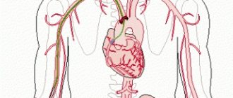
- angiography (x-ray examination of the patient’s blood vessels using contrast);
- laser scanning;
- ultrasound examination (ultrasound);
- perimetry or visual field testing;
- ophthalmoscopy (examination of the patient's fundus using a fundus lens or ophthalmoscope).
Direct ophthalmoscopy
On a note! In addition to the above diagnostic methods, another procedure may be prescribed - electroretinography. This is a special diagnostic method in which the doctor, using special equipment (electroretinograph), measures the electrical potential of the patient’s retina.
Background retinopathy in blood diseases
Blood diseases accompanied by retinopathy include polycythemia, myeloma, anemia, leukemia, etc. At the same time, retinopathy has a certain specific clinical picture. For example, if the pathology occurs against the background of polycythemia, examination of the fundus reveals a particularly bright color of the veins (rich red color), and the fundus has a cyanotic tint. There are signs of vascular thrombosis, as well as papilledema.
In case of anemia, the fundus of the eye is pale, the vessels are pathologically dilated. Retinopathy occurs with frequent hemorrhages (hemophthalmos) under the retina or into the vitreous body. Often the condition of the retina is complicated by a “wet” type detachment.
When retinopathy occurs against the background of leukemia, high vascular tortuosity, swelling of the retina and optic nerve head, small hemorrhages, and accumulation of exudate under the retinal tissue are observed.
Multiple myeloma, like Waldenström's macroglobulinemia, can be accompanied by dilation of retinal vessels (veins and arteries) due to blood thickening, blockage of veins, the appearance of microaneurysms and hemorrhages under the retina.
Treatment of background retinopathy in blood diseases consists of compensating for the malignant process of the underlying pathology and performing a laser coagulation procedure of the retina. The prognosis for the outcome of the disease is usually unfavorable.
Symptoms
Despite the etiological differences, all types of secondary retinopathy are similar in their clinical course. The first stage of vascular changes is most often asymptomatic and is detected only during special examination methods. With the transition from stage 2 to stage 3, retinopathy acquires more obvious signs.
Common symptoms of background retinopathy:
- Photopsia (false feeling of flashes of light or sparks appearing in the field of vision);
- Various color vision disorders;
- Reducing the contrast between the objects in question;
- Decreased visual acuity (in diabetics, as a rule, begins with farsightedness);
- The appearance of “floating” spots and dots before the eyes;
- Hemophthalmos (bleeding into the vitreous body).
Traumatic retinopathy
In case of injury, retinopathy is caused by prolonged compression of the chest, which leads to spasm of the arterioles, causing hypoxia of the retina with the release of edematous transudate inside.
Immediately after the injury, signs of hemorrhages, as well as organic damage to the retina, are detected. The condition is often complicated by optic nerve atrophy. The consequences of traumatic retinopathy (“Berlin opacities”) usually include subchoroidal hemorrhages, swelling of the lower layers of the retina, and leakage of fluid into the space between it and the vascular network.
As treatment, emergency elimination of hypoxia phenomena is used, vitamin therapy and hyperbaric oxygenation are prescribed.
Retinopathy of prematurity
The pathology is observed in premature infants and is caused by underdevelopment of the retina. To complete the formation of the organ, newborns need visual rest and local tissue respiration without oxygen (glycolysis). However, in order to activate metabolism and complete the development of other organs, such children need additional oxygenation, which inhibits the processes of glycolysis in the retina.
As a rule, retinopathy of prematurity is detected in children whose birth occurs before the 31st week of pregnancy and whose weight does not reach 1.5 kg. However, in some cases, the pathology can also occur in infants who have undergone blood transfusions or oxygen therapy due to premature birth.
In this regard, all children at risk are checked by an ophthalmologist one month after birth. His consultation is required every 2 weeks thereafter, until the eye structures are fully formed. These measures are necessary to prevent the occurrence of late complications of this type of background retinopathy - amblyopia, strabismus, retinal detachment, primary glaucoma, etc.
In most cases, retinopathy of prematurity disappears spontaneously without drug treatment, so often the specialist simply chooses a wait-and-see approach. In rare cases, the doctor may prescribe a laser coagulation or cryoretinopexy procedure, and extremely rarely, an operation to remove the vitreous body or scleroplasty.
Prevention of background retinopathy
To prevent the development of background retinopathy in adults, dynamic monitoring and regular examination of patients at risk (patients with hypertension, diabetes, atherosclerosis, etc.) are required.
In order to prevent retinopathy of prematurity, pregnant women with the threat of premature birth are advised to stay in a hospital, which will make it possible to provide early care to the newborn. Children with neonatal retinopathy should be observed by an ophthalmologist until the age of eighteen, even if it resolves favorably in infancy.
By contacting the Moscow Eye Clinic, each patient can be sure that some of the best Russian specialists will be responsible for the results of treatment. The high reputation of the clinic and thousands of grateful patients will certainly add to your confidence in the right choice. The most modern equipment for the diagnosis and treatment of eye diseases and an individual approach to the problems of each patient are a guarantee of high treatment results at the Moscow Eye Clinic. We provide diagnostics and treatment for children over 4 years of age and adults.