What is the aorta and where is it located?
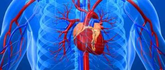
The aorta is considered the largest vessel of the body and has a key role in maintaining normal hemodynamics. It is from here that a large circle of blood circulation begins, which supplies oxygen-rich blood to all structures of the body. It departs from the left ventricle of the heart, is mostly located along the spinal column and ends, diverging into two branches: the right and left iliac.
Structure and departments
It belongs to the elastic type of arteries; histologically its wall is formed by three layers:
- Internal (intima) – represented by endothelium. It is he who is most susceptible to pathological processes, including atherosclerosis. This lining forms the aortic valve.
- Middle (media) - mainly consists of elastic fibers, which, when stretched, increase the lumen of the bed. This allows you to maintain stable blood pressure. It also contains a small amount of smooth muscle fibers.
- External (adventitia) - consists mainly of connective tissue elements with a low content of elastic fibers and high collagen, which gives the vessel additional rigidity, despite the small thickness of the wall.
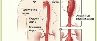
Topographically, the artery consists of three main parts: the ascending section, the arch and the descending section.
The ascending section begins in the area of the third intercostal space, along the left edge of the sternum. The aortic valves are located at the point where the vessel exits the heart. Their second name is “crescentic” because they resemble curved pockets, consisting of three leaflets and preventing the reverse flow of blood after the aorta has left the ventricle. There are also small bulges - sinuses, in which the coronary arteries that supply the myocardium begin. In the same place there is a short expanded section - the bulb. Opposite the articulation of the second right rib with the sternum, the ascending aorta passes into the arch.
The arc turns to the left and ends near the fourth thoracic vertebra, forming the so-called isthmus - a place where the artery is somewhat narrowed. Posterior to this is the tracheal bifurcation (the point at which the breathing tube divides into two bronchi). Branches extending from its upper side supply the upper part of the body:
- brachiocephalic trunk;
- left general carotid;
- left subclavian.
The descending section is the longest part of the vessel, consisting of the thoracic (thoracic) and abdominal (or abdominal) sections. It originates from the isthmus of the arch, is mostly located in front of the spine and ends near the fourth lumbar vertebra. At this point, the aorta diverges into the right and left iliac branches.
The thoracic region is located in the thoracic cavity and extends to the aortic opening of the respiratory muscle of the diaphragm (opposite the 12th vertebra). Along its entire length there are branches that supply blood to the organs of the mediastinum, lungs, pleura, muscles and ribs.
The final, abdominal part, provides blood supply to the abdominal and pelvic organs, the abdominal wall, and the lower extremities.
Normal vessel size indicators
Determining the diameter of the aorta is very important in the diagnosis of many of its pathologies, especially aneurysms or atherosclerosis. This is usually done using x-ray (for example, computed tomography or magnetic resonance imaging) or ultrasound (echocardiography) studies. It is important to remember that this value is very variable, as it changes depending on age and gender.
| Department | Diameter, cm | |
| Men | Women | |
| Root | <4 | <3,7 |
| Rising | 3,4-3,6 | 3,0-3,5 |
| Arc | 3,0-3,5 | 2,7-3,4 |
| Thoracic | 2,5-3,0 | 2,4-2,6 |
| Abdominal | 1,9-2,4 | 1,8-2,1 |
Blood flow, speed and pressure
Also, to study the work of the aorta, its functional indicators are examined. This is usually done using ultrasound or Doppler ultrasound.
Systolic pressure in the vessel is about 120-140 mmHg. Art., diastolic – 70-90 mm Hg. Art. The value may vary slightly depending on age and individual characteristics.
The linear speed of blood flow in the initial sections is about 0.7-1.3 m/s. Distally (i.e. further from the beginning) it decreases to 0.5 m/s.
The normal flow rate is 4-5 liters per minute.
Preparatory process
To prepare for the scan, you simply need to exclude factors that may blur the clinical picture. This includes pharmacological drugs that are designed to help during the course of treatment:
- hypertension;
- heart block;
- angina pectoris.
But only the doctor should note the medicine, since sometimes it is easier to postpone the procedure by a couple of days than to interrupt a pre-agreed therapeutic program in the middle of treatment.
It is also worth holding off on drinking coffee, which can affect your blood pressure.
The procedure begins with the patient being placed on a couch with a cushion placed under his head. Next, the doctor lubricates the sensor with gel, which is designed to improve the conductivity of incoming ultrasonic waves through the subcutaneous fat layer.
The doctor will have to visually assess the area under study in order to correctly distribute the ascending and descending parts, as well as the arc and extensions in the thoracic region. The calculation is made based on the fact that the arc has three branches:
- brachiocephalic trunk, located on the right;
- common carotid artery;
- left subclavian artery.
Using the anatomical principle of zone separation, the condition of the vascular network is assessed using a transducer sensor. To get a clearer picture, the expert can switch examination modes.
How do changes in the functioning of the aorta affect systemic blood flow?
The aorta is the only vessel from which the systemic circulation begins. Any of her diseases cause severe hemodynamic disturbances, including cardiovascular failure.
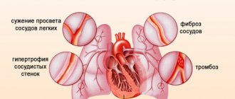
The first thing that suffers is blood pressure. Due to sclerosis and calcification, the arterial wall becomes rigid and loses its elasticity, and this is one of the causes of hypertension. When an aneurysm ruptures, the opposite is true - blood pressure drops sharply.
Aortic valve defects are very dangerous. Failure leads to regurgitation, which is the return of blood to the ventricle, causing it to overstretch, leading to cardiomyopathy. As a result of stenosis, cardiac output is also reduced. However, this is due to the fact that the doors do not open completely. At the same time, blood flow in the coronary arteries is disrupted. This leads to the development of angina pectoris.
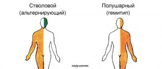
The degree of disturbance of blood flow largely depends on the localization of the pathological process: the closer it is to the beginning of the vessel, the more systemic its effect will be, while damage to only the abdominal region causes hypoxia of a limited area of the body (lower torso).
Acquired pathology
The aorta is a very important and at the same time quite often affected part of the body. The most common problems with the aorta, which arise not as a result of intrauterine development, but during life, are various types of aneurysms and ruptures.
As for ruptures of the aortic wall, it is perhaps the most dangerous condition in medicine. Most often, people who develop a similar pathology cannot be saved. The fact is that the rupture of the largest vessel in the body is accompanied by serious hemorrhage. As a result, the person needs urgent surgical treatment. There is a small chance to save a patient with this pathology only if he is already in a medical institution, and better yet, in a specialized one.
The most common cause of aortic rupture is a significant decrease in the elasticity of its wall, which is observed against the background of the deposition of calcium salts on it.
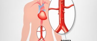
Another serious pathology of the aorta is its aneurysm. Among other things, it can also lead to rupture of the vessel wall. The essence of an aneurysm is that under constant pressure from blood flows, one of the sections of the aorta can expand in a sac-like manner. Most often, this pathology occurs where the vessel wall is at least slightly weakened. The usual location for such changes is the aortic arch and its abdominal region. In this case, compaction of the aorta usually does not manifest itself clinically. In addition to the risk of rupture of the vessel wall, the increased risk of blood clots is also a serious danger. If it forms and begins to move through the bloodstream, this can lead to the most disastrous consequences for a person.
Major diseases and developmental abnormalities
All diseases of the aorta, depending on their origin, are divided into two large classes: congenital and acquired.
The first include genetically determined developmental defects:
- Valve insufficiency - due to the underdevelopment of the valves, they do not close completely, and therefore, during diastole, part of the blood returns to the ventricle. As a result, myocardial hypertrophy develops and the initial part of the aorta expands.
- Valve stenosis is characterized by fusion of the leaflets, which makes it difficult for blood to pass through a narrow opening, which causes a decrease in systolic output and the development of dilated cardiomyopathy.
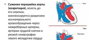
- Coarctation is a narrowing of the thoracic aorta. The modified segment can be from two millimeters to several centimeters in length, resulting in a significant increase in pressure in the area above the narrow part, but a significant drop in the lower sections.
- Marfan syndrome is a genetically determined disease characterized by damage to connective tissue. It is characterized by the frequent occurrence of aneurysms and valve defects.
- A double aortic arch is a defect in which the vessel is divided into two parts. Each of them bends around the esophagus and trachea, as a result of which they are enclosed in a ring. Hemodynamics are usually not impaired, the clinic is characterized by difficulty swallowing and breathing.
- Right-sided aortic arch - with this anomaly, the artery does not go to the left, as it should be normally, but to the right. The disease is usually asymptomatic, except in cases where the aortic ligament forms a ring around the trachea and esophagus, thereby compressing them.
Acquired diseases include:
- An aneurysm is an expansion of a section of a vessel by more than two times, resulting from pathology of the walls. This leads to serious hemodynamic disturbances, primarily to hypoxia of certain organs. Specific symptoms are determined by the location of the lesion.
- Dissecting aneurysm - characterized by rupture of the sclerotic inner membrane, due to which blood flows into the resulting cavity between the walls and causes their further dissection. Over time (usually after a few days), complete destruction of the defect occurs, which causes massive internal bleeding and instant death.
- Atherosclerosis - characterized by the deposition of lipoprotein complexes in the inner layer, which leads to the formation of plaques, calcification and narrowing of the lumen. As a result, oxygen starvation (hypoxia) of organs and tissues occurs, as well as thrombotic complications (including strokes).
- Nonspecific aortoarteritis (Takayasu syndrome) is a vasculitis of autoimmune origin, in which proliferative inflammation develops in the vessel wall, leading to compaction, obstruction, or the formation of aneurysms.
Diagnosis of LV hypertrophy
At the first appointment, the doctor collects anamnesis (patient complaints, information about family diseases). If there are endocrine diseases, hypertension, or heart defects in the family, then hypertrophy of the walls of the left ventricle becomes more than likely.
To clarify the diagnosis, the following procedures are prescribed:
- Chest X-ray. The x-ray will show enlarged shadows of the heart and shadows of the aorta;
- Electrocardiogram;
- 24-hour ECG monitoring;
- Echocardiography;
- Stress echocardiography (ultrasound of the heart before and after exercise);
- Doppler test (checking cardiac blood flow also using stress);
- Laboratory blood test;
- Blood test for hormones;
- Analysis of urine.
To determine the extent of the disease, the doctor will prescribe coronary angiography (an X-ray examination with the injection of contrast fluid into the cardiac bloodstream). This is how they determine how free the lumen of the coronary arteries is.
To accurately visualize intracardiac pathologies, cardiac MRI is performed.
What treatment and correction methods exist and are considered effective?
A peculiarity of aortic pathologies is that their treatment mainly involves invasive surgical intervention. Conservative therapy is used only to support vital signs and relieve symptoms, which allows for safe surgery.
There is now a trend towards minimally invasive endoscopic operations, which are safer and more effective.
Today the following surgical treatment methods are used:
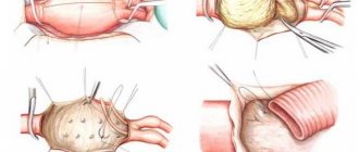
- resection with anastomosis – used for small aneurysms or coarctation;
- prosthetics;
- coronary artery bypass grafting (creation of bypass circulatory pathways) – for occlusive diseases, ischemic heart disease or heart attack;
- implantation of artificial valves, balloon valvuloplasty,
Congenital diseases
All these diseases are very dangerous and in the vast majority of cases require serious surgical intervention. Among the main ailments, coortia of the aorta and Marfan syndrome should be noted.
Aortic coortia is an extremely dangerous congenital disease. It can be suspected by the uneven development of the upper and lower halves of the body. If a person has coortia of the aorta, the muscles of the upper limbs develop normally, but hypotrophy is observed below. In this case, the patient may complain of weakness and pain in the lower extremities, especially after physical exertion.
As for Marfan syndrome, the cause of death in this disease is mainly the pathology of the development of the aorta. Most often it is a dissection of the wall of the largest vessel in the body. The increased risk of this particular pathology is due to the fact that the aortic wall in Marfan syndrome is weakened and is able to withstand much less load than under normal conditions.