Hemodynamic disorders of the central nervous system pose an enormous danger to life. Despite the long-term absence of obvious symptoms, processes of this kind quickly progress, cause neurological deficits, and become the culprits of strokes of varying severity. This is a direct path to disability.
The only way to avoid such an unenviable fate is to study the state of the body in detail.
Ultrasound dopplerography of the BCA is an ultrasound Dopplerography (examination) of the brachiocephalic arteries that supply the brain. The technique allows you to quickly detect all possible problems with nerve tissues and the degree of their blood supply.
Compared to duplex scanning (USD), this procedure is somewhat simpler and not as informative. It is used as a survey technique and creates the basis for further examination of patients.
This method is universal in its way and allows you to diagnose atherosclerosis and other disorders. However, we are also talking about a functional method. There is no need to expect detailed visualization of anatomical structures.
As a rule, diagnostics are prescribed in a system with other measures.
The essence of the survey and what it shows
The basis of ultrasound examination of the brachiocephalic arteries (abbreviated as BCA ultrasound) is the ability to create a clear image of the area of interest. However, this is not as informative and accurate a technique as duplex scanning of the vessels of the neck and head, so its scope of application is somewhat narrower. And the doctor will not receive much information.
On the other hand, the equipment is much cheaper, which makes the measures accessible to a wide range of patients. If necessary, more precise methods are prescribed.
During the action, the specialist has the opportunity to evaluate the following important characteristics:
- Local blood flow velocity. The intensity of brain trophism depends on this indicator. Its quality. But not only. Any deviations clearly indicate either functional changes or the presence of atherosclerosis or arterial spasm. The diagnosis needs to be clarified in other ways.
- The nature of local blood flow. Speed is far from the only indicator, although it is important. It must be assessed in a system with the direction of movement of liquid connective tissue and other points. If necessary, the doctor asks the patient to change the position of the head in order to identify deviations in natural motor activity.
- Condition of vascular structures. Wall thickness, its physical properties. Many abnormalities can also be seen using ultrasound of the BCA, although in this case a highly professional doctor is required.
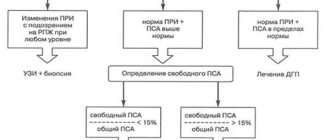
- Doppler ultrasound of the brachiocephalic arteries allows you to detect areas of narrowing of the artery (spasm or stenosis), blocking by foreign objects, be it a blood clot or an atherosclerotic plaque.
On the other hand, this is not enough to obtain comprehensive information, which is the main disadvantage of the study.
Advantages of the method
The advantages of BCA ultrasound are:
- high information content;
- efficiency of research;
- safety and possibility of repeated implementation;
- painlessness of the procedure.
Ultrasound scanning of the BCA is more informative than conventional ultrasound scanning of the brachiocephalic vessels.
During the examination, an image is formed on the monitor, similar to a conventional ultrasound, but against its background the vessel in which the blood flow is formed is clearly visible. Due to the advantages of ultrasound scanning, BCA is considered the gold standard for diagnosing pathologies. A timely vascular ultrasound can save lives and prevent possible disability.
Learn more about the study and how duplex scanning of the vessels of the head is done from the video:
What diseases can be detected
Measures based on the results allow us to identify a group of pathological processes that somehow disrupt the functioning of the brain or are capable of doing so in the short term.
Among them:
- Cerebral atherosclerosis. A classic disease that is identified by diagnostic results. It is a chronic process in which mechanical narrowing of the lumen of the artery occurs.
Usually the culprit is a cholesterol plaque.
Slightly less frequently, the disorder is accompanied by cerebral vascular spasm. Most often this occurs in hypertensive patients, smokers, alcohol drinkers, and people whose professional activities involve intense physical activity.
To clarify the diagnosis, duplex scanning is prescribed. It is suitable for identifying particularly difficult cases.
- Congenital anatomical defects. For example, hypoplasia (underdevelopment of arteries). Patients do not always have an idea of their presence. Asymptomatic course occurs in more than half of clinical situations.
It makes sense to carry out additional measures to clarify the anatomical component. BCA examination provides information about functional impairment. Precise imaging requires an MRI. Together, these methods provide the best diagnostic results.
- Acquired disorders of arterial structure. They occur after suffering vasculitis or other injuries. A typical option is the formation of adhesions. Gross scar defects of the endothelium (internal lining).
This process is accompanied by obvious symptoms. The same as with atherosclerosis. The brachiocephalic arteries are branches of the aorta that supply the brain. The clinical picture is appropriate and is represented by neurological deficits and pain.
- The influence of other pathological processes on blood vessels. Tumors, changes in the musculoskeletal system and others. As a rule, the result is compression of the arteries and impaired blood flow.
It is necessary to assess the severity of the disorder, the exact localization of the transformation of normal trophism into abnormal. For these purposes, ultrasound examination of the brachiocephalic arteries is used.
- Consequences of injuries. Bruises, fractures of the spine, chest and others.
What does the ultrasound scan of the BCA show?
The study is often prescribed to patients with suspected atherosclerotic changes. Ultrasound scanning is a very common examination method because it provides extensive information about the condition of the blood vessels. Ultrasonic scanning of the BCA shows:
- blood flow speed;
- nature of fluid movement;
- permeability;
- anomalies in the development and location of the BCA;
- degree of damage to the walls.
Information is displayed on a special color monitor. With the help of the device, the specialist receives a visual picture; you can examine the vessel from all sides. Ultrasound scanning can be combined with Dopplerography, ultrasound and evaluate the functioning of the arteries along with the tissues adjacent to them.
Indications and contraindications
The list of reasons for prescribing an ultrasound scan is floating. If necessary and at the discretion of the doctor, it can be expanded.
- Hypertension. Stable increase in blood pressure. Moreover, if a person has a long history of the disorder, he receives treatment and the diagnosis has long been established.
- Cervical osteochondrosis. Degenerative-dystrophic problem. Accompanied by compression of blood vessels by growing osteophytes. Hernias. The effect is also provoked by the instability of the pillar.
- Diabetes. Impaired insulin synthesis or decreased tissue sensitivity to it. Regardless of the type of disease.
- Angina and other heart disorders.
- Increased body weight of the patient.
- Age 45+. Over the years, a person takes more and more risks. The likelihood is determined by other factors, but preventive examinations must necessarily include examination of blood vessels.
- Negative family history. Complicated anamnesis. Such patients are at high risk of developing atherosclerosis, cerebral ischemia and subsequent problems.
- Past inflammatory processes on the part of blood vessels. Vasculitis first of all. They are of autoimmune origin.
The result is scarring of the internal structure, the formation of adhesions in the arteries. These mechanical obstacles do not allow blood to move quickly enough, and also lead to dystrophy and gradual destruction of the blood supply structures themselves.
All these diseases have one thing in common. They are factors in the development of atherosclerosis, which in one form or another is identified by ultrasound examination of the BCA.
Such categories of patients are at increased risk, so diagnostics must be performed at least once every six months. As needed and even more often.
There are other indications for prescribing a study:
- Headache. Of unknown origin. And even more so if the cause has already been discovered earlier. Doppler ultrasound in this case involves clarifying the nature of the disorder.
- Sleep disorders.
- Emotional instability. Irritability, aggressiveness. cerebral ischemia leads to a rapid increase in behavioral changes. Negative aspects are emphasized.
- Focal neurological disorders. From vision problems to the gradual decline of mental abilities. In memory. And other changes.
- Dizziness.
Doppler ultrasound of the brachiocephalic vessels (BCV) also has a number of contraindications. Although small.
- Mental inadequacy. If a person is in an acute condition, is unable to control his behavior and is not aware of the specialist’s instructions. Such a patient is unable to sit or lie. At least until normality is restored. It is necessary to correct the changes, only then prescribe a diagnosis.
- General difficult situation of the patient. In some cases it makes research impossible.
- Pronounced hyperkinesis. Especially the head and neck areas. Spontaneous movements create problems with the application of the sensor and interfere with the doctor’s work.
The list is small, such contraindications are rare, and after eliminating the problems, an ultrasound scan can be prescribed.
Indications for ultrasound of the brachiocephalic arteries
The purpose of duplex scanning of the arteries supplying the brain is indicated in the presence of the following patient complaints:
- headache;
- noise, ringing in the ears;
- dizziness;
- feeling of heaviness (“stale head”);
- darkening of the eyes;
- weakened hearing and vision;
- feeling of blood beating in the temples, back of the head;
- impaired coordination of movements;
- unsteady gait;
- attacks of weakness or numbness in the limbs;
- speech disorder;
- episodes of loss of consciousness;
- forgetfulness;
- changes in blood pressure.
The examination is recommended for categories of people without clinical signs, but if they have risk factors for cerebrovascular accident:
- age over 45 years;
- hereditary predisposition to vascular diseases;
- smoking, chronic alcoholism;
- hypertension or secondary hypertension;
- atherosclerosis of the lower extremities or obliterating endarteritis;
- angina pectoris, previous myocardial infarction;
- diabetes;
- lipid metabolism disorders - obesity, metabolic syndrome, high blood cholesterol;
- arterial hypotension;
- osteochondrosis of the cervical spine.
Atherosclerosis of the lower extremities is one of the indications for ultrasound of the brachiocephalic arteries.
Ultrasound of the brachiocephalic arteries is used to diagnose and prescribe treatment for the following diseases:
- congenital anomalies of vascular structure;
- traumatic brain injuries;
- aneurysms;
- inflammatory processes in blood vessels (vasculitis);
- cardiopsychoneurosis;
- diabetic angiopathy;
- consequences and complications of hypertension, poisoning;
- encephalopathy;
- transient cerebrovascular accidents;
- tumor or cystic formations on the neck;
- post-stroke or postoperative period.
Watch the video about ultrasound of the brachiocephalic arteries:
Preparing for the examination
No special events required. If we talk about recommendations:
- About 12 hours in advance, you need to give up drinks and food that change vascular tone. This includes tea, coffee, sweets, pickles, rich foods, some vegetables and acidic fruits.
- For 6 hours, drugs that can artificially affect the condition of the arteries are excluded. Antihypertensive, others. This measure is not always mandatory. It is better to clarify whether or not you can take medications from your specialist. Refusal can be dangerous. Especially if you plan to take the drug at night or in the evening.
- During the same period, 12 hours before, you cannot smoke, drink alcohol, or take a steam bath. In order not to provoke narrowing or artificial expansion of the arteries.
Progress of the procedure
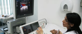
The examination is carried out on an outpatient basis. The patient sits or lies down, depending on the intended diagnostic methods. The doctor places the sensor on it and begins the examination.
During the procedure, you must follow the specialist's instructions. It is possible to change body position, take a deep breath, etc.
Also, the diagnostician often conducts functional tests in parallel. You need to bend your neck and perform certain actions. This is a more informative modification of the study. But it doesn’t always make sense.
Ultrasound of the brachiocephalic vessels of the head and neck lasts about 10-15 minutes. Plus or minus. It happens that it is much less if there are no deviations from the arteries.
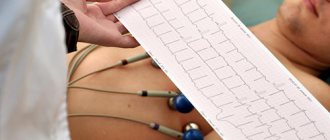
Based on the diagnostic results, the doctor prepares a conclusion. A person receives both the eyeliner itself from a specialist and a full protocol of the study with a thorough description of the characteristics found, changes and normal phenomena. It is possible to provide images, usually in digitized form.
With the results of the diagnosis, you should consult a doctor. Which sent for research or another.
How is ultrasound performed?
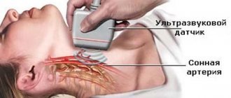
The technique is easy to implement. The subject is placed on a couch with a cushion under his neck. The head is turned away from the device. The area of skin over which the sensor is moved is lubricated with a special gel that improves signal transmission. When duplex scanning of the brachiocephalic arteries, the doctor moves the device, examining each zone of the vessel; if necessary, he may ask the patient to hold his breath.
If altered vessels are identified, all branches extending from the central main trunk are examined. Ultrasound scanning takes place within 25-45 minutes. During the procedure, the patient does not experience painful or unpleasant sensations. At the discretion of the doctor, the study can be carried out using functional tests. To do this, the person is asked to stand or sit.
Decoding the results
Neurologists do the interpretation. You won’t be able to decipher it yourself, since you need to have a certain set of knowledge and specific skills. Experience also plays a big role.
Based on the diagnostic results, the specialist can note the following deviations:
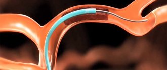
- Stenosis. Narrowing of the lumen of the artery to a certain percentage of the norm, leading to cerebrovascular accident. In this case, there is no blockage of blood vessels.
Read more about the causes of cerebral vasoconstriction and treatment methods in this article.
- Aneurysm. A wall protrusion in the form of a pouch protruding in one or both directions relative to the vertical axis. An extremely dangerous abnormal condition, fraught with violation of the anatomical integrity of the artery. Massive bleeding and death.
Read more about carotid artery aneurysm here.
- Occlusion. It's a blockage of a vessel. Typically a cholesterol plaque. Much less often - a blood clot. Atherosclerosis of the carotid arteries is described in detail in this article.
- Turbulence of blood flow. With turbulence of liquid connective tissue. Occurs as a result of aneurysms and other processes. A reverse movement is possible - this is a consequence of a malfunction of the venous valves.
- Changes in the condition of the vascular wall.
All findings and diagnoses are, one way or another, described by these deviations. Next, you need to determine what disease occurs in this case.
Pros and cons of the study
Among the advantages of the technique:
- Lack of invasiveness. Doppler ultrasound is a completely painless method. It does not create burden or discomfort.
- No lengthy preparation required. It's simple. The event can be carried out without any special action on the part of the patient.
- Minimum contraindications. Since the diagnosis is safe, does not create a harmful load on the body, and does not require invasive measures.
- Availability. The cost, even with a paid passage, is insignificant. It is possible to receive services under a compulsory medical insurance policy.
- Survey speed. Everything takes a few minutes.
- Information content.
Among the disadvantages are the following:
- High requirements for diagnostician qualifications. It takes sufficient experience and a trained eye to assess the condition of the arteries and not lose sight of anything important.
- Relatively narrow scope of application of the survey. Comparatively less than more advanced methods.
Analogues of the method
This can be called duplex scanning as a more sensitive ultrasound examination, and as a complement to MRI diagnostics.
The latter does not convey functional components, but concentrates on a static picture of what is happening and anatomical deviations, which duplex scanning cannot.
Ultrasound dopplerography of the BCS is Doppler ultrasound of the brachiocephalic vessels. A necessary examination for a comprehensive assessment of the condition of the brain and its trophism (nutrition). Prescribed according to indications.
Due to its complete safety and sufficient information content, it is often used.