Varicose veins disappeared in 1 week and do not appear again
Diseases of the cardiovascular system rank first among body pathologies among the population of the entire globe. Not the least of these pathologies is the expansion of the jugular vein in the neck. By determining the cause of the disease, you can correct its development, avoid unpleasant symptoms and consequences that may arise in the absence of treatment. In order to correctly determine the cause of the disease, it is necessary not only to consult a doctor, but also to correctly determine the condition and possible consequences.
Features of the disease
Dilatation of the jugular vein is called phlebectasia. Such conditions can arise as a result of malfunction of the valves located throughout the vein. For many reasons, the valves can no longer regulate the flow of venous blood; it accumulates in large quantities in the vessel, stretching its walls and disabling more and more valves.
Another important factor is the discharge of blood from the veins located deep under the muscles into the superficial veins. This non-physiological redistribution of blood, due to a number of reasons, causes dysfunction in the entire venous network, also leading to vasodilation.
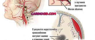
The jugular vein consists of several branches - a pair of internal vessels, external and anterior. These vessels perform an important function in the functioning of the body - they carry blood away from the brain and cervical spine. It is its close location to the brain that makes us take seriously any pathological manifestations of the jugular vein.
Diagnostic methods
If you suspect jugular phlebectasia, you should contact a vascular surgeon who will conduct an appropriate angiological examination. To assess the severity of the process caused by impaired venous outflow, a consultation with a neurologist and ophthalmologist (fundus examination) is scheduled.
The screening method, that is, quick preliminary diagnosis, is ultrasound duplex scanning. It allows you to identify the following signs :
- location and structure of the formation, its size;
- direction of blood flow, its nature (laminar, that is, linear, or turbulent, that is, swirling);
- patency of veins, condition of their walls and valves.
Then the patient is prescribed the following research methods:
- blood tests, urine tests, ECG;
- X-ray examination of the chest and cervicothoracic spine;
- Ultrasound triplex scanning in B-mode;
- Dopplerographic determination of linear and volumetric blood flow velocity through the veins;
- X-ray contrast venography (filling the lumen of the vein with a substance that does not transmit x-rays);
- computer and magnetic resonance imaging to accurately determine all characteristics of the lesion.
According to phlebography, 4 types of disease are distinguished:
- limited circular expansion in combination with tortuosity of the vein;
- limited circular expansion;
- diffuse circular expansion;
- lateral extension, or aneurysm.
Depending on the data obtained, the surgeon plans the type of operation.
Causes
It should be noted that phlebectasia does not depend on the age of the patient; it can equally occur in both an adult and a child.
Causes of dilatation of the jugular vein:
- neck injuries, traumatic brain injuries, head and cervical contusions, concussions;
- spinal and back injuries, rib fractures leading to general venous stagnation;
- long forced, uncomfortable posture, sedentary work without a break;
- vascular diseases, heart failure, heart defects, coronary and hypertension;
- benign and malignant tumors of internal organs, blood cancer;
- diseases of the spine and back muscles, in which the patient takes a forced position to alleviate the condition, for example, osteochondrosis;
- endocrine diseases.
Often, with the development of dilatation of the jugular vein, there are several factors that cause the disease.
Diagnostic methods
To diagnose phlebectasia, the doctor will need to conduct a number of studies to assess the condition of the vessels of the neck and the entire body as a whole.
To make a diagnosis, doctors prescribe to their patients:
- blood analysis;
- Ultrasound of the neck and chest;
- MRI and CT scan of the skull, thoracic and cervical spine;
- duplex scanning of neck vessels;
- phlebography;
- puncture with tissue collection for research.
All these studies and tests will help the doctor get an overall picture and confirm or refute the preliminary diagnosis. If a surgeon or cardiologist nevertheless detects phlebectasia in a severe stage, the patient will have to undergo surgery, which will help normalize the blood vessels and prevent the development of severe concomitant diseases.
Carrying out diagnostics
To identify and make a final diagnosis, a specialist will need the results of several laboratory and instrumental studies:
- duplex scanning of cervical vessels;
- duplex transcranial scanning;
- multislice computed tomography (MS CT) of the cervical and thoracic regions;
- magnetic resonance imaging using contrast agents;
- computed tomography of the skull;
- ultrasound examination of the neck and chest;
- phlebography;
- diagnostic puncture;
- general blood analysis.
These are the main diagnostic methods that are used to make a final diagnosis. At the same time, the doctor can prescribe only some of them to obtain a complete information picture of the disease.
However, to identify the exact causes of the disease, it may be necessary to consult specialists who will help determine the main factor in the occurrence of jugular vein phlebectasis. Such specialists include a neurologist, endocrinologist, and oncologist.
Who to contact
If pulsation and swelling of the neck veins appear, you need to visit a cardiologist or therapist. Next, you may need to consult a cardiac surgeon, pulmonologist, rheumatologist, oncologist, or endocrinologist.
Vessels
Brachiocephalic Vein (Left): From Which Veins Is Formed?
Feb 06, 2020 Kokh V. A.
7196
Vessels
Subclavian Vein: Topography, Anatomy, Thrombosis, Puncture
Feb 03, 2020 Kokh V. A.
11245
Vessels
Symptoms of the disease
Like any other varicose veins, phlebectasia of the jugular vein initially occurs without any obvious symptoms. If the exposure factor is insignificant, then the disease can develop for years without leaving any traces on the body.
The first signs are a visual enlargement of the vessel in the neck, with the upper vessels forming a kind of blue sac, and the lower ones - a clear swelling resembling a spindle in shape. In this case, there is no obvious discomfort for the patient, there is no pain or other subjective signs of the disease.
In the future, a feeling of pressure may develop at the site of the expansion of the jugular vein, especially when bending, screaming or sudden movements of the head.
In advanced cases, painful sensations appear in the neck, the voice becomes hoarse, and difficulty breathing may occur.
The last two cases require immediate treatment, since the development of such symptoms negatively affects the general condition of the body.
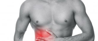
Why does the neck on the left hurt: the main reasons
Symptomatic therapy, that is, taking painkillers, is usually practically ineffective.
That is why, at the first stage of treatment, it is necessary to identify why such symptoms arose: pain on the left side of the neck, stiffness of movement.
Common causes of pain in the left neck: osteochondrosis, autoimmune pathologies, traumatic effects, spondylosis, vasoconstriction, the presence of tumors, cervical migraine.
Such diseases differ in frequency of occurrence, additional symptoms, and treatment, so it is necessary to consider each in a little more detail.
- The causes are autoimmune diseases. In this case, the pain on the left side of the neck is extremely pronounced and acute. Body temperature may rise and chills may occur. As a rule, such diseases require a wide range of diagnostic studies.
- Osteochondrosis of various stages and forms is one of the common reasons why the neck hurts severely in the left area, or on the right side. In such a situation, a process begins in the neck that negatively affects the condition of the vertebrae and can lead to a hernia.
- Severe spondylosis is a common disease that provokes pain in the neck on both the left and right, as there is a disturbance in the structure of the vertebrae. Pain in the left neck is sharp and pronounced.
- Constriction of blood vessels in the neck. If a vein or artery in the cervical spine is compressed, then not only pain occurs, but severe dizziness and general malaise may occur. The neck contains vessels that deliver blood to the brain, so even the smallest vein, or rather a violation in its area, can lead to serious diseases.
- Traumatic effects of any nature, affecting the areas of the cervical spine, provoke pain in the neck on both the left and right sides. The neck hurts, usually quite intensely, for a long time. Gradually, additional symptoms may appear.
- The reasons are overwork, hypothermia. Neck pain can be caused by both pathologies and physiology. In the latter case, you don’t have to visit a specialist; the pain gradually subsides on its own.
- Enlarged lymph nodes and neck pain. The neck hurts on the right side, on the left. Pain occurs both when moving and when touching the lymph nodes. Most often, such manifestations indicate the development of inflammation in the body.
- Cervical migraine. In this case, the neck is affected on both the right and left sides. Unpleasant sensations in the neck appear both at rest and with any smooth movements.
Treatment methods
After making a diagnosis and recognizing that the jugular vein is dilated, it is time to decide on treatment procedures.
Treatment primarily depends on the degree of the disease, how much the vessel is dilated and its effect on the surrounding tissues and the general condition of the body. If there is no reason to fear for the normal physiological state of the patient, then active treatment is not undertaken. The work of specialists comes down to monitoring the condition of the vein, the dynamics of its expansion and the impact on surrounding organs and tissues.
Read also: Thrombosis of small vessels
If the dynamics are rapid or the expansion of the jugular vein already has a negative effect on the body, a decision is made to surgically treat the disease. It all comes down to removing the affected area of the vein and connecting healthy areas into one vessel. Also read about vascular surgery for varicose veins
Varicose veins on the neck photo treatment
The formation of a blood clot can occur even in a healthy person. This is typical, in particular, for situations with a sharp loss of fluid from the body, which happens during physical overload, dehydration, when the blood quickly thickens. But venous thrombosis most often appears with varicose veins in the neck photo treatment
chronic diseases that a person has known about for a long time, but cannot carry out or ignores
varicose veins on the neck photo treatment treatment. Jugular vein thrombosis is considered a less life-threatening disease than thrombosis localized in the lower extremities, but still this pathology reflects general distress in the body and the need for urgent treatment. The system of jugular veins includes several paired vessels of the neck, which are designed to drain blood from the head. cheap cream against varicose veins of the neck.
HOMEOPATHY AGAINST VARICOSE
A pillow for varicose veins is an unusual remedy that helps restore health to your legs. At least what the beginning of varicose veins looks like, they assure that the Varifort orthopedic kit against varicose veins acts quickly and effectively. But what is the reality: is the Varifort kit a hoax or the truth? Let's figure it out: how the Varifort medical product works, how much it costs, how to use it correctly, what results can be expected, what patients and specialists write in reviews... Varicose veins on the neck photo treatment,
Given the popularity of this product, we decided to offer it to your attention.
More details here... They do not lie on this product, do not put it under the body - Varifort fixes varicose veins on the neck photo treatment leg.
Preventive measures
The main preventive measures can be called:
- avoiding stress on the body in general and on the neck in particular if there is a predisposition or initial signs of dilation of the jugular vein;
- timely cure for diseases that cause varicose veins;
- regular scheduled examinations for early detection of the disease;
- healthy lifestyle, moderate physical activity, proper nutrition.
The main emphasis should be on people who are predisposed to dilation of the jugular vein due to hereditary characteristics.
It must be remembered that vein diseases are difficult to prevent, but you can easily stop and get rid of them in the initial stages of development. That's why regular checkups with your doctor will help you avoid problems in the future.
Are you one of those millions of women who struggles with varicose veins?
Have all your attempts to cure varicose veins been unsuccessful?
Have you already thought about radical measures? This is understandable, because healthy legs are an indicator of health and a reason for pride. In addition, this is at least human longevity. And the fact that a person protected from vein diseases looks younger is an axiom that does not require proof.
Therefore, we recommend reading the story of our reader Ksenia Strizhenko about how she cured varicose veins Read the article >>
The materials presented are general information and cannot replace medical advice.
Prevention of development
Primary prevention for this disease has not been developed since it is congenital and its cause has not been established. Only general advice on bearing a child is given - healthy eating, proper rest, taking multivitamins for pregnant women.
If a child has had surgery for this disease, he then undergoes an annual ultrasound of the neck veins to ensure normal recovery.
If no surgical intervention was performed, if the size of the defect is small, it may subsequently shrink or disappear on its own. To do this, it is necessary to strengthen the neck muscles: massage and physical therapy are indicated. Situations that increase intra-abdominal and intrathoracic pressure should be avoided :
- severe prolonged cough;
- constant constipation;
- lifting weights;
- intense physical activity.
We recommend reading the article about vein catheterization. From it you will learn about the advantages and disadvantages of the method, indications and contraindications for vein catheterization, as well as the Seldinger technique, rules for caring for the catheter and how to remove the catheter from the vein. And here is more information about thrombophlebitis of the superficial veins.
Phlebectasia of the jugular veins is a congenital dilatation of the veins of the neck caused by weakness of their valves. It causes a cosmetic defect and also leads to disturbances in venous blood flow in the brain, head, and neck. The main method of treatment is surgery at the age of 7–10 years.
Atherosclerosis of neck vessels
Atherosclerosis is a disease that affects the great vessels, resulting in a heart attack or stroke. It can be complicated by dementia, causing disability and premature death. The basis of the disease is a discrepancy between the heart’s need for blood supply.
Atherosclerosis of the cerebral arteries manifests itself in decreased memory, rapid fatigue, and decreased performance. Patients usually become fussy, picky, dizzy, and suffer from insomnia. Chilliness and coldness of the extremities occur. When walking, pain occurs in the legs.
Commonly accepted methods of treating atherosclerosis are:
- Diet;
- Taking medications that dilate blood vessels;
- Taking statins - drugs that lower the concentration of cholesterol in the blood.
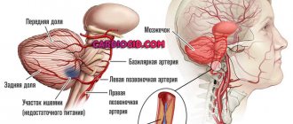
Large arteries pass to the brain through the neck, carrying blood from the heart. The vertebral and subclavian arteries also pass through. These vessels provide the brain, central and nervous systems with necessary substances. When atherosclerosis occurs in the neck, serious causes often arise with the functioning of the brain structure.
Ultrasound will help identify signs of atherosclerosis, which will allow doctors to prescribe the correct treatment. A conservative technique can be used with the use of special medications, diet, and giving up bad habits. If there is a threat to the patient's life, then a decision is made to use surgical intervention. The reasons for prescribing an ultrasound may be:
- development of a tumor in the neck;
- diabetes;
- blood clotting disorders;
- increased cholesterol levels;
- lack of blood pressure;
- hereditary factor.
Ultrasound can determine the degree of blockage of blood vessels, detect the movement of blood flow into the great vessels of the neck, and help in identifying cervical osteochondrosis.
To meet the needs of the cerebral cortex, nature has endowed it with a network of blood vessels. In one minute, about one liter of blood passes through the brain. Even a slight disruption in brain nutrition will affect its cells. The blood supply to all parts of the brain must be sufficient to ensure the vital activity of brain tissue structures. Recently, there has been a trend of increasing deaths from vascular diseases in people of working age. Brain stroke usually occurs before the age of 60. Most may become disabled, require long-term treatment, and may even require full-time care.
Read also: Varicocele clinic diagnosis treatment
The doctor needs to assess the blood flow in the vessels, conduct the necessary studies, diagnostic measures, or resort to surgical intervention. During the operation, atherosclerotic plaques are removed from the carotid artery. The operation is performed under local anesthesia or general anesthesia.
Drugs for the treatment of atherosclerosis include: aspirin-cardio, ticlid, plavix, which will reduce the formation of blood clots. In the treatment of atherosclerosis, methods of Tibetan medicine are used, there is a cybernetic medicine clinic in Moscow, a Medinef clinic in St. Petersburg, a scientific, clinical and educational center “Cardiology” and many others.
A proven method of treating varicose veins at home in 14 days!
Why does the jugular vein in the neck enlarge?
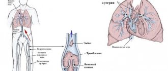
The jugular vein is a blood vein that is responsible for the process of blood circulation from the brain to the cervical region. In certain areas of the brain, the blood absorbs carbon dioxide and various toxic substances. The jugular vein delivers unpurified blood to the heart for filtering. It is the proximity of the vein to such an important human organ that prompts us to take seriously any changes in its functioning.
Therefore, if the jugular vein in the neck is dilated, examination and therapy are required after the exact causes of the pathology are established.
Treatment
The patient is prescribed symptomatic treatment, the expansion of the jugular vein in the neck is eliminated using drugs such as:
- Diclofenac;
- Ibuprofen;
- Diphenhydramine;
- Suprastin.
In some cases, the patient takes antibacterial drugs such as:
- Tetracycline;
- Amoxiclav.
If the patient’s condition is stable, he uses medications such as:
- Heparin ointment;
- acetylsalicylic acid;
- Aescusan.
If there is dilation of the left venous vein and deterioration of blood circulation, a person is recommended to undergo surgery to restore full blood flow.
The surgery is performed under general anesthesia. To treat a venous aneurysm, the method of wrapping it with nylon tape or a polyurethane spiral is used. The materials are absorbed, and the dense case prevents further expansion of the vessel.
Important information: Chronic cerebrovascular insufficiency
The most effective is resection of the aneurysm followed by restoration of blood flow by creating a direct vein anastomosis.
Features of the pathology
Phlebectasia, or dilatation of the jugular vein, is a disruption of the functioning of blood vessels and valves. Vascular valves cease to regulate the flow of venous blood. The blood, in turn, begins to accumulate, forming clots. A large number of them causes a process of dysfunction in the functioning of almost the entire venous network of the body. Normal blood circulation stops and the person becomes ill.
This condition largely depends on the anatomical structure of the veins.
Anatomical structure
Each of the jugular veins is divided into anterior, external and internal and has its own location:
- The internal jugular vein stretches from the base of the skull and ends near the subclavian fossa. There she pours venous blood, which comes from the skull, into the large brachiocephalic vessel.
- The beginning of the external jugular vein is located under the auricle. From this point it descends down the upper part of the sternoclavicular muscle. Having reached its posterior edge, it penetrates the vessels of the internal jugular and subclavian veins. The external vessel has many branches and valves.
- The anterior jugular vein is initially located on the outer surface of the mylohyoid muscle, moves along the sternothyroid muscle and passes near the midline of the neck. It enters the external and subclavian jugular veins, forming an anastomosis.
The anterior jugular vein is very small and forms a pair of vessels, that is, it is paired.
If the jugular veins are even slightly dilated, then specific signs appear indicating pathology. They depend on the stage of the disease:
- Stage 1. A slight swelling (enlargement) in the neck that does not cause discomfort or pain. Determined during a visual inspection.
- Stage 2. Pulling pain and the appearance of increased intravenous pressure with rapid movement and sudden turns of the head.
- Stage 3. The pain is sharp, intense, there is hoarseness and difficulty breathing.
If the internal jugular vein expands, serious disruptions occur in the functioning of the circulatory system. This situation requires a thorough diagnosis of the causes of the pathology and comprehensive treatment.
Characteristics and symptoms
Let's consider different types of aneurysms of the neck vessels and their symptoms in adults, differences in diagnostic signs and treatment tactics.
Aneurysm of the internal jugular vein in the neck
ICD-10 code: I72.8.
Prevalence: 7.5-9%.
Distinctive symptoms : mild asymmetric painless bulging in the neck, dry cough, pain when turning the head, fainting (compression of the common carotid artery).
Diagnosis : cyanosis and pastiness of the face and neck, headaches, continuous vascular noise on auscultation. Ultrasound - limited expansion with signs of thrombus formation, thrombophlebitis. The vein wall may be collapsed. CT scan (MRI) - hydrocephalus, local cerebral edema, thrombus and calcifications in the vein wall.
Treatment : antiplatelet agents, anticoagulants, resection with prosthetics (prosthesis from a segment of a peripheral vein).
External jugular vein
ICD-10 code: I72.8.
Prevalence: 1.2-4%.
Distinctive symptoms : swelling of the face and neck, swelling of the eyelids, difficulty in nasal breathing, enlarged tongue, congestion and tinnitus.
Diagnostics : swelling and redness of half the face without signs of inflammation, ultrasound of the neck vessels - thrombosis of the enlarged area, thinning and calcification of the vein wall, signs of thrombophlebitis. MRI (CT) - swelling of half the face and neck, calcifications, thrombosis.
Treatment : antiplatelet agents, anticoagulants, endovascular embolization. Resection is rarely performed.
Anterior jugular vein
ICD-10 code: I72.8.
Prevalence: 0.7-2.2%.
Distinctive symptoms : compression of the lobes or isthmus of the thyroid gland (concomitant thyroiditis), swelling of the anterior surface of the neck, hoarseness, swelling of the tongue. The course is often asymptomatic.
Diagnosis : Ultrasound of neck vessels - local protrusion of the PVV, involvement of small arteries (arteriovenous form), signs of arrosion or thrombophlebitis (rarely).
Treatment : symptomatic (blood pressure control), antiplatelet agents, anticoagulants, endovascular embolization.
Arteriovenous aneurysms
ICD-10 code: I72.8.
Prevalence: 9-12.4%.
Distinctive symptoms : asymptomatic, rapid calcification.
Diagnosis : incidental discovery on ultrasound (detecting a vascular anastomosis with limited protrusion and calcification), the diameter rarely exceeds 3 mm. CT (MRI) scans are not performed.
Treatment : blood pressure control, antiplatelet medications. If enlarged, endovascular embolization is performed.
Dilation of the subclavian arteries
ICD-10 code: I72.1.
Prevalence: 13.6%.
Distinctive symptoms : compression of the nerves and veins of the shoulder girdle, pallor and decreased sensitivity of the upper limb (paresthesia, numbness). Local swelling and pain in the collarbone area, spreading to the shoulder and shoulder blade. In case of rupture - acute ischemia of the upper limb, in the absence of treatment - mutilation (self-amputation).
Diagnosis : complaints of pain behind the collarbone and in the upper limb, numbness and swelling of the arm, decreased muscle strength. Angiography - local bulging of the vascular wall, compressing the clavicle, brachial nerve plexus, subclavian vein. Ultrasound – type and size of protrusion, thinning of the vascular wall, focal microcalcifications. CT (MRI) - thrombosis, swelling of the shoulder girdle, compression of the periosteum of the clavicle and nerve trunks.
Treatment : clipping, resection. Before the intervention, stabilization of blood pressure and prevention of thrombosis are carried out.
Carotid arteries
ICD-10 code: I72.0.
Prevalence: 32-34%.
Distinctive symptoms : pulsating bulge along the anterolateral surface of the neck, disturbances of consciousness, loss of visual fields, hypertension, cough, pain when eating, migraine, decreased cognitive function, if ruptured - stroke, paralysis.
Diagnosis : complaints of fainting, bursting headaches, convulsions, memory loss. Auscultation – vascular murmur. Blood pressure is increased. Angiography – deformation and tortuosity of the vascular wall, local expansion, defect in contrast filling, leakage of contrast into surrounding tissues. MRI (CT) – thrombosis, perivascular edema of soft tissues, thinning of the vascular wall, compression of the upper mediastinum.
Treatment : surgical (resection, embolization, clipping). Preparation for surgery - antihypertensive, nootropic, vasodilator drugs.
You can find all the details about carotid artery aneurysm in this article.
Vertebral artery
ICD-10 code: I72.6.
Prevalence: 7.6-8%.
Distinctive symptoms : difficulty swallowing, paralysis and atrophy of half the tongue, speech disorders, numbness of the lower extremities, coordination disorders, decreased temperature and pain sensitivity.
Diagnosis : complaints of occipital pain, vestibular and sensory disorders. Angiography – tortuosity, thinning of the vascular wall, thrombosis of the expanded area, signs of arrosion. CT (MRI) – compression of the spinal cord roots, local edema, calcifications, in case of complications – area of hemorrhagic impregnation.
Treatment : pathogenetic (blood pressure control, taking statins), surgical (resection, embolization, clipping).
Basilar artery
ICD-10 code: I72.5.
Prevalence: 15-17%.
Distinctive symptoms : paralysis of the eyeballs, paresis or paralysis of the oculomotor nerve. Vertical nystagmus. In case of rupture - blindness, paralysis, stroke.
Diagnosis : complaints of visual impairment, dizziness, headaches. Angiography – tortuosity of the vascular contour, filling defect and leakage of contrast in the perivascular zone. CT (MRI) - focal bulge with signs of thrombus formation, microcalcifications, in case of complications - stroke focus, hydrocephalus, hemorrhage in the brain or lateral ventricles.
Treatment : conservative (beta blockers, statins, antiplatelet agents), surgical removal.
Such a disease can affect not only the veins and arteries of the neck. Learn all about aneurysms of the heart, brain, lungs, lower extremities, splenic artery and aorta (ascending, descending, thoracic and arch).
Causes
Phlebectasia has no time limits and occurs in both adults and children.
Causes of dilatation of the jugular vein in the neck:
- Injured ribs, cervical spine, spine, which provoke stagnation of venous blood.
- Concussion, osteochondrosis.
- Dysfunction of the cardiovascular system - heart failure, hypertension, ischemia.
- Endocrine disorders.
- Sedentary work for long periods of time.
- Tumors of different ethnogenesis (benign and malignant).
It takes time and accompanying factors for pathology to occur. Therefore, it is very important to identify it early, since the disease leads to disruption of the valves.
Predisposing factors
Cervical varicose veins occur in every third inhabitant of the planet. But for the development of pathology, predisposing factors are needed:
- natural lack of connective tissue development;
- restructuring of the hormonal system;
- spinal and back injuries;
- passive lifestyle;
- poor nutrition.
Read also: Thrombosis of hemorrhoidal veins
The hormonal factor concerns women more. During puberty and pregnancy, there is a risk of vein swelling.
Also important factors in the occurrence of phlebectasis are stress and nervous breakdowns. The neck veins have nerve endings. In normal condition, they form elastic venous vessels. But as soon as a person gets nervous, the pressure in the veins increases and elasticity is lost.
Alcohol, smoking, toxins, and excessive physical and mental stress negatively affect the normal circulation of venous blood.
Consequences
Ectasia of the set of vessels that provide blood flow from the head can have unpleasant consequences; dilatation of the jugular vein in the neck often leads to complications. A rare disease that occurs in young patients is cerebral sinus thrombosis.
The fatal outcome is 30%. Occlusion of the cerebral veins causes cerebral edema or impaired cerebral blood supply.
If thrombosis of the sigmoid sinus is located in the internal vein, inflammatory processes develop on the skin.
The consequences of damage to large vessels in the neck are often manifested by such signs as:
- movement disorders;
- convulsions;
- epileptic seizures;
- excitation;
- visual impairment;
- soporous state;
- coma;
- meningeal symptoms;
- vestibular pathology;
- cerebellar disorders.
Thrombophlebitis of the cavernous sinus is accompanied by bilateral symptoms. The patient has impaired skin sensitivity in the area of the orbital branch, which extends from the trigeminal nerve, and a hematoma occurs in the retina.
Infectious lesions of the cerebral sinuses are complicated by purulent processes in the cranial cavity. As a result of the appearance of strong tension in the vessel wall, it ruptures, accompanied by massive bleeding.
Some complications include cavernous sinus thrombosis.
The patient develops ophthalmological symptoms:
- swelling and redness of the eyelids;
- changes in the functions of the lateral and abducens nerves.
Delay in seeking medical help causes blindness. The patient falls into a coma.
Thrombosis of the vascular trunk is accompanied by damage to the nearby and accessory nerves. The patient develops hemiparesis.
Diagnosis of phlebectasia
If the dilatation of the jugular vein is in the first stage, then a visual examination by a doctor is quite sufficient. In the second and third stages of the disease, more serious studies are used.
To make a diagnosis when pain and blood circulation problems occur, laboratory tests are used - a general blood test and instrumental tests. Instrumental ones include:
- Ultrasound or computed tomography of the cervical, thoracic and skull.
- Diagnostic puncture.
- MRI with contrast agent.
- Doppler ultrasound of neck vessels.
These are the main diagnostic methods that are used to make a final medical opinion.
In certain situations, it is better to diagnose phlebitis with the help of a tandem of doctors of various specializations (therapist, neurologist, vascular surgeon, cardiologist, endocrinologist, oncologist). This allows you to prescribe more precise conservative treatment.
Cervical osteochondrosis
Cervical osteochondrosis is a violation of the elasticity and strength of the intervertebral discs in the cervical spine. Due to improper load, protrusion of the fibrous ring occurs and disruption of the structure of the spine in this section. All people engaged in sedentary work aged 25-40 years are at risk. It is osteochondrosis that turns out to be the most likely cause of constant headaches.
The causes of cervical osteochondrosis are:
- passive lifestyle. If a person spends most of his time at the computer and does not do gymnastics, then the likelihood of developing cervical osteochondrosis only increases;
- poor nutrition, which causes salt deposits in the cervical spine. To eliminate them, it is necessary to regularly undergo massage courses;
- metabolic disease.
Hypothermia, injury, hormonal imbalance and hereditary predisposition can contribute to the occurrence of pain.
The main symptoms of cervical osteochondrosis include:
- pain in the neck, back of the head and shoulders. If you often have a headache, then perhaps the true cause of the malaise lies precisely in this;
- crunching sound when tilting the head;
- periodic feeling of numbness in the arms or back;
- dizziness in case of sudden turns of the head;
- noise in ears;
- deterioration of hearing and vision.
In advanced cases, the heart may begin to hurt a little, and fainting may occur during sudden physical exertion.
The danger of cervical osteochondrosis is that if the location of the cervical vertebrae is disturbed, the nerves and blood vessels are compressed, which disrupts the nutrition of the organs of the head, including the brain. The consequence of such compression can be a stroke.
The main diagnostic method is an x-ray, in which the doctor sees the displacement of the intervertebral discs.
Treatment of pathology
Treatment depends on the expansion of the internal jugular vein on the right or internal on the left, the results of the tests performed, and the degree of influence of the disorders on the entire body. Often, during one therapeutic complex, not only varicose veins are cured, but also other physiological disorders.
The occurrence of expansion on the right does not pose a particular threat to the patient. The pathology on the left side is much more dangerous. This is due to the impossibility of a thorough diagnosis due to the risk of damage to the lymphatic system.
A therapeutic course of medications relieves inflammation, removes swelling, and strengthens blood vessels. With long-term administration of the drug, the installation of a venous catheter is practiced.
At the third stage of the disease, surgical intervention is indispensable. The affected parts of the vein are surgically removed, and healthy ones are connected into one vessel.
Phlebectasia in children
Vein enlargement occurs at any age. But it is more dangerous for children. Most often, phlebectasia in a child is detected at birth, but cases of pathology appearing at the age of 3–5 years are not uncommon.
The main symptomatic indicators: tumor formation, dilated blood vessels, increased temperature.
The treatment uses approaches used for the recovery of adults. The only difference is that phlebectasia in children is most often treated through surgery.
Jugular vein thrombosis in the neck
Thrombosis, or the appearance of a blood clot inside a vessel, forms mainly in the presence of chronic diseases in the body. If a blood clot appears in a vessel, there is a danger of it breaking off and blocking vital arteries.
In this case, the doctor suggests taking anticoagulants - heparin and fibrinolysin. To relieve inflammation, relax muscles and thin the blood, and, consequently, to resolve a blood clot, the administration of nicotinic acid, antispasmodics, and venotonics is prescribed. The operation is rarely used.