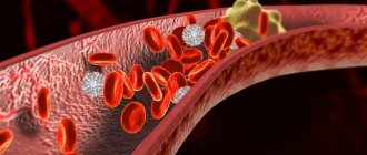
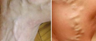
Currently, problems with blood vessels and veins cause discomfort to a large number of people of different ages and genders. A thrombus is a blood clot located on the walls of blood vessels that interferes with normal blood flow. If such a disorder is accompanied by an inflammatory process, a diagnosis of thrombophlebitis is made. In the absence of positive dynamics of conservative treatment, experts recommend removing the blood clot to further restore blood circulation.
Pulling with a probe
If a problem with blood clots is identified, a full examination of the body is prescribed, which will allow us to evaluate all the nuances of the pathology. Depending on the stage of the disease and individual indications, various techniques are used to remove a blood clot from an artery. One method is traction using a probe.
Using ultrasound diagnostics, the exact location of the clot and the extent of soft tissue damage are determined. Then a small incision is made directly into the tissue and artery to install the catheter. A special probe is used to fix the formed clot and pull it out.
The procedure takes no more than 20 minutes, after which blood flow is restored and pain is eliminated. After short-term rehabilitation, the patient returns to his usual lifestyle, following some recommendations of the attending physician.
The essence of the operation to remove a blood clot from the veins of the lower extremities
Thrombectomy requires modern equipment and specially trained doctors. The surgical technique is not particularly difficult and allows you to get blood clots in almost any arteries and veins. There are 2 ways to perform thrombectomy:
- traditional (with excision of the vessel);
- endovascular.
With the traditional technique of performing this operation, a vein is found and incised above the affected area. A special instrument is used to remove a blood clot that is blocking blood flow. The incision is then sutured.
Endovascular thrombectomy removes the clot with a balloon-tipped catheter while maintaining the integrity of the blocked blood vessel except for a small incision to enter the vein.
Surgeons use a catheter to find a blood clot, capture it and remove it. During the operation, the manipulation of the balloon catheter is monitored using x-rays.
The procedure for clearing blood clots from a vein can be performed multiple times. Then the site where the equipment enters the vein is sutured. An elastic bandage is applied to the operated leg.
If a floating thrombus is diagnosed, a vena cava filter can be installed for additional insurance. In the event of a sudden detachment of a blood clot, this device will be able to keep it in the right place and prevent serious complications. After the operation, control arteriography is performed.
This type of surgical intervention is performed in the vascular surgery department and, depending on the complexity, can last from several minutes to 2 hours.
Thrombolysis
The operation is performed under local anesthesia to reduce the pain of the procedure. A catheter is inserted into a deep vessel located in close proximity to the thrombosed vein. Gradually, with the help of angiographic equipment, the instrument is brought closer to the lesion. The blood clot is removed by applying a drug, a thrombolytic, upon contact, during which the resulting clot begins to dissolve.
Ways to fight blood clots
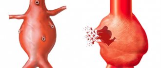
In some cases, they cannot resolve on their own, which leads to them becoming attached to the wall of the venous vessel. This leads to disruption of the blood supply to internal organs, which is fraught with the development of pathologies and complications.
In such a situation, doctors talk about ailments such as thrombophlebitis or vascular thrombosis and give recommendations for treatment that is aimed at dissolving.
Medication method
In order for it to resolve as quickly as possible, drugs from the group of thrombolytic agents are prescribed. Moreover, they are administered only intravenously. Among these drugs are the following:
- Streptokinase makes it possible to restore the function of blood flow. Allows you to destroy its structure. The drug is administered over half an hour using a dropper. The following dosages are administered hourly. Half an hour after the first dropper, it begins to dissolve.
- Urokinase is administered intravenously. Its properties are similar to Streptokinase.
- Alteplase - the drug is administered in a dosage of ten milligrams into a vein for 120 seconds. Next, use the dropper for three hours.
- Tenecteplase. The drug is administered using a syringe. In addition, it is necessary to take aspirin and heparin.
- A nicotinic acid. The drug is administered either intravenously or orally. The product makes the consistency more liquid, and clots are eliminated and dissolved.
Using such means, you can eliminate them in the jugular vein and in the vascular system of the arms and legs.
In addition to thrombolytics, in order to dissolve a blood clot, drugs from the group of heparins and warfarins are used - these are anticoagulants that allow it to resolve. The action of the medication is aimed at reducing the risk of blood clots.
Antiplatelet agents prevent platelets from sticking together, which reduces the likelihood of a blood clot.
New medical advances
Most of the medical products that we talked about above have an extensive list of side effects and contraindications. This suggests that not every person can be cured using these remedies. But chemists have recently developed a new way to eliminate dangerous blood clots.
The new drug belongs to the group of nano-drugs. Its main active ingredient is plasmin, which dissolves blood clots well. The component can move through the blood like a robot that detects blood clots, manages to eliminate them and continues to move through the bloodstream.
This treatment makes it possible to save a large number of people susceptible to heart attacks, since the medication has no side effects.
Hirudotherapy
Doctors who practice hirudotherapy know how to get rid of blood clots. Hirudotherapy is a method of treating vascular diseases using medical-type leeches. These unpleasant-looking creatures can help a person and have a positive effect on his body.
The secretion of leeches contains hirudin - this is a very valuable component that activates the blood thinning process, flushes the vascular system and capillaries.
In addition, hirudin can make vascular walls stronger and more elastic, and starts the process of dissolving blood clots. Leeches are good at removing blood clots. However, the procedures should be carried out in a certified center.
When a leech sucks blood, it also adds a large number of positive enzymes and analgesic components to the human body. All this leads not only to the fact that the clot will dissolve, but also to the relief of unpleasant symptoms of various diseases of the venous system.
Indications and contraindications
The specialist must first assess the need for the procedure and the general condition of the patient, and exclude possible complications. This is due to the fact that surgery to remove blood clots on the legs or other parts of the body involves intervention in the body. And this can lead to negative consequences. The technique is selected taking into account all factors to achieve a positive result.
Important information: How to treat thrombosis (thrombophlebitis) of the deep veins of the lower extremities and its symptoms (signs)
Indications:
- large size of the vessel lesion;
- floating thrombus;
- blockage of large vessels;
- gestation period (except for the last months).
Contraindications:
- mental illness;
- sepsis;
- high level of patient exhaustion;
- gangrene.
Particular attention should be paid to patients with chronic infectious diseases, with renal or gastric failure, with deep veins of the lower extremities, with diabetes mellitus, in old age, etc.
Traditional method of thrombectomy
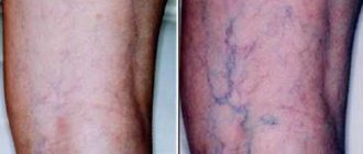
With varicose veins, blood clots often form. They can easily enter the vena cava and lead to blockage of the pulmonary arteries, which leads to negative consequences, including death. Emergency surgery is necessary in cases where there is a high risk of developing gangrene of the legs.
The intervention is performed under general anesthesia. The doctor gains access through an incision in the groin or inferior vein. To preserve the venous valves, catheters with two balloons are used during surgery.
To remove a blood clot with a traditional thrombectomy, an incision is made just above the site of the blood clot. In this case, the blood clot can be removed from any part of the body. Next, the surgeon inserts a special instrument with which the blood clot is removed. The incision site is sutured. The operation is over.
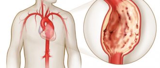
If the blood clot comes off and the lumen of the pulmonary artery is closed, then surgical intervention is carried out immediately. The surgeon gains access to the open heart and opens the chest.
During the procedure, the patient is connected to an artificial respiration apparatus.
Preparation for the event
Before performing blood clot surgery, an accurate diagnosis must be established. When performing diagnostic procedures, the location of the pathology, the degree of damage caused to surrounding tissues, the method of attaching the blood clot to the vessel, and other features of the disease are determined.
Diagnostics involves:
- ultrasound examination (scanning blood vessels to detect the location of blockage);
- venography (assessment of the condition of the veins by administering a contrast agent and performing radiography);
- magnetic resonance imaging;
- studying the results of a blood test.
After this, the day for the operation is set. Clots in blood vessels are removed under local anesthesia, so no special preparation of the patient is required. During this period, it is enough to choose comfortable shoes, stop taking medications and carry out hygienic procedures to remove hair from the operated area.
Thrombectomy of hemorrhoids
Thrombectomy of the hemorrhoidal node is a complication of varicose veins passing in the area of the hemorrhoidal plexus. A blood clot forms when blood circulation in the veins is impaired. When the outflow of blood stops, which is caused by involuntary contraction of the sphincter muscle, the hemorrhoidal node swells, increases in size, and acquires a purplish-black color (due to coagulated blood).
The process is accompanied by acute pain, deterioration of general condition (temperature rises, cold sweat appears, chills or fever develop). Analgesics do not relieve pain, and the symptoms of the disease become more intense. Typically, patients with hemorrhoidal thrombosis are hospitalized urgently, and the operation is unplanned.
Before the operation, which lasts on average 15 minutes, the intestines are washed. Local anesthesia (2% lidocaine solution) completely eliminates pain. The doctor is obliged to check with the patient about the tendency to allergies to medications. If the patient cannot provide reliable information, it is necessary to conduct a test for “antagonism” of the anesthetic. If novocaine drugs are not suitable, other types of pain relief are used.
The area of tissue above the clot is cut with a laser or scalpel, and a saline solution is injected into the wound cavity, softening the clot and having anti-inflammatory properties. The thrombus along with the capsule is carefully removed with a clamp. Drainage is left in the wound, and vascular coagulation is performed to stop bleeding.
Complications (formation of a secondary node, bleeding, wound infection) are recorded extremely rarely in modern medicine.
With proper care, the healing process lasts two days. After discharge, the patient must strictly follow the doctor's instructions regarding diet and hygiene. Lifting heavy objects, as well as bathing procedures and increased physical activity are strictly prohibited for at least a month.
Thrombosis of hemorrhoids, in most cases, is a local disease, that is, the vessels are affected only in a certain limited area (veins in the legs and arteries remain in good condition). The complication of hemorrhoids is a more than common occurrence, which, fortunately, responds well to treatment.
Is it possible to prevent the development of thrombosis of veins and arteries? There is still no clear answer to this question due to insufficient knowledge of the mechanism of clot formation in blood vessels. However, there are a number of effective measures that help maintain normal blood circulation and control blood composition. When the first signs of deviations from the norm appear, a course of therapy is prescribed, which has high therapeutic effectiveness in the early stages. Patients with a hereditary factor should be examined annually by a therapist or phlebologist.
Types of operations
A phlebologist (a specialist in the treatment of veins and blood vessels) or a vascular surgeon prescribes surgery only if conservative methods do not improve the patient’s condition. At an early stage, you can do without surgery, but if left untreated, new cells begin to attach to the inflamed cell, resulting in the lumen in the vessel closing. Blood circulation is impaired, which can lead to complete blockage and death.
Important information: Symptoms and treatment of phlebothrombosis of the deep veins of the lower extremities
Classic surgery method
Traditional thrombectomy involves surgically cutting the affected vessel, after which the resulting blood clot is removed and the damaged tissue is sutured. Before the procedure, you should refrain from eating for 12-15 hours, since the intervention is performed under general anesthesia.
The recovery period takes a fairly long period of time, because the sutures applied require special care to avoid cases of bleeding and infection. After surgery to remove blood clots in the legs, long-term rehabilitation with a course of anticoagulants is prescribed to gradually restore blood circulation.
Minimally invasive methods
Minimally invasive methods involve the use of modern equipment, since the procedure eliminates the dissection of damaged vessels. This is the main advantage over the classic removal of a blood clot from a vein, because the recovery process is faster and easier. The method is also called endovascular surgery.
There are 3 methods:
- aspiration (breaking the clot and pulling it out with a probe);
- thrombolysis (impact on pathology with special substances);
- rheolytic thrombectomy (removal using electric current).
Due to the absence of classical incision, procedures are performed under local anesthesia and constant medical supervision. The duration is about half an hour.
Removing a blood clot with a laser
After local anesthesia, a catheter is inserted into the vein cavity located above the pathological focus. Special laser beams pass through it and glue the venous lumens together. The peculiarity of the procedure is that it affects only the affected areas of the blood vessels, without having a negative effect on healthy areas.
The precision of manipulation is ensured by ultrasound examination for constant monitoring of laser actions. This removal of blood clots is painless for the patient; he experiences only moderate pain from the administration of the anesthetic. Another advantage is the short rehabilitation period; after 5 days, if the dynamics are positive, the patient goes home with a list of recommendations from the doctor. Most often it includes a course of medications and the use of compression garments.
Important information: What is the name of the blood test that needs to be taken to determine blood clots in deep vein thrombosis?
After laser removal, scars on the skin are barely noticeable, and after a few months they completely disappear. But this method is not used for extensive vascular lesions, since it is impossible to achieve the desired result of the procedure.
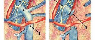
Endovascular thrombectomy method
With venous insufficiency, patients are at high risk of forming blood clots in the vessels and veins. The main method to help diagnose the presence of a clot is duplex ultrasound scanning. It not only allows you to find a blood clot, but also determines its density, the presence of moving fragments, and the degree of overlap of the vascular lumen.
Latest information: Varicose veins of the lower extremities: clinical recommendations
The danger of the pathological process is that in 99% of cases it occurs without symptoms if the blood passes without resistance. It is also necessary to determine the “age of the thrombus”; it is determined by the number of bypass paths.
Removal of a blood clot in the leg is performed in the hospital. It is still preferable to use the endovascular method for a blood clot in the legs, since it allows you to save the blood vessel. Using angiography, the location of the pathological area is determined.
The intervention consists of several stages:
- The surgeon determines the location of the blood clot.
- The wall of the blood vessel is incised.
- An empty balloon catheter is inserted through the incision.
- The doctor slowly moves the catheter to the clot, while simultaneously monitoring its position in the vessel using x-rays.
- When a blood clot comes into contact with the catheter, saline solution is pumped into the device.
- The instrument with the balloon is slowly pulled out.
The stretched catheter catches the blood clot and “leads” it to the incision site. It is the use of a catheter that allows you to reduce the likelihood of heavy bleeding to a minimum.
Rehabilitation period
After thrombectomy of the lower extremities, regardless of the method performed, the patient must adhere to medical instructions, which will shorten the recovery process and reduce the risk of relapse of the disease. These include the following:
- If pain persists, analgesics can be taken in recommended doses.
- To prevent inflammation of a blood clot in the leg, a course of non-steroidal anti-inflammatory drugs is prescribed.
- To thin the blood, it is necessary to take anticoagulants and antiplatelet agents.
- In accordance with the doctor’s recommendation, the period of wearing compression garments is determined.
- To prevent the recurrence of a blood clot on the leg, you should avoid any degree of physical activity (even long walks) for a selected period, stop drinking alcohol and tobacco, and follow a diet.
- Do not carry out hygienic and cosmetic procedures that involve heating the skin (sauna, steam bath, hot bath, thermal wraps, etc.).
The rehabilitation period is assigned individually, so after removal it is important to regularly visit a specialist to monitor and evaluate the positive dynamics.
Prevent trouble
Doctors are trying to prevent a possible catastrophe, the threat of which remains for several postoperative weeks, in different ways: by wrapping the patient’s legs with elastic bandages, as well as by shortening the period of bed rest. According to new surgical standards, even after a complex heart operation, they try to get the patient back on his feet almost on the day of the operation or the next day.
Postoperative treatment of patients with anticoagulants (blood thinners) has also become a great help for doctors. Until recently, the gold standard for such therapy was injections of low molecular weight heparin, after which the patient was prescribed so-called vitamin K antagonists, which, however, had a number of disadvantages, such as the need for constant laboratory monitoring of blood and dose adjustment due to the possible risk of bleeding.
Today, patients at high risk of thromboembolism have an alternative - a new generation of oral anticoagulants, which include direct thrombin inhibitors. Unlike vitamin K antagonists, this new, more effective class of drugs does not require constant monitoring of blood counts. And most importantly, this therapy significantly reduces the risk of dangerous bleeding, which has already been appreciated in many Russian hospitals, where direct thrombin inhibitors are successfully used to prevent thromboembolic complications in patients who have undergone knee or hip replacement. Experts are convinced that new oral anticoagulants have a great future.
Article on the topic
Heart attack: typical symptoms and surgical treatment options
How a blood clot occurs, causes and elimination
To begin to treat a blood clot, you will need to determine the symptoms and cause. Let's look at the key causes and remedies:
- Difficulties with blood vessels are congenital: insufficient patency of venous valves, difficulties with the elastic membrane, varicose veins from birth, fistulas in the deep veins. If abnormalities are found in the veins, this leads to stagnation of blood, platelets stick together and new blood clots constantly appear.
When the wall of a blood vessel is damaged, the cells in the veins are disrupted, causing the vessel to become damaged and blood to accumulate, which quickly clots to form a blood clot. People who exhibit increased blood clotting suffer - elastic fibers quickly stick together and increase blood viscosity. The reason may be hidden in a decrease in blood speed, this often happens when people remain in bed for a long time.
Indications for surgery
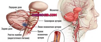
The formation of a blood clot in the leg, regardless of the cause, is a real risk for blockage of large vital vessels of the heart and lungs. Deep vein thrombosis is especially dangerous when, with minimal symptoms and the absence of external changes, an intravascular clot breaks off and enters the pulmonary artery through the bloodstream: in most cases, thromboembolism in the lungs is the cause of sudden death.
If occlusion in the legs is detected in a timely manner, it is necessary to conduct a full diagnosis and remove the blood clot from the vein.
The main indications for surgery are:
- the presence of a large clot detected during duplex scanning;
- a mobile (floating) thrombus that can break off at any time and cause acute occlusion;
- complicated variant of varicose veins of the legs and pelvis;
- lack of effect from drug therapy for thrombophlebitis and phlebothrombosis;
- high risk of blockage of the pulmonary artery or coronary vessels.
Observation after treatment
After removing blood clots and eliminating the causes of their occurrence, the patient needs to be monitored by a vascular surgeon. While in the hospital, the surgeon evaluates circulatory compensation and blood flow velocity using Doppler ultrasound.
Upon discharge, the patient is prescribed antithrombotic therapy, depending on the causes of thrombosis. These can be drugs such as Plavix, warfarin, aspirin.
A month after discharge, it is necessary to conduct a control duplex ultrasound scan of the arteries to assess the condition of the restored segments and circulatory compensation. It is also necessary to evaluate possible sources of thromboembolism (cardiac cavities, aorta) using echocardiography.
Subsequently, the patient must repeat ultrasound examinations and undergo blood clotting tests (APTT, TV, platelet aggregation) twice a year.
Along with drug therapy, other treatment methods are also used. Minimally invasive interventions are often used, in which all manipulations are carried out through a small puncture in the blood vessel. In this case, no anesthesia is required.
Possible complications during thrombectomy
- Rupture of the inner wall of the artery with its dissection and secondary thrombosis
- Tearing off the probe head and leaving it in the lumen of the artery
- Perforation of an altered artery wall with a probe with internal bleeding
- Complications associated with access (hematoma, lymph accumulation, wound suppuration)
Such complications are quite rare. Their frequency decreases as clinical experience with thrombectomies accumulates. In our clinic, we have not observed such complications over the past five years.
- Dissection of the arterial wall due to rough passage of the probe
- Transfer of pieces of blood clots to small arteries below the site of thrombosis
- Formation of hematoma along the artery
Such complications are usually identified during the operation and are usually corrected immediately.