The formation of a blood clot in a vessel is a natural reaction of the body to damage to the walls. It is aimed at restoring integrity and preventing bleeding. Normally, it dissolves after completing the task. In this way, only small blood clots can disappear if the blood circulation is in good condition.
With large formations and slow blood movement, recanalization of the blood clot : connective tissue and cells of the inner lining of the vessels grow into its thickness. They form peculiar passages through which blood moves. In addition to natural recanalization, there is also medicinal or surgical restoration of vascular patency.
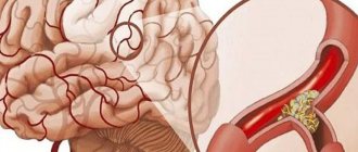
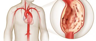
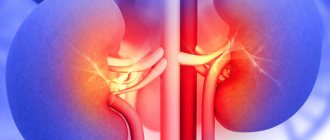
If the blood clot is not dissolved or removed from the vessels, then when the artery is blocked, there is a risk of infarction of the internal organs or brain . Venous thrombosis leads to obstruction of blood outflow and the formation of chronic venous insufficiency.
The most severe consequence is the detachment of a blood clot or part of it and its movement into the pulmonary artery system. The resulting thromboembolism is life-threatening.
Options for thrombus recanalization:
- Natural. Fibrinolysis prevents accelerated and widespread thrombus formation and is able to dissolve small blood clots. This option is optimal, since blood flow is restored. A large or even small thrombus formed in a place of weak blood flow is recanalized over time.
In this case, blood flow cannot be completely restored, since the formed pathways allow much less blood to pass through. Therefore, clinical manifestations during natural recanalization in most cases remain unchanged or become less pronounced.
- Medication. The use of drugs is justified in the first hours after blockage of the vessel if there is no threat to the patient’s life. If the risk is high and the blood flow has completely stopped, surgery is needed. Fibrinolytic therapy can be local (drugs are administered to the site of the thrombus) or general with the medication entering a vein.
Three generations of thrombolytics are used . The first is represented by natural enzymes - “Streptokinase” and “Urokinase”. They have a serious disadvantage: they do not stay in the blood for long, which requires constant administration, and with increasing dosages the risk of severe bleeding increases.
Therefore, any therapy to dissolve a blood clot is carried out only in a hospital setting . The plasminogen activator Actilyse and its genetic engineering modification Metalise are more selective, less likely to lead to uncontrolled blood loss and penetrate deeper into the blood clot.
Thrombolytics are prescribed for:
- acute coronary syndrome;
- myocardial infarction;
- pulmonary embolism;
- blockage of the arteries of the lower extremities with the threat of gangrene.
After using fibrinolytics, to prevent recurrent thrombosis, anticoagulants are recommended , for example, Heparin, Clexane, Warfarin, Sinkumar, as well as antiplatelet agents that inhibit platelet joining.
Action of anticoagulants
To accelerate the recanalization of a blood clot and protect blood vessels from blockage, the drug "Wessel Due F" is used . It is first administered intravenously, and then the patient is transferred to tablets. The minimum course lasts a year . This therapy restores the structure and function of the inner lining of blood vessels, improves blood flow, and prevents postthrombophlebitic syndrome.
Xarelto is recognized as an equally effective remedy ; it is often combined with venotonics, angioprotectors and vasodilators.
In case of acute blockage of the arteries, which threatens a vascular catastrophe, and there is no result from fibrinolytic therapy, surgery is indicated to restore blood flow. Surgical recanalization involves:
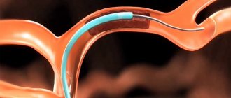
- blood clot extraction using the intravascular method: instruments are passed through the inserted catheter, which captures and removes blood clots;
- creating a bypass path for blood flow (bypass) using your own vein or a synthetic prosthesis;
- collateral training: a suture is placed on the vessel above and below the blockage, which leads to the opening of additional branches of the bloodstream, bypassing the affected artery or vein;
- balloon expansion and installation of a stent (frame) that prevents the walls from collapsing.
Surgery to remove a blood clot
Over the course of several days or 2-3 weeks after the formation of a blood clot and blockage of a vessel, the blood clot goes through the following stages :
- melting: partial or complete fibrinolysis with reduction or disappearance of the thrombus;
- organization: ingrowth of connective tissue fibers, thickening of the thrombus and its movement into the vascular wall;
- recanalization: formation of capillary channels to restore blood flow;
- vascularization: restoration of blood movement through a thrombus;
- remodeling: since the movement of blood through the thrombus is weak, some of the channels close, and the thrombus becomes covered with vascular epithelium and turns into a fibrous lump, changing the outline of the vein or artery;
- Enzymatic destruction: inside the organized blood clot, enzymes remaining in the white blood cells and platelets that are stuck in the blood clot are released. Because of this, its central part may collapse, and if there is a microbial infection in this place, the vascular wall may collapse.
Stenting and balloon dilatation of the vessel
in the mode of duplex or triplex scanning of vessels is most often prescribed . It includes conventional echolocation, blood flow analysis and blood flow mapping - creating a color portrait of a vein or artery. This helps to quickly install:
- how tightly the thrombus blocked the vessel;
- is there recanalization;
- where the bypass routes are located and how they function;
- How effective is thrombolysis or surgery?
The accuracy of diagnosing venous thrombosis using an expert-class device approaches 99%. Less commonly, patients are prescribed rheovasography of the lower extremities or radionucleoid venography. If there is a blood clot in the artery, angiography is performed with X-ray or tomographic control.
Read more in our article on thrombus recanalization.
Vein recanalization. What is this? Classification.
Recanalization is the process of restoring the patency of a vessel whose lumen is closed by a thrombus. Vein recanalization occurs in one of three ways:
- natural;
- medicinal;
- surgical
Natural recanalization is a physiological process. It occurs under the influence of aseptic fibrinolysis. The clot resolves on its own in almost half of the cases. In addition to the destruction of the blood clot, its revascularization is possible: it grows with microvessels and collagen structures. The patency of the vessel is restored and the destruction of the clot is accelerated. Independent removal of blockage is possible at the stage of a “loose” blood clot. When it is overgrown with connective tissue, fibrinolysis is difficult.
In healthy people, recanalization occurs under the influence of the protein plasmin a few days after the formation of the blockage - at the initial stage of development.
However, sometimes the body cannot cope with the dissolution of a blood clot: blood thickening, increased clotting activity, genetic predisposition, and impaired venous circulation complicate this process. The resulting clot blocks the lumen of the vessel and can cause embolism, a life-threatening condition. In these cases, medical recanalization is indicated.
Stages of recanalization and its timing
Over the course of several days or 2-3 weeks after the formation of a blood clot and blockage of a vessel, the blood clot goes through the following stages:
- melting: partial or complete fibrinolysis with reduction or disappearance of the thrombus. Old thrombotic masses cannot be dissolved due to the presence of bonds between fibrin strands. Therefore, fibrinolytics are effective only in the first hours;
- organization: ingrowth of connective tissue fibers, thickening of the thrombus and its movement into the vascular wall;
- recanalization: formation of capillary channels to restore blood flow;
- vascularization: restoration of blood movement through a thrombus;
- remodeling: since the movement of blood through the thrombus is weak, some of the channels close, and the thrombus becomes covered with vascular epithelium and turns into a fibrous lump, changing the outline of the vein or artery;
- Enzymatic destruction: inside the organized blood clot, enzymes remaining in the white blood cells and platelets that are stuck in the blood clot are released. Because of this, its central part may collapse, and if there is a microbial infection in this place, the vascular wall may collapse.
Stages of thrombus recanalization
Vascular diagnostics
The need for medical recanalization is determined using instrumental research methods. The condition of blood vessels and the degree of blood flow disturbance are assessed using the following methods:
- duplex scanning - determination of blood flow speed through the vessel under study and anatomical defects of veins and arteries;
- angiography is an x-ray method that uses the introduction of contrast to detect obstacles to blood flow: blood clots, narrowings;
- computer and magnetic resonance imaging are high-precision diagnostic methods that make it possible to detect pathological processes in all organs and tissues, including blood vessels.
Indications for thrombus recanalization
Recanalization of a blood clot is necessary to restore circulatory function in the problem area and prevent dangerous complications, which include atrophy, stroke, coronary artery disease, and even death. The procedure is carried out immediately after detection of a venous clot. This allows you to normalize the patient’s condition and prevent pathological processes.
The main indications for its implementation are as follows:
- thrombosis of the lower extremities;
- formation of a clot in the jugular vein;
- blockage of the vessels of the cavernous sinus;
- portal vein thrombosis;
- pathological damage to the vessels of the central retinal vein.
To diagnose the pathological process, you should visit a therapist. He will conduct a survey that will allow you to understand which doctor to go to. Most often, vascular diseases are dealt with by a phlebologist. In some cases, the help of highly qualified specialists is required.
Independent removal of blockage is possible at the stage of a “loose” blood clot.
The following procedures help make an accurate diagnosis:
- Duplex scanning evaluates the vessel, identifying the area of deformation.
- Ultrasound with Doppler allows you to analyze the quality of blood circulation.
- Angiography is an x-ray method for detecting a blood clot. The procedure is performed by injecting a contrast agent into a vein.
- CT and MRI can detect pathological processes not only in the circulatory system, but also in other organs.
If symptoms of thrombosis occur, you should not resort to traditional medicine and ignore the problem. You should immediately consult your doctor for advice.
Drug recanalization
Conservative, or medicinal, recanalization is carried out if there is no threat to the patient’s life. It is based on the use of drugs that promote the process of restoration of the vascular bed and destruction of the blood clot. Drug recanalization is carried out within the first 3 days from the moment of clot formation, and the greatest effectiveness is achieved when therapy is carried out in the first 6 hours. The following types of drugs are used:
- anticoagulants;
- antiplatelet agents;
- thrombolytic agents;
- angioprotectors.
Thrombolytic therapy
Thrombolytic drugs (fibrinolytics) start the process of resorption of blood clots. Their action is based on the transformation of plasminogen into plasmin. This protein, in turn, breaks down fibrin, the basis of the blood clot. The use of fibrinolytics has a number of contraindications, non-compliance with which can provoke life-threatening conditions, therefore they should be taken only as prescribed by a doctor.
Fibrinolytics of 3 groups are used:
- 1st generation (Streptokinase, Urokinase);
- 2 generations (Alteplase, Prourokinase, Actylase);
- 3 generations (Retaplase, Tenecteplase).
1st generation drugs are effective, but they often cause allergies, and they also have an effect on fibrinogen and prothrombin, blood clotting factors. This creates a risk of bleeding. The shortcomings of the first drugs are smoothed out in the 2nd and 3rd generation drugs. They have a selective effect and influence the blood clot. The blood thinning effects are not as pronounced as with Streptokinase and Urokinase.
Use of anticoagulants
Anticoagulants are used to limit the growth of blockage in the vessel. Their use is advisable specifically to stop the increase in the diameter and length of the clot; the effect on resorption is not as pronounced as that of thrombolytics. According to the mechanism of action, anticoagulants are classified into direct and indirect agents. Indirect drugs reduce the synthesis of vitamin K in the liver, as a result of which thrombin formation is inhibited. These include: dicoumarins, monocoumarins and indanediones. Direct anticoagulants affect coagulation factors. Among them: heparin, hirudin, sodium hydrogen citrate, danaparoid.
The choice of drug group depends on the severity of the patient’s condition, concomitant diseases and coagulation status. To assess the latter, there is an analysis - a coagulogram; it displays the activity of the blood system during treatment with anticoagulants.
Antiplatelet and angioprotective therapy
Antiplatelet drugs block platelet aggregation. Acetylsalicylic acid (aspirin) is used for these purposes. This is one of the inexpensive and common drugs used to prevent the formation of new clots and stop the growth of existing ones.
Angioprotectors (Pentoxifylline, Parmidine) increase venous tone and reduce capillary permeability. As a result, blood stagnation decreases and its return to the heart increases, capillary swelling decreases, their stability increases, microcirculation in the affected tissues is normalized, thrombus formation is reduced and the rheological properties of blood are improved.
Natural restoration of blood flow
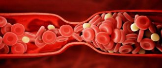
Blood clots form due to blood's natural ability to clot. This is necessary to prevent excessive blood loss in the event of injury. As a result of certain abnormalities in the body, blood clotting increases. A plug forms in the vessel, formed from individual elements of blood cells. It does not allow blood to circulate in the right direction.
Despite this ability of the body, doctors try to eliminate the blood clot until it resolves itself. The fact is that there is a high risk of blood clot detachment. This will cause it to move through the circulatory system. If a blood clot clogs a vessel in a vital organ, the person will die.
According to statistics, up to 150 cases of venous thrombosis are registered every year. In every eighth person with this disease, this condition is worsened by pulmonary embolism, which in 50% of cases leads to death.
Surgical recanalization
If there is a possibility of a blood clot breaking off, when a vein of a vital organ is blocked, when waiting for the effect of taking medications is unacceptable, surgical recanalization of the veins is performed. Surgery is also prescribed if drug therapy is ineffective. In this case, anticoagulants are first discontinued, since taking them can provoke bleeding.
The following types of surgical recanalization are performed:
- Removal of a blood clot is carried out minimally invasively, using endovascular therapy methods. An incision is made under local anesthesia, a catheter is inserted into the damaged vessel and, under the supervision of the operating physician, it is brought to the location of the thrombus. Next, the clot is captured and removed from the vein.
- Bypass surgery is used when the clot cannot be removed. A bypass path of blood flow is formed. The material used is your own vessels - veins taken for plastic surgery, or synthetic analogues.
- Ligation involves applying a ligature above and below the site of the clot, blood flow is redistributed among small arteries and veins.
- Stenting is the insertion of a balloon that dilates the vessel. Blood circulation improves and platelet aggregation on the affected wall decreases, but such an operation is advisable only if thrombus formation is gradual.
Surgery is not always performed to restore blood flow. Recanalization of the umbilical vein is performed to provide access to the liver and gallbladder in case of their pathology. Infusion solutions are administered through a catheter; in case of purulent damage to organs, antibacterial agents are delivered to the site.
Venous thrombosis of the lower extremities occurs in 10-20% of the population. This disease is manifested by circulatory failure and trophic disorders. For more complete recanalization of deep veins, complex therapy is necessary: taking medications, following a regimen, using compression stockings, and, if necessary, surgical treatment.
Recanalization of a thrombus is often a lengthy process that requires the attention of not only doctors, but also patients. To achieve a better result, as well as to prevent further thrombus formation, the patient must reconsider their lifestyle, follow a diet and promptly consult a doctor at the first symptoms of the disease. Advances in modern medicine can significantly increase the likelihood of a full recovery.
Features of thrombus recanalization
When thrombosis occurs due to blockage of a vessel, circulatory disturbance occurs.
This leads to gradual atrophy of the organ and associated complications. To restore the function of the affected organ and prevent the development of undesirable processes, measures are taken to expand the lumen of the vessel. These include thrombus recanalization. In a healthy body, it is carried out physiologically under the influence of plasmin. If this does not happen, the clot is removed by surgery or with the help of medications. In this article we will tell you:
How is thrombus recanalization performed?
Thrombus recanalization is the process of restoring the patency of arteries or veins that have undergone thrombosis (the formation of blood clots that impede blood flow through the vessels).
This manipulation can be carried out through lysis of blood clots (dissolution using various medications) with their further vascularization (plasty of new blood vessels) or surgical intervention. But nevertheless, specialists currently do not have the opportunity to completely restore the function of venous patency, therefore, after thrombosis, a person remains with its consequences for the rest of his life in the form of a pathological process such as chronic venous insufficiency.
Causes of blood clots
The ability of blood to clot is the most important quality developed in the process of evolution, preventing blood loss in the event of minor injuries. As the blood thickens, it forms a kind of plug, blocking the damaged vessels and thus stopping the bleeding. If a person is healthy and his vascular system is in order, then as they are no longer needed, they “self-liquidate” - they dissolve under the influence of the fibrinolysis mechanism. But when various pathologies occur, thrombosis processes are activated, and self-destruction becomes more difficult, and the protective factor takes on the exact opposite meaning.
Read also: Contraindications for varicose veins on the legs in women
There may be several reasons:
- changes in the structure of the vascular wall (thinning, fragility);
- excessive blood thickness;
- disruption of normal blood flow speed.
When the formation of a clot occurs as a result of atherosclerotic phenomena, the clumping red blood cells that make up its “body” settle on the surface of cholesterol plaques that appear in the bloodstream. Sometimes the walls of the arteries become so thin that biological substance begins to leak through them, and the body, trying to eliminate bleeding, increases thrombosis.
Stagnation of blood flow is promoted by a sedentary lifestyle, work that requires long-term static postures (sitting and standing), as well as bad habits: smoking, poor diet, and excessive coffee consumption.
In addition, thrombosis can be caused by:
- Toxicological and chemical poisoning.
- Surgery – the body tries to eliminate the blood loss that occurs during this process, increasing the formation of “patches”.
- Phlebeurysm.
In all these cases, constant monitoring by a specialist and diagnosing the condition of the vascular system is necessary. The consequences of possible complications of the situation in the form of thromboembolism in the absence of timely diagnosis and medical care are extremely tragic, even death.
Removing a blood clot using medication
After diagnosing thrombosis of the pulmonary or cerebral vessels, as well as the coronary arteries, the patient needs immediate medical care. The outcome of events depends on the speed of therapeutic manipulations. The main danger of untimely treatment is the increased likelihood of death.
In order to expand the blood arteries that supply the brain with nutrition, intravenous drugs are prescribed, which include Cavinton and Cinnarizine.
In case of development of coronary heart disease, organic donors of nitric oxide and nitrates are used to enlarge the coronary veins, among which are sodium nitroprusside and nitroglycerin. To prevent thromboembolism, it is also necessary to take angioprotectors and antiplatelet agents.
Angioprotectors slow down the further progression of varicose veins, reduce the intensity of postthrombophlebitis syndrome, which manifests itself in increased capillary permeability and the formation of swelling and trophic ulcers.
Sulodexide is administered intravenously or taken orally. Enoxaparin and other low molecular weight heparins are used under APTT monitoring. Therapy with indirect anticoagulants, which include Warfarin, is carried out under the control of INR, since it prevents the synthesis of blood coagulation factors in the liver.
The main advantage of Sulodexide is considered not only to stop the process of thrombus formation and recanalization of problematic vessels, but also to the possibility of using it to prevent relapses, as well as eliminate chronic venous insufficiency.