The essence and causes of occurrence
The causes of subclavian vein thrombosis are the following factors:
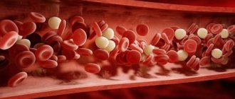
- chaotic blood movement or significant slowdown;
- impaired blood clotting (under the influence of any factors, due to diseases of a genetic and hereditary nature);
- The subclavian vein may be compressed by a large and abnormally formed bone spur, which may result from a clavicular fracture or the appearance of an uncharacteristic cervical rib.
Subclavian vein thrombosis can only rarely be the result of a blood clot formed in other areas of the body. This phenomenon is associated with the organization of the human circulatory system. Thrombosis of the upper extremities occurs as a result of a blood clot breaking off in the heart muscle. In most cases, subclavian vein thrombosis practically does not manifest itself at all if the process occurs slowly.
Thrombosis of the subclavian vein - treatment at the Innovative Vascular Center: symptoms and causes
Treatment under the compulsory medical insurance policy is free!
Paget-Schroetter syndrome is a thrombosis of the subclavian vein, extending to the subclavian and axillary veins, as well as to the veins of the shoulder, which leads to impaired venous outflow in the arm.
The syndrome is also called traumatic thrombosis or effort syndrome. Despite its relative rarity, it has become increasingly diagnosed in the last decade.
The main complaints are swelling of the tissues of the upper limb and bursting pain.
The first case of the syndrome was described by James Paget in 1875, and in 1894 von Schrötter identified vascular injury as a potential cause of the disease. The term Paget-Schroetter syndrome was first used in 1948.
Causes and risk factors
Subclavian vein thrombosis is associated with compression of the thoracic outlet.
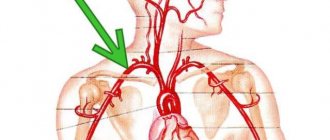
The subclavian vein originates from the first rib from the axillary vein, and at the level of the sternoclavicular junction with the jugular vein it forms the brachiocephalic vein.
Compression of the vein walls in the area between the collarbone and the first rib leads to slower blood flow and thrombosis. It can be considered the venous equivalent of thoracic outlet syndrome.
One of the causes of thrombosis is muscle hypertrophy (increase in the volume or mass of skeletal muscles). As a result, the subclavian vein can become compressed between the ribs (in front of it), the muscle (behind it), and the collarbone (above it).
Another reason is that a person has a congenital small anatomical space between the collarbone and the first rib. In this case, compression of the subclavian vein is possible even without major muscle hypertrophy.
Other causes of thrombosis include:
- incorrect posture,
- bone pathology (in the subclavian region),
- collarbone fractures
- use of subclavian catheters
- incorrect sleeping position
- thoracic outlet syndrome
- Risk factors include excessive physical activity. People who undergo intense physical activity and athletes (wrestlers, weightlifters or bodybuilders) have a higher risk of developing thrombosis due to repeated damage to the subclavian vein from frequent mechanical compression of the vessels between the collarbone, first rib and joint.
The average age of patients with Paget-Schroetter syndrome is 30-40 years, and the male to female ratio is approximately 2:1. It is more common on the right side, probably due to the frequency of right hand dominance, and 60% to 80% of patients have this condition. who performed vigorous exercise involving the upper limbs.
Course of the disease
The course of the disease is divided into two stages: acute (lasts about three weeks; at the initial stage, symptoms appear during physical activity) and chronic (symptoms last for more than two months).
The acute phase is characterized by increasing swelling of the arm, pain, and a feeling of fullness. The patient's ability to work decreases. Gradually, tense saphenous veins develop, which take on the function of outflow of venous blood and contribute to the subsidence of the process.
The chronic phase is the consequences of thrombosis. With inadequate treatment, occlusion of the subclavian vein persists and chronic Paget-Schroetter syndrome develops. It is characterized by the development of a powerful collateral network of saphenous veins around the shoulder joint. Patients are concerned about the increase in volume of the sore arm, sometimes there is pain and increased fatigue.
Forecast
Life-threatening complications are rare. Pulmonary embolism is observed in only 2% of cases of subclavian thrombosis.
Even with immediate intervention, some patients do not fully regain arm function and may have residual symptoms or require continued treatment. Severe venous insufficiency of the arm usually does not occur.
Thrombosis of the subclavian vein does not pose a serious threat to life. After adequate treatment, the edema decreases significantly, but complete patency is rarely restored.
If the causes for subclavian thrombosis persist, relapses of the disease may occur, so it is necessary to identify and eliminate them.
As a reminder of the history of venous thrombosis, the patient is left with an increase in the volume of the shoulder and a network of visible saphenous veins in the area of the shoulder joint.
Advantages of treatment in the clinic
Effective thrombolysis – dissolution of blood clots
Eliminating the causes of subclavian vein thrombosis
Angioplasty and stenting
Source: https://angioclinic.ru/zabolevaniya/tromboz-podklyuchichnoy-veny/
Symptoms
Thrombosis of the subclavian vein occurs as a result of excessive physical activity and stress. This factor plays a major role in the formation of a blood clot. In some cases, a blood clot can break off regardless of the degree of physical activity, but this happens in very rare cases. Blockage of the subclavian vein has either increasing or disappearing symptoms, manifested in influxes. There are no serious consequences from thrombosis, because blood circulation is replaced by other vessels. However, this blood is not enough to fully supply the tissues of the upper extremities. In ICD-10, subclavian vein thrombosis has code I82.8.
Reasons for the development of subclavian thrombosis
Depending on the type of vessel affected (vein or artery), etiological factors may differ.
Blockage (occlusion) of an artery
Most often, atherosclerosis leads to blocking the lumen of the subclavian artery. With this disease, a plaque forms in the inner layer of the vessel, which can be complicated by thrombosis.
The occurrence of blood clots is provoked by the following pathologies:
- high blood pressure;
- increased levels of low and very low density lipoproteins and triglycerides in the blood;
- nicotine addiction;
- diabetes;
- heart failure;
- rhythm disturbances;
- inflammation of the artery with a pronounced increase in the muscle layer (obliterating endarteritis);
- autoimmune damage to the aorta with blockage of its segments (Takayasu arteritis);
- external compression from tumors, injuries, scars;
- structural anomalies.
Venous thrombosis
A high risk of thrombus formation in the subclavian vein area is observed in the presence of the following conditions:
- long-term catheter stay;
- installed pacemaker or cardioverter-defibrillator;
- malignant tumors;
- compression by overdeveloped muscles in athletes.
Primary acute vein thrombosis may be a manifestation of Paget-Schroetter syndrome, which occurs due to anatomical features of the structure of the cervical and thoracic skeleton (additional cervical rib, narrow space from the clavicle to the first rib). Provoking conditions for compression of the vein are sports or heavy physical labor, heavy lifting, and stooping.
Damage and subsequent thrombus formation also occurs after injuries to the chest and shoulder girdle.
Other symptoms of pathology
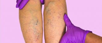
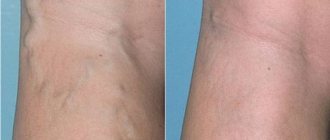
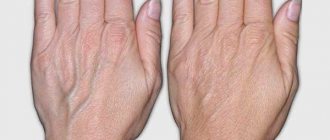
The appearance of a venous pattern on the arm is difficult to miss, especially for white-skinned people. The diameter of the veins will depend on the size of the clot and the increasing hypertension of the thrombus.
The pain process is usually observed during physical activity. The pain can be constantly present, throbbing, “bursting”, but in all cases it is quite intense. Basically, the pain is felt throughout the arm, in the area of the shoulder and collarbone, and in some cases also on top of the chest and back.
Swelling occurs throughout the entire arm. If you press on the swollen area, the hole does not remain in this place. The hand acquires unusual heaviness and hardness. If the swelling process has been going on for quite a long time, blood circulation is disrupted and becomes reactive, as a result of which thrombosis of the subclavian vein only intensifies.
Neurological disorder affects everyone differently. For many, the fingers of the limb twitch, there is a tingling sensation in them, a burning sensation. The affected limb may have limited movement.
If acute thrombosis of the subclavian vein becomes chronic, the clinical picture of the disease becomes blurred and not so pronounced. Swelling and vein patterns practically disappear. Most often, there remains a low response of the injured limb to external stimuli, restrictions in motor activity, muscle atrophy and pain during severe physical exertion. In some cases, disability is prescribed for thrombosis of the subclavian vein.
Symptoms, causes
Signs of the disease are usually limited to the upper extremities. The reasons may be:
- Hypercoagulation. The process represents manifestations that are characterized by hereditary as well as autoimmune diseases. Blood clotting is accelerated by chemotherapy, hormone therapy, and radiation surgery.
- Damage to blood vessels. Thrombophlebitis tends to be located in places where there was one or another injury or operation. This is where wall deformation begins.
- Blood stagnation, in most cases, occurs due to abnormal heart function or a sedentary lifestyle. The duration of treatment or diagnosis may vary.
Diseases that activate the process of disease development:
- Certain diseases from the field of oncology
- Diabetes
- Improper metabolism
- Fractures
- Kidney dysfunction
- Atherosclerosis
- Hormone imbalance
In the case when the venous column is blocked by a blood clot, the functioning of the entire venous system is disrupted. Typically, such moments are also accompanied by intoxication syndrome, as a result of which one can observe swelling, cyanosis, and swelling. Also, the patient will feel heaviness in the lower extremities. If the disease exists, but there are no symptoms, you need to worry, since such moments can lead to death.
Thrombosis of the lower extremities is a disease that very often has complex outcomes. Typically, this disease affects the thigh area and also produces pain in the affected areas where a blood clot has formed. The pain may be accompanied by cramps and numbness. The intensity of pain increases when walking, as well as when climbing stairs. At the most “advanced” stages, you feel complete distension of the leg and a blue color of the skin.
- As a result of impaired brain function, a stroke very often occurs. Typically, it develops after blockage of blood vessels in the brain and can cause hemiparesis.
- Pain after exercise
- Swelling of skin tissue
- Blue skin color
- Formations in the form of pits
- Impaired blood flow to the hand
- Enhancing vein pattern features
Such symptoms differ in their individual characteristics and are individual in nature. It is worth noting that the disease can be easily suspected without special examinations.
Latest information: The danger of parietal thrombosis of the heart and aorta
Obvious symptoms of the disease
The venous pattern can be noticed not only by the patient, but also by other people. Moreover, it is pronounced on skin that has a light shade and is not characterized by a tan. In this case, the veins simply swell and begin to expand. The intensity of such manifestations may indicate the stage of the disease, as well as the size of the blood clot.
Pain can occur after certain physical activities, but it is not permanent. The pain is throbbing and has a pronounced intensity. Such sensations can cover not only the area that is affected, but also spread to other parts of the body - the collarbone, back.
A clear symptom is swelling, which can be seen throughout the upper limb. If a medical representative examines the patient’s skin, he will notice that small pits have appeared in such places, and the swelling itself is tense. In some situations, such edema can even cause complete disruption of arterial blood flow. Such a moment only aggravates the course of the disease.
There are also symptoms in the field of neurology. The patient will feel a burning sensation in the affected areas of the body. Also, movements may be slightly limited, and tendon reflexes will increase.
The chronic stage of the disease is distinguished by its own symptoms, which are not so clearly expressed. At this stage, symptoms that are neurological in nature are more prevalent:
- Movement restrictions
- Decreased all reflexes
- Atrophic changes in muscles
- Diagnosis and treatment
To diagnose this disease, the following steps are taken:
- X-ray and MRI of the shoulder girdle. Using this operation, the cause of thrombosis is determined
- contrast radiography
- Doppler - research
Diagnostic measures
Diagnostic actions begin with collecting anamnesis - that is, the doctor must question the patient in detail about the symptoms that are troubling him and about when and what types of physical activity could lead to such consequences. This is necessary in order to find out how long the thrombosis process has been occurring.
To diagnose chronic or acute thrombosis of the subclavian vein, the following diagnostic methods are used:
- radiography and magnetic resonance imaging to determine the cause of the pathology and identify the location of the blood clot;
- duplex scanning of the subclavian vein;
- assessment of blood circulation in the injured vein - Dopplerography;
- contrast x-ray;
- Ultrasound examination (ultrasound) of deep veins;
- venography;
- computed tomography (CT) of the shoulder girdle.
Causes
The primary etiology is referred to as effort-induced thrombosis or Paget-von Schrötter syndrome. This usually occurs due to overuse of the involved hand by predisposed individuals. A secondary etiology is subclavian vein catheterization, especially in cancer patients.
Mortality rates for untreated pulmonary embolism may be as high as 10%, which is comparable to that observed for VT originating from the lower extremity. The long-term consequences of venous occlusion cause significant morbidity associated with persistent pain and swelling over long periods.
These symptoms are sometimes severe and may be aggravated by physical activity, especially with prolonged use of the affected arm. Consequently, this syndrome can be disabling and can negatively affect the patient's quality of life.
With the development of endovascular technologies, there is increasing evidence that these methods are the method of choice when encountering a patient with thrombosis of the upper extremities. Anticoagulant or thrombolytic therapy was insufficient. Today, endovascular stenting can address both benign and malignant causes of upper extremity thrombosis. This method allows for significantly faster recovery, while reducing morbidity and fewer complications in the long term.
Treatment
If the disease is a consequence of the action of the catheter, then it must be removed. If the vessels are slightly clogged, then local therapy is resorted to. The limb should be in so-called functional rest, and no elastic bandages or complete bed rest are needed here. In a horizontal position, the hand should be raised slightly above the heart, and in a vertical position, it should be suspended, bent at the elbow, using a bandage or scarf. For local treatment the following drugs are used:
- alcohol-based compresses (about 50%);
- “Gepatrombin”, “Liotongel” - ointments containing heparin;
- gel ointments containing troxevasin and rutoside;
- non-steroidal anti-inflammatory drugs, for example, Indomethacin ointment, Indovazin, Diclofenac.
Symptoms
Patients may describe a history of trauma or, more commonly, heavy use of the hand (>50% of cases). Common accelerating actions include repeated hyperabduction and external rotation of the arm or backward and downward rotation of the shoulder. Causal activities may include participation in cricket, tennis, wrestling, weight lifting, water polo, gymnastics, baseball or chopping wood.
Because the symptoms of subclavian stenosis are quite severe, most patients present to the emergency department immediately, usually within 24 hours. They may report a dull ache in the shoulder or armpit, and the pain is often worsened by activity.
Conversely, rest and elevation often relieve pain. Patients with catheter-associated axillary subclavian vein thrombosis report similar symptoms in the ipsilateral arm or shoulder with an indwelling catheter. Patients may describe a history of trauma or, more commonly, heavy use of the hand (>50% of cases). Common accelerating actions include repeated hyperabduction and external rotation of the arm or backward and downward rotation of the shoulder. Causal activities may include participation in cricket, tennis, wrestling, weight lifting, water polo, gymnastics, baseball or chopping wood.
Because the symptoms of subclavian stenosis are quite severe, most patients present to the emergency department immediately, usually within 24 hours. They may report a dull ache in the shoulder or armpit, and the pain is often worsened by activity. Conversely, rest and elevation often relieve pain. Patients with catheter-associated axillary subclavian vein thrombosis report similar symptoms in the ipsilateral arm or shoulder with an indwelling catheter.
A little more about drug treatment
If the pathology becomes acute and is accompanied by extremely painful symptoms, the patient is admitted to a hospital. There treatment consists of the following medications:
- fibrinolytic drugs - “Fibrinolysin”, “Streptokinase”, “Urokinase”, etc.;
- antiplatelet agents;
- angioprotectors;
- medications against blood clotting (for the first few days it can be “Heparin” and “Fibrinolysin”, and then they use “Phenilin”, “Sinkumar”, “Fraxiparin”);
- non-steroidal anti-inflammatory drugs.
The main task of treating subclavian vein thrombosis with the help of medications is to restore impaired blood circulation in the subclavian vein,
To summarize, drug treatment comes down to two types of drugs:
- antithrombosis medications that help destroy blood clots and prevent the formation of new ones (heparin is one of these);
- drugs that help improve the metabolism of vein walls, painkillers and anti-inflammatory drugs.
Causes
The primary etiology is referred to as effort-induced thrombosis or Paget-von Schrötter syndrome. This usually occurs due to overuse of the involved hand by predisposed individuals. A secondary etiology is subclavian vein catheterization, especially in cancer patients.
Mortality rates for untreated pulmonary embolism may be as high as 10%, which is comparable to that observed for VT originating from the lower extremity. The long-term consequences of venous occlusion cause significant morbidity associated with persistent pain and swelling over long periods.
These symptoms are sometimes severe and may be aggravated by physical activity, especially with prolonged use of the affected arm. Consequently, this syndrome can be disabling and can negatively affect the patient's quality of life.
With the development of endovascular technologies, there is increasing evidence that these methods are the method of choice when encountering a patient with thrombosis of the upper extremities. Anticoagulant or thrombolytic therapy was insufficient. Today, endovascular stenting can address both benign and malignant causes of upper extremity thrombosis. This method allows for significantly faster recovery, while reducing morbidity and fewer complications in the long term.
When is surgery scheduled?
Typically, drug therapy lasts from one to several months. If during this period the blood clot does not resolve, then surgical intervention must be resorted to. If thrombosis of the subclavian vein (left or right) lasts too long, tissue necrosis of the upper limb may occur. In such a situation, an operation is also prescribed, during which it is necessary to remove tissue that has died.
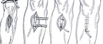
To remove a blood clot, a special device called a laparoscope is used. It passes into the vein, captures the clot and pulls it out. For this purpose, a small incision is made in the armpit area, which allows you to get close to the subclavian vein; a puncture is also made in it - this is where the laparoscope passes. In case of minor injury to the vascular walls, a special catheter is inserted there. In some cases, a special shunt is inserted into the affected area of the vein.
It is also important to note that if the patient experiences a feeling of warmth, sharp pain, there is swelling and the process of redness or blueness of the shoulder, then it is urgent to start a new course of treatment. All of these symptoms may indicate a pulmonary embolism. If a patient is diagnosed with a stomach ulcer or gastritis, he is prescribed suppositories. Also, the usual “Aspirin” is excluded from the course of treatment; instead, it is necessary to select drugs of the same properties, but soluble in the intestines. To avoid relapses and repeated exacerbations, antihistamines are used as a prophylactic agent.
The severity of the pathology of the main veins, the peculiarities of the formation of blood clots and the formation of bypass circulatory pathways influence how seriously the venous outflow is disrupted and how intensively the characteristic symptoms and signs of the disease appear. If a patient has idiopathic thrombosis, then in most cases he will require a special course of treatment for life.
Treatment of subclavian artery thrombosis
If, when an artery is blocked, insufficient blood flow to the brain, hand and fingers is detected, then surgical treatment is indicated for patients. It includes the following types of angiosurgical methods for restoring blood flow:
- removal of a blood clot along with part of the inner membrane - endarterectomy;
- removal of the thrombosed part of the vessel and installation of a prosthesis;
- suturing the subclavian artery to the carotid;
- creation of a shunt between the damaged vessel and the aorta, bypassing the thrombus, between the paired subclavian arteries;
- balloon angioplasty and stenting;
- recanalization (restoration of patency) using ultrasound or laser radiation.
Balloon angioplasty
Surgical treatment of arterial thrombosis is not safe, since the area of the subclavian artery is part of the vascular network that supplies the brain, and there are many nerve plexuses next to it. Therefore, frequent consequences are:
- strokes during surgery;
- damage to the fibers of the peripheral nervous system;
- decreased diaphragm movements;
- swallowing disorder;
- swelling of brain tissue.
In case of venous thrombosis, thrombectomy is prescribed to remove the clot. It is used in patients with severe blood outflow disorders, acute inflammatory processes, and compression of arterial vessels. Complications of the operation include the risk of pulmonary embolism, often fatal.
For postoperative treatment of the patient, anticoagulant therapy is used under the control of coagulation parameters. It is recommended to administer low molecular weight heparins (Fraxiparin, Flenox, Tsibor) for a week, followed by a transition to indirect anticoagulants (Warfarex or its analogues). Taking the pills continues for at least 6 months.
Thrombosis of the subclavian artery is associated with atherosclerosis, inflammation of the walls or external compression. Venous thrombosis occurs when the integrity of the inner lining is compromised, blood flow is slow, and coagulation activity is increased.
Manifestations of the disease can be in the form of pain, insufficient blood supply to the brain, ischemia of the tissues of the upper limb due to blockage of the artery. Blood clots in the veins cause swelling, dilatation of the superficial veins and cyanosis of the skin. For treatment, it is necessary to restore blood flow surgically, and then carry out anticoagulant therapy.
Latest information: Causes of varicose veins on the buttocks
Surgical intervention
If blood circulation through the veins is seriously impaired, and thrombosis of the subclavian vein has become chronic, then the following two types of surgical intervention are resorted to.
To restore the outflow of venous blood. This operation includes several stages:
- removal of the blood clot itself, that is, thrombectomy or recanalization;
- venous grafting: transplantation of a section of a vein or bypass surgery;
- phlebolysis, that is, the isolation of a vessel from the nearest scar tissue, and scalenotomy, that is, the absolute intersection of the muscles that surround a bundle of vessels and nerves, or even the removal of sections of individual ligaments and muscles.
To improve the outflow of venous blood. This also includes several stages:
- removal of mechanical barriers, in particular bone growths;
- effects on the sympathetic nervous system, for example, perivenous sympathectomy.
If the clinical picture is pronounced and the patient experiences extremely painful symptoms, then surgery can be prescribed approximately 3-4 days after the pain and swelling have decreased slightly, but before blood clots form and attach to the vein wall. Circulation is usually restored after throbectomy. But the result of this procedure cannot always be predicted. Quite often, thrombosis occurs again, and the vein in the area where the surgical intervention took place becomes narrower. Once the thrombus has been removed, it is necessary to eliminate those factors that can cause injury to the subclavian vein. For this purpose, the central portion of the subclavian muscle, clavicle or process of the first rib is removed, the costocoracoid ligament and the anterior scalene muscle are excised.
Thrombosis of the subclavian vein: causes, symptoms, treatment
Thrombosis of the subclavian vein often occurs in young men with well-developed muscles. Among representatives of the fair sex, isolated cases of the disease are observed.
Experts attribute this to the weaker development of the muscles of the shoulder girdle in women and the fact that they are less engaged in heavy physical labor.
What is Paget–Schretter syndrome
Paget-Schroetter syndrome is also called “effort thrombosis.” Damage to the deep veins at the level of the exit from the chest predisposes to its development.
During heavy physical activity, the vessels are constantly compressed by the surrounding muscles, which subsequently leads to disease.
The syndrome affects exclusively the deep tubular structures of the shoulder - usually on the side of the working arm.
Forms
There are three forms: acute, subacute and chronic.
The disease has an acute onset of varying severity (mild, moderate, severe) depending on the width of the closure of the vessel lumen by a thrombus.
The acute form is characterized by a sharp appearance of clinical manifestations.
With proper therapy, a complete cure is possible.
If medical care was not provided in a timely manner or the patient self-medicated, a transition to subacute may occur first. And then - the chronic form, which occurs with exacerbations.
Causes
Reasons include physical activity.
Constant overexertion of the arm leads to disruption of the integrity of the vascular wall and its damage.
The walls thicken, the venous lumen narrows, which creates conditions for acute thrombosis.
Symptoms
Swelling is an early sign of the disease and is accompanied by severe pain. It develops on the first day of vessel occlusion.
At first, the swelling is dense, and there are no fingerprints left on it when pressed. Later it becomes loose.
Swelling may involve the entire arm from the fingertips to the collarbone. In this case, the thickening of the edematous limb is easily determined by eye.
Due to the overflow of blood and lymph in the venous and lymphatic vessels below the site of blockage by a thrombus, an unpleasant dull pain of a bursting nature appears in the arm.
Subsequently, when the swelling begins to subside, the pain decreases.
Blue discoloration of the skin often occurs in the acute stage and can involve the entire arm - but this does not have to be the case. Subsequently, when the process subsides, cyanosis may persist in the hand area.
If the disease becomes chronic, the subcutaneous venous vessels in the shoulder and chest area become easily distinguishable. This occurs a week after the blockage.
This symptom indicates that the blood bypasses the occlusion through the subcutaneous vessels (collaterals), and the acute stage has passed.
Diagnostics
To accurately diagnose subclavian vein thrombosis, the doctor must conduct the following types of studies:
- Laboratory tests: General and clinical blood test, blood biochemistry and blood clotting test. These tests are necessary to decide whether to prescribe anticoagulants and fibrinolytics to the patient. And also in severe cases they help determine the possibility of surgical treatment of the patient.
- Phlebomanometry. The venous pressure will be increased in the arm affected by the thrombus. It will be different from the pressure in a healthy hand. By the difference in venous pressure between a healthy and diseased limb, one can judge the severity of the disease.
- X-ray of the cervical and thoracic spine. Allows you to exclude intrathoracic processes (for example, benign and malignant neoplasms) leading to compression of deep vessels. An X-ray of the cervical spine may reveal a cervical rib that is exerting constant external pressure. Therefore, the cervical rib can be attributed to the factors provoking the disease.
- Phlebography. Helps to assess the extent of the thrombotic process and its location in the vascular bed.
- Ultrasound with Dopplerography. The most accurate non-invasive research method. You can evaluate the structure of the blood clot and judge its age. Doppler makes it possible to evaluate post-thrombotic changes in the walls of blood vessels and the destruction of venous valves, which helps to correctly judge the chronicity of the process.
Which doctors should I contact?
at the first symptoms: swelling of the upper limb, pain and blue discoloration, you should immediately call an ambulance or go to the vascular surgery department yourself.
Not all cities have such departments, so the option of going to the emergency department of general surgery is possible.
in case of mild form, it will be enough to visit a phlebologist or vascular surgeon.
Compression methods
Compression methods improve blood flow and reduce the severity of swelling. These methods include bandaging with elastic bandages and compression hosiery.
In case of swelling of the entire limb, a compression sleeve with a shoulder pad and a fixing belt is suitable.
Compression therapy during the period when subclavian vein thrombosis is being treated is considered auxiliary and does not play the role of the main method of treatment.
Doctors also prefer to prescribe compression sleeves to prevent relapses when the main stage of treatment has been completed.
Medicines
Conservative therapy begins immediately upon admission of the patient to the hospital.
Anticoagulants and antiplatelet agents are used to improve blood flow in the affected limb.
Fibrinolytics may be prescribed to break up the clot.
Hirudotherapy
If anticoagulants are poorly tolerated, leeches may be prescribed to improve the patient's condition.
When bitten, a leech injects hirudin into the patient’s body, which improves blood properties and prevents blood clots.
After the session, patients' swelling subsides and pain decreases.
Surgical intervention
Surgical treatment is carried out only if the outflow of venous blood is severely impaired. The operation of choice is catheter thrombolysis.
With catheter-guided thrombolysis, a fibrinolytic is injected into the thrombus, which completely restores the patency of the vessel.
However, patients also need operations to restore patency and eliminate compression of tubular structures.
Thrombolysis alone is ineffective - relapses are possible.
During the first week after thrombolysis, if internal stenosis of the subclavian vein is detected, bypass surgery or patch angioplasty should be performed.
For decompression, resection of the first rib is performed.
Phytotherapy
Callisia fragrant. Better known as the “golden mustache”. Used in the form of tea, tinctures, decoctions, ointments. Thins the blood, accelerates systemic blood flow. It is not recommended to take golden mustache products while taking anticoagulants, as this may provoke bleeding. It can be used to prevent relapses after completing the course of primary treatment.
Ginger. It is also a natural anticoagulant and significantly improves blood circulation. To achieve a healing effect, it is enough to drink a mug of ginger tea every day.
Making tea at home doesn't take much time. Fresh ginger root should be grated or finely chopped, pour boiling water over it, let it brew - the tea is ready to drink.
Nettle, cabbage leaf, and hazel are also suitable for treating a chronic process at home.
Other natural traditional medicines
Honey. Buckwheat honey is more suitable. Improves the elasticity of the vascular wall due to easily digestible proteins in its composition. It is recommended to use fresh, non-crystallized honey, as its healing properties are higher.
Linseed oil. Take a teaspoon before each meal. Helps to restore the injured venous wall, improves blood flow.
Complications
The most common consequences of subclavian vein thrombosis include chronic venous insufficiency.
Cases of severe complications, such as pulmonary embolism and gangrene of the limb, practically do not occur.
: Thrombosis of the subclavian vein on ultrasound
Source: https://venaprof.ru/tromboz-podklyuchichnoy-veny/
Preventive measures against thrombosis
Thrombosis of the subclavian vein is a problem from which no one is immune; it can occur in the life of every person. However, factors that may influence the development of this disease can be excluded. There is a risk of subclavian vein thrombosis due to an abscess, which must be eliminated in a timely manner. In many other cases, it is almost impossible to avoid the disease. But there are some recommendations that can significantly reduce the risk of blood clots. Such measures include daily exercise and physical activity, regular walks in the fresh air, a nutritious balanced diet and timely treatment of all diseases.
Subclavian venous thrombosis: pathophysiology, causes, diagnosis, treatment
The subclavian veins run along the first rib and behind the collarbone; the artery lies higher and closer to the vein.
Sir James Page first described thrombosis of the subclavian veins in 1875. He coined the name gouty phlebitis to describe spontaneous thrombosis of the veins draining the upper limb. Paget noted that the syndrome was accompanied by pain and swelling of the affected limb, but he incorrectly attributed the syndrome to vasospasm.
In 1884, von Schrötter proposed that this syndrome was the result of occlusive thrombosis of the subclavian and axillary veins. In 1949, in recognition of the work of these pioneers, Hughes coined the term Paget von Schrötter syndrome.
A related condition is subclavian vein thrombosis, which is caused by the presence of indwelling catheters. The incidence of this condition has increased significantly over the past two decades due to the widespread use of central venous catheters (CVCs) in patients with cancer and other chronic diseases.
Demand for more standardized treatment of subclavian venous thrombosis is growing. However, until randomized data from well-designed studies are available, absolute statements about treatment cannot be made.
Prevention with folk remedies
To strengthen the vascular walls and maintain the normal functioning of blood circulation, it is recommended to regularly drink tinctures of St. John's wort, cranberry or rose hips. And most importantly, as a preventative measure, it is worth visiting a doctor at the first painful manifestations in the area of the upper extremities, since thrombosis in the initial stages is treated much faster and easier than in the advanced stage. It is better to prevent the development of the disease in time than to deal with its consequences later. If you want to know about subclavian vein thrombosis in the ICD, then this disease has the code I82.8.
Symptoms
Patients may describe a history of trauma or, more commonly, heavy use of the hand (>50% of cases). Common accelerating actions include repeated hyperabduction and external rotation of the arm or backward and downward rotation of the shoulder. Causal activities may include participation in cricket, tennis, wrestling, weight lifting, water polo, gymnastics, baseball or chopping wood.
Because the symptoms of subclavian stenosis are quite severe, most patients present to the emergency department immediately, usually within 24 hours. They may report a dull ache in the shoulder or armpit, and the pain is often worsened by activity.
Conversely, rest and elevation often relieve pain. Patients with catheter-associated axillary subclavian vein thrombosis report similar symptoms in the ipsilateral arm or shoulder with an indwelling catheter. Patients may describe a history of trauma or, more commonly, heavy use of the hand (>50% of cases). Common accelerating actions include repeated hyperabduction and external rotation of the arm or backward and downward rotation of the shoulder. Causal activities may include participation in cricket, tennis, wrestling, weight lifting, water polo, gymnastics, baseball or chopping wood.
Symptoms of the disease
In the clinic of the acute form of Paget Schrötter syndrome, the main symptoms are swelling of the affected area and the upper chest. It is rare to see thrombosis widespread in the forearm areas.
When pressing with a finger on the swelling, a hole does not appear (as a result of the expansion of lymphatic and venous vessels, where fluid flows into the subcutaneous tissue from the vascular bed). In this case, patients complain of a feeling of fullness, tension, weakness and rapid fatigue in the limb.
Another sign of the disease is subcutaneous dilatation of the brachial veins and vessels in the anterior zone of the chest. Such symptoms are detected during the chronic stage. In the acute form, subcutaneous vasodilation may be absent or mild.
Patients note heaviness, burning, cooling and distension of the limb. Sometimes cyanosis of the skin appears.
Compared with thrombosis of the lower extremities, the symptoms of shoulder pathology are associated not with reflux, but with improper venous patency.
It is often possible to detect the occurrence of deep vein thrombosis of the shoulder without conducting a study based on severe symptoms:
Latest information: Treatment of varicose veins with magnetotherapy
- an enhanced pattern of veins, the boundaries of which may correspond to the spread of edema;
- constant pain that becomes pronounced, throbbing and bursting;
- swelling of soft tissues;
- the appearance of a dense, glossy and tense surface of the shoulder.
Thrombosis of the axillary vein: how dangerous?
Thrombosis is a fairly serious disease, which is characterized by various types of problems leading to temporary disability.
However, with thrombosis of the upper extremities, the prognosis is much more favorable than in other places. Thrombosis of the subclavian vein can occur as a result of playing sports or significant stress on the upper shoulder girdle due to the specifics of work. It is described as Paget's syndrome. If thrombosis of the right subclavian vein is diagnosed, then the person is often right-handed and loads this particular arm. Such diseases lie in wait for fairly young men.
What other problems lead to blood clots?
- Thrombosis can also be caused by thrombophilia. It occurs with genetic and autoimmune diseases. This leads to blood clotting disorders.
- The cause may be injury, surgery, infection, or other factors.
- Vessels can also thrombose due to problems with the cardiovascular system, high hemoglobin and other factors.
- In addition, a sedentary lifestyle also leads to the formation of blood clots;
- Age-related problems, when the vessels are low-elastic, and there may be blood retention in them due to lipid plaques.
What is the difference between venous thrombosis of the upper extremities
Venous thrombosis of the upper extremities differs significantly from other types of this disease in many ways. In addition, subclavian vein thrombosis symptoms increase gradually, without revealing the real picture of the disease. The picture may be blurry and does not indicate danger. If you do not consult a doctor in time, the problem may not be identified as thrombosis.
For example:
- Pain and swelling of the hand of a glossy and dense nature;
- Pronounced vein pattern;
- Neurological symptoms: pain after physical activity is intermittent and spreads to the entire chest on the affected side;
- Burning, tingling, numbness;
- Limited range of motion;
- The symptoms that arise are characteristic of the side of the arm where exactly the blood clot has formed, while the second arm remains unchanged in appearance.
In general, subclavian vein thrombosis has a good prognosis; during treatment, peripheral vessels develop, and blood flow can bypass the deep veins. There are three stages of the disease: acute thrombosis, subacute and chronic.
One of the most basic symptoms when making a diagnosis will be the absence of a dimple when pressing on the swelling with a finger. In addition, there is the effect of swelling and rapid fatigue of the hand. Possible cyanosis on the affected side. Thus, acute thrombosis of the subclavian vein is characterized by cyanosis, which is located throughout the arm.
In the chronic stage, only on the hand. If there is redness, blueness, sharp pain and swelling in the shoulder, or a feeling of warmth, urgent surgical intervention is required, as acute thrombosis of the subclavian vein may occur, which leads to pulmonary embolism.
Thrombosis of the ulnar vein develops in the same way after exercise or sports. The thrombus causes compaction at the site of manifestation and weakness in the arm. In women, thrombosis of the axillary vein, in addition to pain, can cause cyanosis of the mammary gland.
How to recognize thrombosis
- The temperature may rise;
- Pain, redness and swelling along the vein;
- On palpation, a compaction is felt.
When symptoms of axillary vein thrombosis are diagnosed, not only a dense cord is palpated, but also the cyanosis intensifies when the fingers are clenched into a fist.
The acute stage ends by the end of the third week. At this time, a blood clot has already formed, and the disease enters the chronic stage.
Recovery therapy
Treatment of thrombosis of the ulnar, subclavian, and axillary veins is carried out in similar ways and only in a hospital. Full resumption of blood flow can be restored if the correct diagnosis is made within the first two weeks.
Diagnosis is made using ultrasound and venography. There are two treatment options for Paget's syndrome:
- Conservative;
- Operational.
A thrombus in the cubital vein often resolves on its own and does not require special treatment. Anticoagulants, antiplatelet agents, fibrinolytics, and anti-inflammatory drugs are usually used. Iontophoresis may be prescribed. They tie up the hand and give it rest.
My patients used a proven remedy that allows them to get rid of varicose veins in 2 weeks without much effort.
During surgery, thrombectomy is performed. It allows you to restore normal blood flow in the subclavian, brachial and axillary veins. Sections from the saphenous vein of the thigh can be used as a transplant.
Experts believe that subclavian vein thrombosis and surgical treatment must be carried out in the first 72 hours from its formation, before the clot takes hold in the venous wall. This allows you to remove it with minimal risk and less traumatic.
In case of chronic disease, operations are performed only in cases where the venous outflow is very impaired. It is carried out just below the collarbone and parallel to it.
Thus, rehabilitation therapy after thrombosis of the subclavian vein requires a special approach. It is necessary to take medications that promote venous and lymphatic blood flow for at least a month.
To reduce the factors of thrombosis, it is necessary to follow a diet low in cholesterol, take drugs that lower blood lipids, nicotinic acid, vitamin K antagonists. Failure to treat thrombosis leads to blockage of the vessel, tissue death and other sad consequences.
A proven method of treating varicose veins at home in 14 days!
There are contraindications. Consult your doctor.
Source: https://lechenieus-varikozus.ru/%D1%82%D1%80%D0%BE%D0%BC%D0%B1%D0%BE%D0%B7-%D0%BF%D0%BE% D0%B4%D0%BC%D1%8B%D1%88%D0%B5%D1%87%D0%BD%D0%BE%D0%B9-%D0%B2%D0%B5%D0%BD%D1 %8B-%D0%BD%D0%B0%D1%81%D0%BA%D0%BE%D0%BB%D1%8C%D0%BA%D0%BE-%D0%BE/