Most pathologies of the cardiovascular system, one way or another, affect the ECG picture. The main disorders that are recorded are various types of arrhythmias associated with the course of the main disease. At the same time, it is almost impossible to determine the nature of the deviation and the degree of its danger from just one picture of such a study, not counting egregious cases.
Early ventricular repolarization syndrome is an incidental finding on the ECG graph. It is not considered a disease and does not have its own code in the ICD classifier.
We are talking about a very mysterious, quiet state, which until recently was not given due attention due to its apparent harmlessness and comparative safety.
Indeed, pathological deviation from the norm is common: according to research. Approximately 10-30% of adults on the planet suffer from this syndrome. Others point to more modest figures (no more than 8%).
But this does not mean there is no threat. Against. It became known that under certain conditions the condition progresses, causing cardiac arrest and death.
No treatment as such is required. Since this is just an indicator on a graph, you need to eliminate the root cause.
Mechanism of development of the disorder
Early ventricular repolarization syndrome or EVR is of metabolic origin and is caused by improper movement of potassium, calcium, and magnesium ions into and out of myocytes. Hence the deviation of electrical activity in the structures of the heart.
Normal physiology involves two phases of muscle fiber activity.
At the first stage, complete contraction occurs, depolarization occurs, that is, the implementation of the charge received from the sinus node. The second phase is repolarization, observed at the moment of relaxation of structures. At this stage, a violation occurs.
The accumulation of impulse is noted too early. The heart contracts repeatedly and is unable to rest. Muscle relaxation does not occur. The organ is working hard, which became clear relatively recently.
Over time, the ability of the myocardium to function normally is lost. Failure is a logical result of the heart being in this position for a long time.
However, this is not an axiom. The heart has a significant margin of safety. Because here everything is akin to playing roulette. If you are lucky, the condition will not make itself felt for many years.
What is early ventricular repolarization syndrome?
Changes in the ECG (electrocardiogram) that have no obvious cause are called early (or accelerated, premature) ventricular repolarization syndrome (ERVRS). The pathology has no specific clinical signs and is detected after undergoing an electrocardiograph examination in both patients with diseases of the cardiovascular system and healthy people. Disease code according to ICD-10 (international classification of diseases) – I 45.6. Diseases of the circulatory system. Premature arousal syndrome.
Causes
Heart contractions occur as a result of changes in the electrical charge in cardiomyocytes, during which potassium, calcium and sodium ions move into the intercellular space and back. The process goes through two main phases, which alternately replace each other: depolarization - contraction, and repolarization - relaxation before the next contraction.
Early repolarization of the ventricles of the heart occurs due to a disruption in the conduction of impulses along the conduction paths from the atrium to the ventricles, activation of abnormal pathways for transmitting electrical impulses. The phenomenon develops due to an imbalance between repolarization and depolarization in the structures of the apex of the heart and the basal regions, when the period of myocardial relaxation is significantly reduced.
The reasons for the development of pathology have not been fully studied by scientists. The main hypotheses for the occurrence of early repolarization are the following assumptions:
- Changes in the action potential of cardiomyocytes associated with the mechanism of potassium release from cells, or increased susceptibility to a heart attack during ischemia.
- Disturbances in the processes of relaxation and contraction in certain areas of the myocardium, for example in type 1 Brugada syndrome.
- Genetic pathologies are mutations of genes responsible for balancing the processes of ions entering cells and their release outside.
According to statistics, accelerated repolarization syndrome affects from 3 to 10% of healthy people of different ages. This pathology is more often found in young men aged about 30 years, athletes or people leading an active lifestyle. Among nonspecific risk factors, doctors note the following phenomena:
- Long-term use or overdose of certain medications (for example, adrenergic agonists).
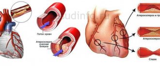
- Congenital hyperlipidemia (increased fat content in the blood), which provokes the development of atherosclerosis of the heart.
- Changes in the connective tissue of the cardiac ventricles, in which additional chords are formed in them.
- Acquired or congenital heart defects.
- Hypertrophic cardiomyopathy.
- Malfunctions of the autonomic nervous system.
- Neuroendocrine problems.
- Electrolyte imbalance in the body.
- High blood cholesterol levels.
- Excessive physical activity.
- Hypothermia of the body.
Classification
Early ventricular repolarization syndrome in children and adults can have two development options regarding the functioning of the heart, blood vessels and other organs involved in the functioning of the system - with and without damage to the cardiovascular system. Based on the nature of the pathology, a distinction is made between transient (periodic) and permanent SRR. There is a classification into 3 types depending on the location of ECG signs.
How dangerous is the disease?
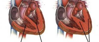
The consequences of the development of early ventricular repolarization syndrome are characterized by complete or partial, gradual loss of contractility of the muscular organ.
This is not a one-time process; it takes more than one year to fully develop. The duration varies among patients. It all depends on individual characteristics.
Complications are associated with disruption of the normal ejection of blood from the left ventricle into the aorta and its movement in a large circle. Both distant organs and systems and the myocardium itself suffer.
Possible consequences include:
- Heart attack. Acute necrotic process. Cardiac structures die, and even with successful treatment, sclerosis occurs. Replacement of normal active muscle tissue with scar compounds. They are not able to work like myocytes. Therefore, the patient’s companion becomes constant coronary artery disease with the prospect of early death from a relapse of the emergency condition.
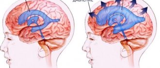
- Stroke. A similar phenomenon, but affecting the nerve tissue of the brain. The ischemic variety is typical. That is, an acute malnutrition of a separate area of cerebral structures.
- Heart failure. Without clear prospects for resuscitation and restoration of vital functions. Because there are fundamental anatomical violations. Even if the organ can be “started”, it is highly likely that it will stop again.
- Cardiogenic shock. The culmination of a decline in the contractility of muscle structures. It consists of insufficient supply of the body with nutrients and oxygen at a general level.
Blood pressure drops, critical arrhythmia occurs. Recovery from the onset of this condition is almost impossible; mortality is close to 100%.
The patient has extremely low chances even with successful recovery from the acute process. Over the course of a couple of years or less, relapse and death occur.
Read more about the condition and first aid measures here.
- Vascular dementia. As a result of insufficient blood flow to the brain. Stroke is not the only possibility. Changes may also be a consequence of it.
The possibility of life-threatening results has recently been discovered. Only after the unpleasant discovery did active research into the problem begin.
The risk is currently assessed as moderate. In the presence of a mass of concomitant pathologies and negative prognostic factors - pronounced.
Diagnosis of the syndrome
In addition to the electrocardiogram, there are other methods that can detect repolarization.
Heart ultrasound is one of the methods for diagnosing the disease.
Among them, the most famous are:
- Ultrasound of the heart;
- echocardiography;
- electrophysiological study.
For a more detailed study, the patient may be prescribed an ECG with physical and medicinal stress, additional blood and urine tests, and daily Holter monitoring. In addition, the doctor may ask for EGC to be done regularly, to ensure that the results are not erroneous, and to identify the persistence of characteristic changes. Also, the patient should be observed by a doctor at certain intervals: in the presence of radiating pain behind the sternum, myocardial infarction may occur.
According to the ECG, early repolarization is similar to myocardial infarction, however, an experienced doctor will distinguish changes in the cardiogram, and in the absence of pain, one can confidently determine that the patient has only this phenomenon.
Causes are pathological
The moments of development of the process are diverse. The basis is made up of groups of objective phenomena. They are not under the control of the patient himself.
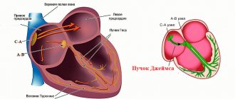
- Bundle branch block. It is characterized by the development of obstruction of the electrical impulse from the sinus node along special fibers.
The condition is rarely complete. Patients with total blockade do not live long. This is also not an independent disease, but a consequence of third-party pathological processes.
In the complex, the doctor receives good food for detective research: to discover the factor in the formation of repolarization, which itself has an indirect origin. Read more about the right bundle branch block here, and the left one here.
- Hypertrophic cardiomyopathy. A disease accompanied by the growth of the muscle layer of the organ. This is a very common condition. It develops more often in representatives of the stronger sex. Apparently, this is due to genetic factors or bad habits. Also reduced resistance (resilience) to negatively influencing factors. Read more about the disease in this article.
- Systemic, often autoimmune diseases of connective tissue. Lupus erythematosus, rheumatoid arthritis and others. Cause gradual destruction of cardiac structures.
The condition has irreversible consequences and is therefore considered extremely dangerous. Early repolarization of the ventricles is a clinical variant; it develops against the background of disruption of normal metabolism resulting in cardiosclerosis—replacement of tissue with scar tissue.
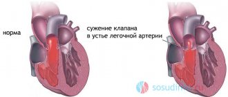
- Congenital and acquired heart defects. The condition is genetic or caused by disturbances in the prenatal period.
Both options lead to myocardial deformations. The former are accompanied by a host of other symptoms, not only of cardiac origin.
Deformations of the maxillofacial area, distant organs and systems are possible.
Recovery is carried out surgically. But this is not a guarantee that the syndrome will stop. Medicinal correction with drugs is also required.
- Excessive physical activity. As relevant medical statistics clearly demonstrate, the main contingent of patients with early ventricular repolarization syndrome are professional athletes and also fanatical lovers of outdoor activities.
Apparently, this is due to the increasing phenomena of degeneration of cardiac structures and metabolic abnormalities.
The approximate number of sick athletes is 60-70%. Perhaps the number is more significant. Because close attention to SRRH was paid relatively recently. Previously, people turned a blind eye to such deviations.
- Neuroendocrine diseases. A common variant is hypothalamic syndrome of the same type.
Develops in childhood and adolescence and is represented by gross metabolic disorders at a generalized level and brain symptoms.
Causes a danger to the reproductive and cardiovascular systems, increases the risk of developing endocrine conditions.
- Puberty (puberty). The most dangerous moment determines the onset of early repolarization syndrome, especially often (in approximately 20% of adolescents, ECG is detected on the ECG). This is temporary, but it is strongly recommended to be observed by a cardiologist so as not to miss the right moment.
Subjective factors
In addition to the reasons already mentioned, a group of phenomena has a subjective origin.
- Smoking. Patients with physiological dependence on nicotine suffer from SRRD more often than others. This is due to a partial disruption of normal metabolism. This is a dangerous process. As the body gets used to the harmful substance, there is an increase in resistance to therapy.
After 5-10 years of constant tobacco use, quitting the habit is no longer enough. A long rehabilitation period under the supervision of a cardiologist will be required. Drug therapy.
- Alcohol abuse. It works in the same way.
- Excess of oral contraceptives.
The syndrome of premature myocardial repolarization is well masked; it is extremely difficult to determine its true cause. This is not a matter of one day, perhaps not a week.
Fortunately, the process leads to dangerous consequences relatively late. There is a possibility that they will never come.
Symptoms
Early ventricular repolarization syndrome has no manifestations as such. This is not a diagnosis, but a clinical finding.
The main list of manifestations is determined by the disease that caused a disruption in the normal movement of the charge.
Possible signs include:
- Chest pain. Low intensity. It is possible to differentiate cardiac discomfort from muscular discomfort, neuralgia, using a typical feature. They do not increase with breathing or changing body position. Usually, unpleasant sensations occur in attacks and waves. Relatively little continues.
- Arrhythmias. By type of bradycardia most often. An increase in the number of heartbeats per minute is possible, and in about 30% of situations it is observed. In addition to this type, related ones also arise. They directly indicate a disruption of normal metabolism in the myocardium. Fibrillation and ventricular extrasystole are common.
- Dyspnea. In a state of complete rest or against the background of intense physical activity. Depends on the underlying pathological process.
Cyanosis of the nasolabial triangle and pallor of the skin are also possible. The list of manifestations is determined by the main diagnosis. The most likely ones are presented.
Attention:
There may be a complete absence of typical signs. This is the most dangerous clinical option.
Causes of repolarization disorders
The repolarization process can be influenced by many factors, including:
- Diseases of the myocardium itself (for example, myocarditis, ischemia, infarction, infiltrative process).
- Medicines (eg digoxin, quinidine, tricyclic antidepressants and many other drugs).
- Electrolyte disturbances in the concentration of potassium, magnesium and calcium.
- Neurogenic factors (for example, ischemic or hemorrhagic stroke, traumatic brain injury, brain tumor).
- Metabolic factors (eg, hypoglycemia, hyperventilation).
- Disturbances in the conduction of electrical signals in the ventricles.
- Pathological rhythm, the source of which is in the ventricles.
Secondary abnormalities in myocardial repolarization are normal ST segment and T wave changes that develop solely due to changes in the ventricular firing sequence. Such changes are most often focal in nature, that is, they are observed only in part of the ECG leads. These include:
- Changes characteristic of His bundle block.
- Changes that occur in Wolff-Parkinson-White syndrome.
- Changes characteristic of premature ventricular contractions, ventricular arrhythmias and ventricular rhythm.
Primary disturbances of repolarization processes are changes in the ECG that do not depend on uncoordinated ventricular activation, but may be the result of a diffuse or focal pathological process affecting ventricular relaxation. These include:
- Effect of drugs (for example, digoxin or quinidine).
- Electrolyte disturbances (eg, hypokalemia).
- Ischemia, heart attack, inflammation (myocarditis).
- Neurogenic factors (for example, subarachnoid hemorrhage can cause prolongation of the QT interval).
Diagnostics
The survey presents certain difficulties. It is carried out under the supervision of a cardiologist. The duration depends on the severity of the condition. In non-critical cases, it is indicated on an outpatient basis.
Sample list of events:
- Oral questioning of the patient. In most cases, there are no pronounced manifestations or they are nonspecific, which does not provide an accurate indication of the possible condition.
- Anamnesis collection. Lifestyle, family history, duration of course and other points, also previous illnesses.
- Measurement of blood pressure and heart rate. Both indicators are changed, how depends on the root cause.
- Daily monitoring. Registration of specified levels within 24 hours. Appointed as needed.
- Electrocardiography. Basic examination technique. Actually, it is based on the results of the ECG that repolarization syndrome is determined.
- Echocardiography. To assess organic abnormalities in the heart.
- General blood test, biochemical, hormones. Plays an important role in assessment. Makes it possible to diagnose endocrine and metabolic pathologies.
As part of the expanded technique, MRI is used.
Symptoms of deviation
This condition is not a disease in the strict sense of the word. SRR is an electrocardiographic phenomenon that occurs in absolutely healthy people and during myocardial infarction. Moreover, it may first appear in the acute period and disappear at the time of scarring, be present before a heart attack, and stop as it develops. Some patients do not have SRGC in the acute stage, but the symptoms resume when the ECG normalizes.
In some cases, clinical symptoms resemble paroxysmal tachycardia:
- attacks of rapid pulse,
- dizziness,
- compression in the heart area,
- noise in the head
- nausea,
- fainting states,
- drop in blood pressure,
- darkening of the eyes.
Signs on ECG
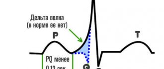
Early repolarization, also known as a “J wave” or “J-spot elevation,” is an electrocardiographic abnormality consistent with an increased connection between the end of the QRS complex and the beginning of the ST segment in two adjacent leads.
Among the typical features of pathological deviation from the norm:
- Widening of the T wave, growth of the peak in height.
- ST interval shift upward.
- Serrated R shape.
- Presence of J-wave.
- Raising point J above the isoline.
It is also possible for your heart rate to speed up or slow down. The signs are typical enough to make a diagnosis (if you can say so about the described condition).
The syndrome of early ventricular repolarization on the ECG has typical features. But accurate decoding requires high qualifications. In addition, specific abnormalities may be confused with other findings.
The phenomenon of SRRH never exists in isolation. The main points are found in one, two or all leads. Based on this criterion, they speak of three types of condition.
| Type | Description |
| Type 1 | Signs of early repolarization were found only in the chest lateral ECG leads. The risks of complications are minimal. |
| Type 2 | The presence of deviations in the inferolateral and inferior leads (II, III, aVF). The risks of sudden death are increased. |
| Type 3 | Abnormal complexes are present in all leads. The risk of cardiovascular complications is highest. |
Treatment and prevention
Single SRGC, without adjacent cardiac pathologies, is not subject to special drug therapy. In order not to complicate the situation, the patient is recommended to follow a set of preventive measures, including:
- rational physical activity. Physical activity and sports training must be adjusted taking into account the characteristics of the heart, and are carried out under cardiac supervision (measurement of pulse and blood pressure);
- giving up harmful addictions. Alcohol and nicotine should be excluded as companions of cardiovascular diseases;
- changing eating habits. Fatty foods high in “bad” cholesterol must be eliminated from the diet, replacing them with healthy vegetables, fruits, and herbs;
- visits to a cardiologist on a regular basis to monitor cardiogram parameters;
- systematic course use of plant-based cardiac dietary supplements (in the absence of allergic reactions to herbal medicines);
- compliance with the work and proper rest regime. Overvoltage must not be allowed;
- maintaining a stable, calm psycho-emotional state. You must try to avoid conflicts and stress.
Nicotine and alcohol negatively affect the functioning of the heart; if there are abnormalities in cardiac activity, it is necessary to avoid their consumption
In cases where SRHR is not the only abnormal phenomenon and the patient has other heart diseases, treatment is prescribed by a doctor. Symptomatic therapy of the underlying disease is carried out, adjusted for the presence of the syndrome. A radical measure is surgery to implant a cardioverter-defibrillator. However, such intervention is often based on other complications. If preventive measures are taken, the prognosis is always favorable.
Treatment
Therapy is conservative or surgical. The main disease must be eliminated. It all depends on the primary diagnosis. Medicines:
- Antiarrhythmic. Amiodarone or Hindin as needed in small courses.
- Cardioprotectors. Mildronate and others. Supports the heart.
Other medications are provided as part of additional assistance.
Surgical therapy consists of implanting a defibrillator, which brings the rhythm back to normal and prevents the stop of the muscle organ.
A significant role is played by lifestyle changes. Under no circumstances should you smoke, consuming alcohol even in minimal doses is impossible due to the likelihood of complications, all the more so you need to give up drugs.
It is sometimes impossible to do this on your own; the assistance of a narcologist is required. Now there is a developed system of anonymous help, so there is no need to be afraid of the stigma.
Whenever possible, the diet is adjusted. Fats are minimized, of artificial origin completely, of natural origin - partially. Fast carbohydrates are not needed. More vitamins and protein.
A diet correction is indicated in accordance with treatment table No. 10. There is no need for such a strict diet, but you can take the specified menu as a starting point.
Physical activity is kept to a minimum, but cannot be completely eliminated. This will lead to aggravation of the pathological process.
Treatment of abnormalities
In most cases, therapy is aimed at preventing rhythm disturbances. For this, metabolic agents (Mildronat, Kudesan, Preductal, Cardonat) and drugs containing magnesium (Magnicum, Magnevit), antiarrhythmic medications (Cordarone, Ritmilen) can be used.
In the presence of fainting conditions, installation of a cardioverter-defibrillator is indicated. If additional pathways are found, then cauterization with radiofrequency waves (ablation) of a section of the myocardium is prescribed.
Forecast
The syndrome of early repolarization of the ventricles of the heart is dangerous due to premature death from stopping the functioning of the muscular organ. The likelihood of such an outcome is determined by the main diagnosis.
- Defects, previous heart attack, defects due to cardiomyopathy are more likely to lead to death.
- Potentially treatable conditions contribute to full recovery, but also with some conditionality.
- In later stages, anatomical defects are possible that are irreversible in nature.
Conclusion - the best prognosis is determined by timely initiation of treatment.