The list of cardiac pathologies includes CLC syndrome. This congenital cardiac anomaly has many other names - premature ventricular excitation phenomenon, short PQ interval syndrome. In the English-speaking world of cardiology, the abbreviation LGL is used.
We use the abbreviation SLS, made up of the first letters of the names of the doctors who first described the pathology in 1938.
Conducting system of the heart: normal and abnormal – James bundle
Clerk Lewy Cristesco syndrome is a congenital disorder in the structure of the conduction system of the heart, which leads to premature contraction of the ventricles. It is an additional inclusion of specific conductive tissue (pictured above) between one of the atria, usually the sinoatrial (CA) node, and the atrioventricular (A-V) node, which is absent in normal myocardium.
What is the disease
Clerk-Levy-Christesco syndrome or CLC is a congenital pathology of the heart structure, one of the types of premature ventricular excitation syndrome. The anomaly is due to the presence of a James beam. Thanks to this bundle, the atrioventricular node is connected to one of the atria. This feature is accompanied by the transmission of a premature impulse to the ventricles.
Pathology can be diagnosed using an ECG. Often, CLC syndrome occurs accidentally in completely healthy people, and the patient does not have any symptoms. There are known cases of this disease being detected in children.
Even in the absence of symptoms, one should not assume that the pathology is harmless. Ventricular preexcitation syndrome can cause arrhythmia. The condition is accompanied by increased heart rate (sometimes over 200 beats per minute). This is especially felt in older people; young patients tolerate arrhythmia more easily.
Treatment of CLC syndrome
In the absence of attacks of arrhythmia, treatment for this condition is not carried out. If a rapid heartbeat occurs, exceeding 140 beats per minute, then vagal tests can be used (if recommended by a doctor).
If there is any doubt about the diagnosis or serious condition, you should immediately call an ambulance, as this may be a sign of myocardial or cerebral ischemia.
For drug relief of rhythm disturbances, the administration of ATP, Isoptin, Cordarone or Novocainamide is used intravenously. If restoration of normal rhythm is not achieved, cardioversion with electrical impulses is used. In some cases, destruction of the accessory pathway can be achieved with radiofrequency ablation (cauterization) through a catheter.
Features of the pathology
Pre-excitation of the cardiac ventricles can be asymptomatic, in which case we are talking about the “pre-excitation phenomenon”. When signs of pathology appear in a patient, the disease is classified as “pre-excitation syndrome.”
There are several types of disease:
- Breschenmash (atriofascicular) - here the right atrium is connected to the trunk of the His bundle;
- Maheima (nodoventricular) - in this case, the right side of the interventricular septum is connected to the atrioventricular node;
- Kent (atrioventricular) - here the atria and ventricles are connected bypassing the atrioventricular node;
- James (atrionodal) - impulses pass between the lower part of the atrioventricular and sinoatrial node.
Important! Sometimes there are several paths of abnormal conduction of impulses. The number of such patients is no more than 10% of all cases of the disease.
CLC syndrome is clearly monitored by performing an electrocardiogram
What happens with short PQ syndrome?
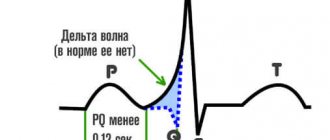
The PQ interval is a purely electrocardiographic criterion that allows one to evaluate the transmission time of an electrical impulse from the sinus node in the atrium to the contractile fibers located in the ventricles. In other words, it reflects the operation of the atrioventricular junction, a kind of “switch” that redirects electrical excitation from the atria to the ventricles. Normally, it is no less than 0.11 seconds and no more than 0.2 seconds:
example of shortening PQ to 0.03 s
- An increase in the interval beyond the specified time indicates a slowdown in conduction through the atrioventricular node,
- Shortening means that the excitation is carried out too quickly. In fact, there is more frequent impulse of the ventricles, with the so-called “reset” of excitation.
The shortening of this interval is due to the presence of additional conduction bundles in the conduction system of the heart. It is through them that additional pulses are reset. Therefore, at certain moments the ventricles receive double impulses - physiological in a normal rhythm (60-80 per minute), and pathological, through bundles.
There may be several pathological bundles, and all of them are named after the names of the authors who first discovered them. Thus, the Kent and Maheim bundles are characteristic of the SVC syndrome, and the James bundles are characteristic of the CLC syndrome. In the first case, the pathological discharge of impulses goes from the atria directly to the ventricles, in the second, the James bundle passes through the atrioventricular node, that is, the node is stimulated first, and then the ventricles. Due to the “throughput” capacity of the AV node, part of the impulses conducted to the ventricles returns through the same bundle to the atria, therefore such patients have a high risk of developing paroxysmal supraventricular tachycardia.
main types of pathological pathways of additional conduction through the heart
What Causes CLC?
In order to understand how CLC syndrome occurs, you should remember the anatomy. For the normal filling of the ventricles of the heart with blood and their contraction after contraction of the atria, there is a special filter located between them in the structure of the organ. This filter is called the atrioventricular node. When an impulse is transmitted to this node, the excitation passes slowly, after which it is transmitted to the ventricles. Thanks to this process, blood enters the aorta and pulmonary trunk.
With some congenital pathologies, a person may have bypass pathways for conducting cardiac impulses. Often the James beam and some other channels act in this way. In this case, the ECG shows minor deviations from the norm or severe disturbances, for example, with serious deformation of the ventricles. The syndrome of premature excitation of the ventricles in medical practice is called WPW.
Causes of CLC syndrome
The first manifestation of premature excitation of the ventricles can occur at any age, or the patient is unaware of it throughout his life. It can be congenital or acquired.
The child has
In the fetal heart there are additional fibers of the conduction system, which cease to function after 21 weeks of pregnancy. If this does not happen, then such pathways lead to disruption of myocardial rhythm. There is a hereditary predisposition to the disease, and familial forms of CLC have been noted.
Most patients have only an accessory bundle of James, but combinations of this pathology with signs of connective tissue diseases (Marfan syndrome, mitral valve prolapse), and congenital cardiomyopathy have been described.
During pregnancy, in adults
The acquired form of the syndrome can also occur in adult patients; this is associated with increased automaticity of the atrioventricular node . This condition is caused by:
- chronic inflammatory processes – myocarditis, rheumatism;
- ischemia of the heart muscle - angina pectoris, heart attack;
- metabolic disorder – dismetabolic cardiomyopathy;
- high blood pressure.
Metabolic disorders due to insufficient intake of vitamin B1, as well as excessive thyroid function, can cause or provoke clinical manifestations of the congenital phenomenon of CLC.
If only electrocardiographic signs are detected in pregnant women, no specific restrictions or treatment are required. More often, such a deviation does not lead to significant hemodynamic disturbances. To prevent arrhythmia, vitamin complexes are prescribed.
How does the disease progress?
Pathology can manifest itself at any age of the patient. For a long time, the disease can occur without obvious signs and symptoms. The CLC phenomenon is accompanied by a shortening of the PQ interval during an ECG. At the same time, the patient’s well-being is not impaired, the person leads a full-fledged lifestyle.
Signs of pathology often include attacks of tachycardia and some other manifestations
With CLC syndrome, many patients experience sudden attacks of strong heartbeat - paroxysmal tachycardia. The following symptoms are observed:
- the appearance of an attack of rapid heartbeat. Heart rate is up to 220 beats per minute. The onset of an attack is accompanied by a strong shock in the sternum or neck;
- dizziness;
- noise in ears;
- increased sweating;
- bloating;
- sometimes nausea occurs, accompanied by vomiting;
- copious amounts of urine at the beginning or end of an attack, sometimes involuntary urination.
Less commonly, the pathology is accompanied by atrial fibrillation, a rapid heartbeat of an irregular nature.
First aid during an attack
Patients with CLC syndrome who frequently experience an attack of increased heart rate are advised to adhere to the following recommendations during an attack:
- Perform carotid sinus massage. Impact on this area between the division of the carotid artery into the external and external helps to normalize the heart rate.
- Gently press on the eyeballs.
- To alleviate the condition, you can lower your face into a container of cool water, holding your breath for 5 to 10 minutes.
- Take a deep breath, strain, hold your breath for a few seconds, exhale slowly.
- Sit down several times, tensing your whole body.
Important! If attacks occur frequently, do not delay visiting a doctor. Early diagnosis of pathology will help prevent complications and bring the disease under medical control.
Treatment
In most cases, CLC syndrome does not require specialized intervention. During attacks of palpitations, the patient can independently stop them with the help of a special massage, cooling the face with water or straining while inhaling, that is, performing Valsalva maneuvers. If these methods do not help, you must call an ambulance.
Also, when dealing with attacks of tachycardia caused by CLC syndrome, you can resort to the services of a cardiologist, who should prescribe special medications, for example, Verapamil or Amiodarone.
In cases where attacks of tachycardia greatly affect the patient’s life, an operation is performed to destroy the James bundles, which prevents the occurrence of a circular course of excitation. Such operations are not dangerous, and afterward the patient quickly recovers.
Diagnostics
The main method for diagnosing CLC syndrome is an electrocardiogram. An ECG can detect the following disorders:
- reduction of PQ intervals;
- D-wave in the presence of an ascending part of the R-wave with a simultaneous decrease in the Q-wave;
- the appearance of an extended QRS set;
- deviation of the CT segment and T-wave in the opposite direction from the D-wave.
Much attention is paid to the spatial vector electrocardiogram. Using the method, it is possible to accurately determine the location of additional conductive channels of the heart.
Timely diagnosis of CLC syndrome allows you to take the pathology under medical control and prevent its complications
For a more detailed study, magnetocardiography is used. The method allows you to give a complete assessment of the activity of the heart muscle. This happens thanks to equipment that records the magnetic components in the electromotive force of the heart.
Instrumental research methods such as electrophysiological examination of the heart (EPI), endocardial and epicardial mapping can provide an accurate assessment of the activity of the heart muscle.
The phenomenon of clc cardiology what is it
Clerk-Levy-Christesco syndrome is a special case of the syndrome of premature excitation of the ventricles of the heart, which is characterized by the conduction of excitation to the ventricles along additional pathways. The human heart is designed so that the ventricles contract later than the atria, this is necessary for them to be sufficiently filled with blood.
The functioning of this mechanism is ensured by the atrioventricular node, which is located between the ventricles and atria; in it the impulse travels much more slowly, which ensures a delay in the contraction of the ventricles. However, some people have congenital abnormal pathways that bypass the atrioventricular node, such pathways include the bundles of James, bundles of Kent, and Maheim fibers.
Thanks to these pathways, the pulse travel time is reduced and the CLC phenomenon occurs. This mechanism can be seen by analyzing the ECG. The phenomenon itself does not affect the functioning of the heart in any way and appears only on the cardiogram. But sometimes there are cases when a circular course of excitation occurs.
The phenomenon and syndrome of CLC are congenital; the exact cause of the appearance of these anomalies is unknown. There are suggestions that this is due to disturbances in the development of the fetus at the stage of heart formation. It should also not be ruled out that the cause may be genetic disorders.
The occurrence of short PQ syndrome
Shortened pq syndrome is diagnosed if the ECG shows that the travel time of the electrical signal between the P and Q waves is no more than 0.11 s.
This is possible in two cases:
- with Wolff-Parkinson-White syndrome (WPW);
- with Clerk-Levy-Christesco syndrome (CLC).
In the first case, in addition to the shortened PQ interval, the ECG shows an additional wave between the Q and R waves and a sharp depression of the ST region. This pattern indicates the presence of additional nerve fibers (bundles of Kent) that transmit a premature electrical signal to the ventricles, forcing them to contract more often.
WPW syndrome manifests itself as tachycardia and tachyarrhythmia and usually occurs in people with congenital heart defects. The pulse rate can reach 200 or more beats per minute.
Its manifestations: dizziness, short-term loss of consciousness, a feeling of the heart jumping out.
According to statistics, most patients with this diagnosis have a mild form of the disease, not aggravated by circulatory disorders, and require timely prevention.
Clerk-Levy-Christesco syndrome occurs when there is an additional conduction bundle between the atria (James bundle). This nerve connection is a kind of shunt of the His bundle (atrioventricular node). Therefore, the impulse passes without a normal delay and causes rapid contraction of the heart muscle.
In cardiology, clc syndrome and clc phenomenon are considered. In both cases, the patient’s cardiogram shows a shortened pq interval without the appearance of a delta wave.
Diagnoses differ in their manifestations. Those who are diagnosed with the phenomenon may not experience any symptoms and may not even suspect until a certain point that the cause of periodic palpitations is shortened pq interval syndrome. For such people, prevention is extremely important:
- periodic examination by a cardiologist;
- healthy lifestyle;
- rejection of bad habits;
- developing the ability to withstand stress and anxiety.
The clc syndrome, unlike the phenomenon, can become a threat to the patient's life.
Cardiogram analysis
ECG analysis helps identify this syndrome. It is characterized by a shortening of the PR (PQ) interval.
This interval shows the time it takes for excitation to reach the ventricular myocardium through the atria and atrioventricular junction.
In people over 17 years of age, an interval of 0.2 s is normal, but a shortening of this interval, which can cause tachyarrhythmia, may also be a prerequisite for the diagnosis of CLC syndrome.
In addition to the reduction in the mentioned interval, in the presence of CLC syndrome there are no other changes on the ECG. The ventricular complex (the QRS complex is the most significant deviation on the cardiogram, showing the time of passage of excitation inside the ventricles) does not look abnormal. CLC syndrome most often occurs in people whose hearts are normal.
Main manifestations
Symptoms of the pathology are periodic. Attacks without systemic treatment are repeated again and again, become prolonged, their strength increases, although in the intervals between them the patient does not experience any unpleasant sensations. The most dangerous manifestation of the syndrome is supraventricular paroxysmal tachycardia.
The following reasons should be the reason to contact a cardiologist for treatment:
- SLS syndrome detected on ECG;
- a feeling of tightness or burning in the left side of the chest;
- cold hands and feet;
- sudden redness or, conversely, paleness of the face;
- lack of oxygen (a person takes frequent breaths in an attempt to breathe in air), accompanied by a state of panic;
- frequent weakness and fatigue;
- tachycardia is accompanied by arrhythmia.
By taking good care of your health and the health of your loved ones, you can prevent the development of the disease. To do this, do not miss the appointment for a preventive examination with a cardiologist. This way, changes will be detected in time, which will allow you to take the necessary measures.
What is CLC syndrome and why does it occur?
CLC syndrome is a congenital disorder
CLC is also known medically as Clerk-Levy-Christesco syndrome. This is not an acquired disease, but a congenital disease, in which there is a violation of the structure of the heart muscle. The pathology is a type of syndrome of premature excitation of the myocardial ventricles.
The disease develops as a result of an anomaly of the James bundle, due to which the atrioventricular node unites with one of the atria. Thus, a premature node to the ventricles occurs.
It is difficult to diagnose the disease in the initial stages, since it is practically asymptomatic. The syndrome is diagnosed randomly during an electrocardiogram. The pathology is diagnosed in adults, but in medical practice there have been cases when CLC syndrome was also found in children.
As a result of disruption of the impulse transmission process, arrhythmia may occur, which is accompanied by an increase in heart rate by more than 200 beats per minute. To a greater extent, this disorder is felt by older and older people. Patients under 35 years of age tolerate the disease much easier.
Very often the syndrome does not cause any symptoms
The disease is congenital. At the initial stages of its development and for quite a long time, it may not manifest itself. The syndrome is diagnosed when an electrocardiogram is performed at any age.
The pathology may not show symptoms for many years and proceed secretly. This is precisely where its danger lies. The patient does not feel any change in health and leads a normal lifestyle.
But in certain cases, attacks of paroxysmal tachycardia occur. The condition is characterized by the sudden onset of palpitations. In this case, patients often complain of the following symptoms:
- At the beginning of the attack, a strong shock is felt in the neck and chest.
- The frequency of SS increases. Indicators can exceed 200 beats per minute.
- Dizziness of varying intensity.
- Increased sweating.
- Noise in ears.
- Bloating.
- Nausea, in some cases accompanied by vomiting, which does not bring relief.
- Copious urination at the beginning of the attack and after its completion. The process is often uncontrollable.
In exceptional cases, as noted by experts, an attack may be accompanied by rapid heartbeat, the nature of which is nonspecific, and the development of atrial fibrillation.
Diagnosis of pathology
Symptoms characteristic of this disease are also found in other diseases. Therefore, only an in-person examination for cardiac problems is not enough. Additional information about the state of the heart muscle is obtained after reading an ECG, ultrasound of the heart or electrophysiological study (EPS).
If short pq syndrome is suspected, the patient is usually prescribed electrocardiography to confirm the diagnosis.
The procedure involves obtaining a graphical recording of the electrical activity of the heart from the surface of the body. Today this is the most common research method in cardiology.
Recording electrical processes in individual muscle cells of the organ allows us to obtain complete information about the work of the heart as a whole.
An experienced specialist, comparing different segments, will definitely see a shortened pq interval, analyze the result taking into account the general condition of the patient, determine the severity and, if necessary, prescribe treatment.
An experienced specialist, comparing different segments, will definitely see a shortened pq interval, analyze the result taking into account the general condition of the patient, determine the severity and, if necessary, prescribe treatment.
How can it be diagnosed?
ECG results may reveal the presence of the syndrome
The main diagnostic method for suspected Clerk-Levy-Christesco syndrome is electrocardiography.
Using this technique, it is possible to identify a number of the following violations:
- Changes in P-Q intervals. They become significantly smaller.
- Decreased Q wave. In this case, the ascending part of the R wave is noted.
- The appearance of the QRS complex. It has an expanded character.
- The CT segment, together with the T wave, deviates in the opposite direction from the D wave.
The patient is also prescribed a spatial vector electrocardiogram. The technique allows you to determine the exact location of additional heart channels that conduct electrical impulses.
Magnetocardiography is used to obtain more reliable data. The diagnostic method is used to assess the performance of the myocardium. The procedure is carried out using special highly sensitive equipment that detects magnetic components in the electromotive force of the heart muscle.
Electrophysiological studies, epicardial and endocardial mapping are also used to determine the functionality of the myocardium. Based on the results of diagnostic measures, the cardiologist establishes an accurate diagnosis, determines the degree of the disorder and prescribes the necessary course of therapy.
Complications and possible consequences
To avoid complications, people with CLC syndrome need to take care of their health
Lack of treatment in the presence of CLC syndrome causes the development of tachycardia and sudden death.
In certain cases, a recurrent course of overexcitation of the ventricles of the heart muscle is noted. That is why, if unpleasant symptoms occur and CLC syndrome is established, you should not delay treatment.
Clerk-Levy-Christesco syndrome is a dangerous myocardial disease that can lead to serious consequences. The disease is most often diagnosed randomly during an electrocardiogram. In order to diagnose the disorder in a timely manner, experts recommend regularly visiting a doctor for a preventive examination.
Select it and press Ctrl Enter to let us know.
A condition in which the impulse from the atria to the ventricles is transmitted twice as fast as normal is called short PQ interval syndrome. This cardiac abnormality is asymptomatic or manifests itself as tachycardia.
The PQ interval is one of the main parameters of the electrocardiogram, which indicates the speed of transmission of the electrical impulse between the zones of the heart. The normal value is approximately 0.2 s. Small deviations from this figure are not considered an anomaly. They are explained by the characteristics of the body and do not affect the physical condition of a person.
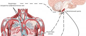
Contractions of the heart occur under the influence of electrical impulses coming from a collection of nerve fibers in the sinus node (right atrium). Thanks to this, blood is pushed from the atria into the ventricles.
Nature made sure that the ventricles were completely filled with blood. Passing through the atrioventricular node at the border between them, the electrical signal slows down and, only after leaving this node, it accelerates again, causing the ventricles to contract.
In this case, blood is pushed into the aorta.
However, some people are born with additional pathways for electrical impulses - nerve bundles that carry the signal bypassing the atrioventricular junction. The impulse travels the entire path without slowing down, sometimes returns and again follows the initial trajectory. The ventricles, not having time to fill with blood, contract, and the heart rhythm fails.
On the cardiogram, the presence of nerve bundles is indicated by a reduction in the PQ interval by 0.09 s. and more. The pathology may not manifest itself for a long time, but if symptoms develop, diagnosis and assistance from a doctor are required.
The impetus may be the active growth of the child or adolescence. More often - lifestyle and chronic diseases. Experts identify a number of factors that can aggravate the condition and provoke the development of symptoms:
- hard physical labor;
- frequent stressful situations;
- excessive drinking, drug use and smoking;
- the influence of high temperatures (prolonged exposure to the sun, hot food, visiting a sauna);
- overeating (especially in the evening);
- pregnancy and childbirth;
- hypertension, thyroid disease, colds.
Source: https://moeserdce.net.ru/fenomen-l-kardiologiya-hto-eto/
Features of pathology treatment
We advise you to read: WPW syndrome on ECG
Patients diagnosed with asymptomatic pathology do not require special treatment. Attention is paid to patients who have a history of sudden death in their family. People involved in heavy physical work, athletes, divers, and pilots are also at risk.
If a patient experiences paroxysms of supraventricular tachycardia, therapy consists of prescribing medications to relieve the attack. In this case, the nature of the heart rhythm disturbance and the presence of concomitant diseases of the cardiovascular system must be taken into account.
To get rid of orthodromic reciprocal supraventricular tachycardia, blockades are used aimed at inhibiting the conduction of impulses in the atrioventricular node.
Drug therapy includes the following drugs:
- Adenosine;
- Verapamil.
Often, to reduce the manifestation of pathology, beta-blockers are prescribed, which have the ability to dilate the bronchi, arteries and veins. Drugs in this group activate the production of glycogenolysis by the liver and skeletal muscles and stimulate insulin secretion.
Amiodarone is a medication often prescribed for CLC syndrome.
In the absence of the desired therapeutic result, doctors use Novocainamide, which helps block impulses passing through the accessory atrioventricular canal. The medicine is relatively safe and has a good effect in the treatment of many cardiac pathologies.
In addition to those described above, the following medications are used:
- Amiodarone;
- Sotalol;
- Propafenone;
- Flecainide.
Important! The use of medications is permitted strictly as prescribed by the attending physician. Many of them have serious contraindications.
Non-drug treatment methods include transthoracic depolarization and atrial pacing. Surgical treatment of accessory pathways is used in special cases when conservative therapy does not produce a positive effect. The operation is performed using the open method in emergency cases when there is a serious threat to the health and life of the patient. The method involves destroying additional pathways of the heart.
A positive prognosis for patients with impaired conduction of cardiac impulses is possible with strict adherence to the doctor’s recommendations
Pathologies of the circulatory system
Paragraph No. 42 of the resolution on military medical examination lists heart diseases of rheumatic and non-rheumatic etiology that caused heart failure. These illnesses require consideration of unfitness for service. Citizens belonging to column IV who have all of the following diseases are assigned category D (unfit). Whether someone with arrhythmia is accepted into the army or not depends on the type of disease.
The degree of heart failure is confirmed by a complete examination in the hospital.
Severe deficiency
For all groups of citizens with such pathology, group D is assigned.
The list also includes the following diseases:
- Heart diseases (heart attacks, dilated and hypertrophied cardiopathy, heart disease, coronary heart disease), leading to heart failure of FC category 4, in which it is impossible for a person to perform any physical activities without the appearance of general malaise. Signs of deficiency are also felt at rest.
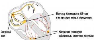
- Persistent arrhythmias with atrioventricular block of 2 and 3 degrees.
- All types of paroxysmal tachycardias.
- Polytopic ventricular extrasystole (with several ectopic pacemakers).
- Weak sinus node syndrome, which resulted in the development of sinus arrhythmia of the heart.
Moderate degree of deficiency
For the first and second groups of citizens, category D is assigned, for the third - B.
- Heart diseases that led to heart failure of group 3 FC. In this case, a person in a calm state does not feel signs of heart failure, but at the slightest load all the symptoms (weakness, shortness of breath, tachycardia) sharply worsen.
- Citizens with persistent cardiac arrhythmia, with persistent pathology of the conduction system.
Important! Arrhythmias are considered persistent if they last longer than 7 days with constant daily Holter ECG monitoring. In terms of severity, the arrhythmia should lead to clinically noticeable circulatory disorders and require mandatory treatment, and after the cessation of antiarrhythmic therapy, the arrhythmias resume.
- Paroxysmal (sudden) tachyarrhythmias.
Wolf-Parkinson-White WPW overexcitation syndrome
WPW syndrome is a congenital pathology in which overexcitation of the ventricles occurs. The reason is the presence of an abnormal Kent bundle, which acts as an additional flow of electrical impulses between the atria and ventricles. Often patients diagnosed with WPW syndrome do not experience discomfort. But when the Kent bundle is involved in the conduction of excitation, patients begin to have orthodromic (in 95% of cases) or antidromic reciprocal supraventricular tachycardia. The wpw syndrome differs from tachycardia with circuits in the AV node by a higher heart rate (more than 200). WPW syndrome is determined by ECG or EPI (removal of potentials from different parts of the heart using catheters). On the ECG the RP interval is shortened. Once differentiated and diagnosed, WPW syndrome is successfully treated with radiofrequency ablation.
Sinus node weakness
Acquired sinus node weakness as an independent disorder is rare and usually appears in old age. This dysfunction is caused by infiltration of myocardial cells due to amyloidosis and other organic lesions of the myocardium.
In other cases, sinus node dysfunction is provoked by other factors and is transient. With hypothyroidism, liver diseases, and infectious diseases with high intoxication, sinus slowing of the heart rate occurs. With hyper- or hypovagotonia, sinus bradycardia or tachycardia appears.
Sinus arrhythmia of autonomic origin is a normal condition for young people and athletes who have increased vagal tone.
Mild deficiency
For the first three groups, category B is assigned.
- Heart failure 2nd degree FC. There are no symptoms at rest, but when performing normal activities, all the symptoms of heart failure appear.
- Primary (congenital) prolapse (bending) of the mitral, bicuspid, left valve, separating the left atrium and ventricle.
- Myocardial cardiosclerosis, which caused persistent arrhythmia.
- Previous operations to eliminate heart defects and install a pacemaker. In this case, there may be no symptoms of heart failure.
Left ventricular pathology with no symptoms
For the first three groups, category B is assigned.
- Previous heart diseases, cardiosclerosis, congenital heart valve prolapse, as a result of which group I heart failure was diagnosed.
Important! Previously suffered myocarditis of non-rheumatic origin, which did not develop into myocardiosclerosis and did not cause persistent arrhythmia, is not a reason for exemption from military duty and allows a citizen to enter military educational institutions.