Short description
Open foramen ovale - interatrial communication that continues to function after 2 years of life without blood shunting;
minor anomaly of heart development. Code according to the international classification of diseases ICD-10:
- Q21.1 Atrial septal defect
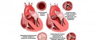
The oval window is a hole in the interatrial septum with a valve that connects the right and left atria during fetal development and normally closes after birth. In 50% of children under 1 year of age, the oval window continues to function; its anatomical closure occurs by 1–2 years.
Causes • When the newborn's lungs expand with air and pulmonary blood flow increases, the pressure in the left atrium increases and contributes to the closure of the oval window • With connective tissue dysplasia, alcoholic embryopathy, prematurity, physiological closure of the oval window does not occur.
Functional significance • May occur with congenital heart disease due to stretching of the atrium walls; the shunt unloads the right parts of the heart and facilitates the natural course of the defect • In primary pulmonary hypertension, the presence of a patent foramen ovale is a prognostically favorable sign that increases life expectancy • In newborns, a patent foramen ovale can be accompanied by respiratory distress syndrome; in adolescents it occurs latently.
Open oval window - causes, symptoms and treatment
Patent foramen ovale (PFO) is a gap in the wall formed between the right and left atria. Normally, such an open gap functions during embryonic development and completely closes during the first year of the child’s life. If this does not happen, we begin to talk about an anomaly, which is assigned code Q21.1 in ICD 10.
On the left atrium side, the opening is covered by a small valve, which is fully matured by the time of birth.
When the baby's first cry occurs and the lungs open, there is a significant increase in pressure in the left atrium, under the influence of which the valve completely closes the oval window.
Over time, the valve firmly adheres to the wall of the interatrial septum, so the gap between the atria closes.
Most often, in half of children, such valve growth occurs during the first year of life. This is the norm.
But if the valve size is insufficient, the gap may not close completely, that is, some hole will remain, the dimensions of which are determined in millimeters. Because of this, the atria are not isolated from each other.
Then the child is diagnosed with an open window, which is otherwise called MARS syndrome.
Cardiologists classify it as a minor anomaly of cardiac development.
In some cases, when there are no severe symptoms that affect the quality of life, this syndrome may be perceived as an individual feature of the cardiac structure.
But it often happens that such an anomaly becomes known by chance.
For an adult, this may come as a surprise. They get scared, thinking that this is a serious vice and their life will soon end. Some young people believe that because of this they will not be allowed into the army.
Are there reasons for such concerns? To understand this, you need to understand the causes, symptoms and other factors associated with LLC.
Causes
So, a patent foramen ovale is an opening, measured in millimeters, that forms between the atria. Through it, blood can flow from one atrium to another. Most often it comes from the left atrium to the right. This is due to the fact that the pressure in the cavity of the left atrium is higher. When a diagnosis is made, the following formulation is often given: LLC with left-right reset.
But LLC is not an atrial septal defect, although in accordance with ICD 10 they are assigned one code. A defect is a more serious pathology. MARS syndrome is not a congenital heart defect or a septal defect. And the differences are not only in the structure and development of the heart, but also in the causes, symptoms, treatment and other factors.
The reasons for this condition of the oval window are not always precisely known. It is believed that a hereditary predisposition can lead to this condition.
Of course, it’s unlikely that anything can be done about this factor.
But there are other reasons that largely depend on the woman who carries a new life within herself; their presence becomes especially important during the period of carrying a child in the womb:
- smoking;
- malnutrition;
- toxic poisoning with drugs;
- alcoholism and drug addiction;
- stress.
Unfortunately, today more and more women are starting to lead a bad lifestyle and continue to do so even during pregnancy. At the same time, they do not think at all that their baby will suffer. A patent foramen ovale is just one consequence, which can be considered not very serious compared to others, which can be, for example, a heart defect.
A patent oval window may develop due to poor environmental conditions.
LLC can develop for other reasons: poor environmental conditions, congenital heart disease, connective dysplasia, prematurity. If these reasons occur at a time when a woman becomes pregnant, you need to be prepared for the consequences that relate to the development of the baby or the organs of his body.
It has been noted that MARS syndrome often occurs with other cardiac malformations. These include open aortic disease, as well as congenital defects of the mitral and tricuspid valves.
Several other factors can contribute to window opening:
- very strong physical activity, which is especially true for athletes who engage in weightlifting, diving, and strength sports;
- manifestations of pulmonary embolism in those patients who have thrombophlebitis of the lower extremities and pelvis.
Symptoms
Although the abnormality is often detected in adults during testing for other conditions, it is best to do this early because other heart problems may be detected.
Thanks to the identified symptoms, an adult or a child’s parents can seek medical help in time, undergo an examination, after which a diagnosis will be made: LLC with left-right discharge, and a code will be noted in accordance with ICD 10.
If the size of the defect is small, from two to three mm, there is no particular reason to worry, since this is a common situation for a small child. Therefore, there are no special manifestations.
By the way, all children under one year of age are prescribed a heart ultrasound, which makes it possible to identify LLC. If the size of the defect is more than three mm, most likely, some signs will be observed that allow certain conclusions to be drawn:
- cyanosis of the nasolabial triangle or lips in a child when he cries a lot or screams;
- frequent colds, bronchitis, pulmonary inflammation;
- slowdown in psychological or physical development, which may even indicate that the oval window is open even by two or three mm;
- attacks of loss of consciousness;
- fast fatiguability;
- feeling of lack of air.
The latter signs are observed when the size of the anomaly exceeds three mm. If the doctor suspects that a child has PFO, he will refer him for examination by an experienced cardiologist and an ultrasound scan. This way the dimensions of the defect are clarified and it turns out that they exceed three mm. All this allows you to understand whether there is cause for concern. By the way, the size of an open window can reach 19 mm.
Cyanosis of the nasolabial triangle may indicate a patent oval window measuring more than three mm
There are practically no specific symptoms in adults. A person may complain of severe pain in the head area.
A preliminary diagnosis in accordance with ICD 10 can be made on the basis of almost the same signs that were listed above.
There may also be impaired mobility of body parts and periodic numbness of the limbs.
It is important to understand that an open oval window is not a death sentence! The heart is still functioning well, of course, it all depends on what concomitant diseases, heart defects, and so on are present, but in itself PFO does not pose a very serious danger, although the consequences can be very unpleasant, but this will be discussed later. To diagnose PFO with left-right shunting and designate the code according to ICD 10, it is necessary to conduct an examination.
Diagnostics
First, the doctor collects general data about the patient’s health, anamnesis, and complaints. This will help identify the causes and possible complications. A physical examination is also carried out, that is, the doctor examines the skin, determines body weight, measures blood pressure, and listens to heart sounds.
Then a general blood test, urine test, and biochemical blood test are prescribed. These tests help identify comorbidities, cholesterol levels, and other important factors.
In addition, the picture is clarified by such studies as coagulogram, ECG, echo CG, transesophageal, contrast echocardiography, chest x-ray.
All this allows you to accurately assess the patient’s health status, his heart, determine the size of the anomaly in millimeters, and so on.
Thanks to such important studies, the doctor makes an accurate diagnosis and determines the code in accordance with ICD 10. What treatment is prescribed if an open foramen ovale with left-right collection or another similar diagnosis is detected?
Treatment
What to do if you suspect problems with the oval cardiac window? Go to the doctor immediately! This rule applies to everyone who discovers at least some health problems. What to do after visiting a doctor? Follow his recommendations and appointments.
The scope of treatment measures is determined depending on the symptoms and concomitant diseases. Although the ICD 10 abnormality code is atrial septal defect, patent foramen ovale with left-to-right shunt is a different condition.
If there are no obvious disturbances in cardiac function, the doctor gives the patient recommendations that are aimed at properly organizing the daily routine, limiting physical activity, and observing nutritional rules. Taking medications for asymptomatic anomalies is not advisable. General strengthening procedures may be prescribed, such as exercise therapy, treatment in sanatoriums and others.
For minor complaints about the functioning of the heart and blood vessels, vitamins and means to strengthen the heart muscles may be prescribed.
If there are minor complaints about the functioning of the heart and blood vessels, treatment based on taking vitamins and drugs that strengthen the heart muscle can be prescribed. At the same time, it is important for the patient to limit himself in terms of physical activity. If, in a PFO with a left-to-right shunt and a significant size of the anomaly in millimeters, the symptoms are clearly expressed and there is a risk of blood clots, the following may be prescribed:
- disaggregants, anticoagulants, these drugs prevent the formation of blood clots;
- endovascular treatment, when a patch is applied through a catheter to the oval window, stimulating the opening to close with connective tissue; this patch resolves on its own after a month.
After surgery, antibiotics are prescribed to prevent the possible development of infective endocarditis. Thanks to endovascular treatment, a person returns to a full life, in which there are practically no restrictions.
Under no circumstances should you prescribe medications yourself. Each remedy has contraindications and side effects. For these and other reasons, every prescription should be made by a physician. When a diagnosis is made: a patent foramen ovale in the heart, in accordance with ICD 10, it is important for the patient to know what complications there may be.
Complications and prevention
Of course, the likelihood and form of complications depends on many factors. But it is important to understand that complications are rare. In fact, the following diseases can develop:
If an open foramen ovale is detected, it is necessary to regularly see a cardiologist and perform an ultrasound of the heart.
This occurs because a paradoxical embolism develops. If we talk about forecasts, then in most cases everything is favorable.
Those who have been diagnosed with LLC in accordance with ICD 10 need to be regularly monitored by a cardiologist and undergo an ultrasound of the heart. It is necessary to abandon sports that constantly subject the body to very strong physical stress.
It is important for every woman who is planning to have a baby or has already become pregnant to remember that she can prevent her unborn child from developing a heart abnormality. You cannot smoke, drink, take drugs or do anything that could somehow affect the health of the fetus in the womb.
As a result, we can say that LLC is an anomaly, which in itself does not pose a very serious danger, unless we are talking about the fact that there is an accompanying defect or other serious defect. It all depends on various factors. But the health of every person is very often in his hands! Every day you need to think about your health and your loved ones!
Source: https://cardio-life.info/zabolevaniya-serdca/otkrytoe-ovalnoe-okno.html
Diagnostics
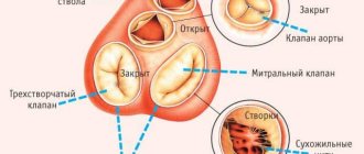
Diagnosis • ECG - there are no specific changes, incomplete blockade of the right branch of the His bundle occurs no more often than in the general population (unlike atrial septal defect) • EchoCG. Signs of an open oval window in the differential diagnosis with a secondary atrial septal defect in the first weeks of life •• Inconsistency of visualization of the defect in the area of the oval window •• Visualization of the oval window valve in the cavity of the left atrium •• Cross-discharge of blood (with color Doppler examination) depending on the clinical condition of the newborn.
Open oval window :: Symptoms, causes, treatment and code according to ICD-10
Name: Open oval window.
Anatomical features of atrial septal defect
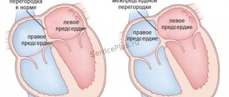
Open oval window.
Incomplete closure of the foramen ovale in the interatrial septum, which normally functions during the embryonic period and closes in the first year of the child’s life.
An open foramen ovale can be manifested by cyanosis of the nasolabial triangle, slow physical development, shortness of breath and tachycardia, sudden fainting, headache, frequent acute respiratory viral infections and bronchopulmonary diseases.
Diagnosis of an open foramen ovale includes an ECG (at rest and after exercise), conventional and Doppler echocardiography, radiography, and probing of the cardiac cavities. With an open foramen ovale, anticoagulant therapy can be used, and, if necessary, surgical treatment (endovascular occlusion of the defect).
A patent foramen ovale is a congenital communication between the right and left atria and is a residual element of the foramen ovale of the fetal heart. The interatrial foramen with a valve is formed in utero and is a necessary condition for the functioning of the cardiovascular system during this period of development. Thanks to the open foramen ovale, part of the placental, oxygenated blood flows from the right atrium to the left, bypassing the undeveloped, non-functioning lungs, and ensures normal blood supply to the neck and head of the fetus, the development of the brain and spinal cord. In healthy full-term children under normal developmental conditions, the patent foramen ovale usually closes and ceases to function within the first 12 months after birth. But its closure occurs individually for each person: on average, by the age of 1 year, the oval window remains open in 40-50% of children. The presence of an open foramen ovale after 1-2 years of a child’s life is classified as minor anomalies of cardiac development (MARS syndrome). In mature patients, a patent foramen ovale is detected in approximately 25-30% of cases. The fairly high prevalence of patent foramen ovale determines the relevance of this problem in modern cardiology. The open foramen ovale is located at the bottom of the fossa oval on the inner left wall of the right atrium, often has a small size (about the size of a pinhead) and a slit-like shape. The size of the open foramen ovale is on average 4.5 mm, but can reach 19. The open foramen ovale, unlike an atrial septal defect, has a valve structure that ensures the variability of the interatrial communication, the possibility of blood discharge in only one direction (from the pulmonary circulation to the systemic circulation). ). The clinical significance of a patent foramen ovale is controversial. A patent foramen ovale may not cause hemodynamic disturbances and may not have a negative impact on the patient’s health due to its small size and the presence of a valve that prevents blood from shunting from left to right. Most people with a patent foramen ovale are unaware of this anomaly and lead normal lives. The presence of a patent foramen ovale in patients with primary pulmonary hypertension is considered prognostically favorable in terms of life expectancy.
However, excess pressure in the right atrium compared to the left with an open foramen ovale leads to the periodic occurrence of a right-to-left shunt, which allows a certain volume of blood to pass through and leads to hypoxemia, transient cerebrovascular accidents (TIA), and the development of life-threatening complications: paradoxical embolism, ischemic stroke , myocardial infarction, kidney infarction.
All children are born with a patent foramen ovale. After the first independent breath, the newborn’s pulmonary circulation turns on and begins to fully function, and there is no need for an open foramen ovale. An increase in blood pressure in the left atrium compared to the right leads to the closure of the oval window valve. In most cases, the valve closes tightly and is completely overgrown with connective tissue - the open oval window disappears. Sometimes the hole closes partially or does not close at all, and under certain conditions (during excessive coughing, crying, screaming, tension in the anterior abdominal wall), blood is discharged from the right atrial chamber to the left (functioning oval window). The reasons for incomplete closure of the oval window are not always clear. It is believed that a hereditary predisposition, prematurity, congenital heart defects, connective tissue dysplasia, exposure to adverse environmental factors, smoking and alcohol consumption by a woman during pregnancy can lead to an open foramen ovale. Due to genetic characteristics, the diameter of the valve may be smaller than the diameter of the oval opening, which will prevent its complete closure. A patent foramen ovale may be accompanied by congenital defects of the mitral or tricuspid valves or a patent ductus arteriosus. Risk factors for the opening of the oval window valve may include significant physical activity in athletes involved in weightlifting, wrestling, and athletic gymnastics. The problem of an open foramen ovale is especially pressing for divers and divers who dive to significant depths and have a 5 times higher risk of developing decompression sickness.
In patients with thrombophlebitis of the lower extremities or pelvis with a history of episodes of pulmonary embolism, contraction of the pulmonary vasculature can cause increased pressure in the right side of the heart and the appearance of a functioning patent foramen ovale.
A patent oval window does not have specific external manifestations; in most cases it occurs latently, and can sometimes be accompanied by scant symptoms. Indirect signs of an open oval window may be: severe pallor or cyanosis of the skin in the area of the lips and nasolabial triangle during physical stress (crying, screaming, coughing, straining, bathing a child); tendency to frequent colds and inflammatory bronchopulmonary diseases; slower physical development of the child (poor appetite, insufficient weight gain), low endurance during physical activity, combined with symptoms of respiratory failure (shortness of breath and tachycardia); sudden fainting and symptoms of cerebrovascular accident (especially in young patients, with varicose veins, thrombophlebitis of the lower extremities and pelvis).
Patients with a patent foramen ovale may experience frequent headaches, migraines, postural hypoxemia syndrome - the development of shortness of breath and a decrease in arterial blood oxygen saturation in a standing position with improvement when moving to a horizontal position.
Studying the medical history and physical examination of the patient often does not immediately allow one to determine the presence of an open foramen ovale, but can only assume the possibility of this anomaly of the interatrial septum (cyanosis of the skin, fainting, frequent acute respiratory viral infections, developmental delay in the child). Auscultation helps identify the presence of a heart murmur as a result of an abnormal shunt of blood from a higher-pressure chamber to a lower-pressure chamber. To establish an accurate diagnosis of an open foramen ovale, instrumental studies and visualization methods are used: ECG (at rest and after exercise), conventional and Doppler echocardiography, chest radiography, probing of the cardiac cavities. When the foramen ovale is open, changes appear on the electrocardiogram indicating an increased load on the right parts of the heart, especially on the right atrium. In older people with a patent foramen ovale, radiological signs of enlargement of the right chambers of the heart and an increase in blood volume in the vascular bed of the lungs may be detected. In newborns and young children, transthoracic two-dimensional echocardiography is used, which makes it possible to visually determine the presence of an open oval window and its diameter, obtain a graphic image of the movements of the valve leaflets over time, and exclude an atrial septal defect. Doppler echocardiography in graphical and color mode helps to clarify the presence and size of an open oval window, identify turbulent blood flow in the area of the oval foramen, its speed and the approximate volume of the shunt. In older children, adolescents and adults, a more informative transesophageal echocardiography, supplemented by a test with bubble contrast and a test with straining (Valsalva maneuver), is used to diagnose a patent foramen ovale. Bubble contrast improves the visualization of the open oval window, allows you to determine its exact dimensions, and assess the pathological blood shunt. The most informative, but more aggressive method for diagnosing an open foramen ovale is cardiac catheterization, which is performed immediately before surgical treatment in a specialized cardiac surgery hospital.
Patients with varicose veins, thrombophlebitis, cerebrovascular accident, chronic lung diseases, who are at risk of developing paradoxical embolism, must be examined for the presence of an open foramen ovale.
With an asymptomatic course, a patent oval window can be considered a normal variant. Patients with a patent foramen ovale and a history of a transient ischemic attack or stroke are prescribed systemic therapy with anticoagulants and antiplatelet agents (warfarin, acetylsalicylic acid) to prevent thromboembolic complications. The method for monitoring anticoagulant therapy is the international normalized ratio (INR), which should be in the range of 2-3 when the foramen ovale is open. The need to eliminate a patent foramen ovale is determined by the volume of shunted blood and its effect on the functioning of the cardiovascular system. If there is a small discharge of blood, there are no concomitant pathologies and complications, surgery is not required.
In case of pronounced pathological discharge of blood from the right atrium to the left, low-traumatic X-ray endovascular occlusion of the open foramen ovale is performed. The operation is carried out under X-ray and echocardioscopic control using a special occluder, which, when opened, completely plugs the hole.
For patients with a patent foramen ovale, regular monitoring by a cardiologist and echocardiography are recommended. Endovascular occlusion of the patent foramen ovale allows patients to return to their normal rhythm of life without restrictions.
In the first 6 months after surgical treatment of an open oval window, antibiotics are recommended to prevent the development of bacterial endocarditis.
The greatest effect from endovascular closure of the patent foramen ovale is observed in patients with platypnea who had a pronounced right-to-left shunt.
42a96bb5c8a2ac07fc866444b97bf1 Content moderator: Vasin A.S.
Source: https://kiberis.ru/?p=32024
Treatment
Treatment of concomitant pathology. Concomitant pathology • Prematurity • Other congenital heart diseases • Neonatal respiratory distress syndrome • Hereditary connective tissue diseases.
The course and prognosis are favorable. Synonyms • Functioning foramen ovale • Patent foramen ovale
ICD-10 • Q21.1 Atrial septal defect
Excludes: acquired cardiac septal defect (I51.0)
Coronary sinus defect
Unclosed or preserved:
- foramen ovale
- secondary hole (type II)
Venous sinus defect
Common atrioventricular canal
Endocardial defect at the base of the heart
Atrial septal defect (type II)
Ventricular septal defect with pulmonary stenosis or artesia, aortic dextroposition and right ventricular hypertrophy.
Aortic septal defect
Eisenmenger's defect
Excluded: Eisenmenger
- complex (I27.8)
- syndrome (I27.8)
Septal defect (heart) NOS
ICD-10 alphabetical indexes
External Causes of Injury - The terms in this section are not medical diagnoses, but rather a description of the circumstances under which the event occurred (Class XX. External Causes of Morbidity and Mortality. Heading Codes V01-Y98).
Medicines and chemicals - table of medicines and chemicals that have caused poisoning or other adverse reactions.
In Russia, the International Classification of Diseases
10th revision (
ICD-10
) was adopted as a single normative document for recording morbidity, reasons for the population’s visits to medical institutions of all departments, and causes of death.
ICD-10
introduced into healthcare practice throughout the Russian Federation in 1999 by order of the Russian Ministry of Health dated May 27, 1997 No. 170
The release of the new revision (ICD-11) is planned by WHO in 2022.
Abbreviations and symbols in the International Classification of Diseases, 10th Revision
NOS
- without other instructions.
NEC
— not classified in other categories.
†
— code of the main disease. The main code in the dual coding system contains information about the underlying generalized disease.
*
- optional code. An additional code in the double coding system contains information about the manifestation of the main generalized disease in a separate organ or area of the body.
A patent foramen ovale is a cardiac structural feature, which according to the ICD 10 classification is classified as a defect of the septum dividing the atrium. It is present in all babies at the time of birth and closes as they grow due to uselessness. But there are reasons why the window does not disappear and leads to the development of consequences.
Anomaly factors
Maternal smoking during pregnancy can lead to the appearance of a patent foramen ovale in the heart.
The exact reason for the oval window not closing has not yet been clarified. One of the main factors is considered to be hereditary predisposition.
Experts also identify several reasons that relate to the period of gestation and can lead to the appearance of such an anomaly:
- Smoking
- Deficiency of nutrients and vitamins in the diet
- Drug poisoning
- Alcohol abuse
- Addiction
- Severe stressful experiences
- Living in an environmentally unfavorable area
- Premature birth
- Prematurity of the baby
- Congenital heart defects
Many women lead a destructive lifestyle and, when pregnancy occurs, do not take any measures to change it. They don’t even think about the consequences of this for the child.
Scientists have found that this gap can also be present in an adult who has heart defects. Among them are defects of the aortic, mitral or tricuspid valve.
At-risk groups
A woman's poor nutritional status during pregnancy is a risk factor for a patent foramen ovale in the baby's heart.
A window remains open for many reasons. In addition to genetic inheritance, there are factors that contribute to the opening of the gap:
- Physical activity with a high load, this applies to weightlifters, divers or strength athletes
- Thromboembolism of the pulmonary artery, more often in patients with vascular occlusion of the lower extremities
- Dependence on bad habits during pregnancy
- Poor nutritional status of a woman while expecting a baby
- Severe toxin poisoning during pregnancy
- Premature birth
- Low immunity of women
- Harmful environmental conditions
The role of the oval window
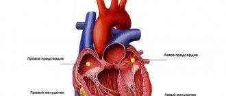
The open window in the heart is an oval hole, which is located in the interatrial septum and during the development of the embryo ensures the normal functioning of the cardiovascular system.
Blood moves in the fetus along a special path. During intrauterine development, the baby has fetal structures that provide access to oxygen-enriched blood into the systemic circulation.
The baby does not yet breathe air, since his lungs have not formed. Blood enters the child's body through the umbilical veins. One of them communicates with the liver, which performs a hematopoietic function, and the other with the vena cava through the ductus venosus. Arterial blood from the liver mixes with venous blood and enters the right atrium. An open window in the heart allows this blood flow to reach the left atrium. The remaining blood passes through the aortic duct into a large circle.
Possible signs
A baby's heart murmur may indicate a patent foramen ovale.
Signs of an open window are almost invisible. In adult patients, the pathology is diagnosed during an examination of the body for other complaints.
With a window length of 2-3 mm, there are no disruptions in the functioning of the heart and blood flow. The patient feels well.
Complications may occur with a length of more than 4 mm. Signs of the anomaly vary depending on the age category of the patient.
Symptoms in children:
- Blueness of the triangle between the lips and nose when crying or active games
- Frequent colds
- Physical or mental underdevelopment
- There is a heart murmur
- Frequent fainting occurs
- Insufficient blood circulation to the brain
- Attacks of shortness of breath during active activities
If the pathology is not detected in childhood, it remains for life. An adult patient may not even be aware of his heart condition for a long time.
The symptoms are similar to those in children, may appear to a small extent and resemble other pathologies.
Signs in adults:
- Blueness of the triangle between the lips and nose after physical activity or during colds
- Frequent occurrence of fainting
- Blood flow in the brain is impaired
- Varicose veins or thrombophlebitis
- Attacks of shortness of breath
- Frequent colds
- Attacks of tachycardia
- Migraine
- Severe condition after physical activity
- Increased amount of blood in the lungs
- Numbness of the limbs
Stages
The parameters of the oval window determine whether the patient requires surgical intervention
A patent foramen ovale is considered a developmental defect of the heart. The stages of the anomaly differ in the degree of damage to the organ and the severity of the symptoms.
The anomaly is included in the group of MARS syndrome (small cardiac development syndromes). Its location is the atria.
Any stage of development of the anomaly is the result of connective tissue dysplasia. Window parameters and role in blood flow determine the severity of the anomaly.
Divers
An open oval window in an adult facilitates the transformation of gases dissolved in water into bubbles when diving to depth. An embolism develops, which, in the absence of timely assistance, threatens death. Therefore, professions such as diver and diver are prohibited for people with LLC.
Children with this anomaly are also not advised to engage in scuba diving. A sharp change in pressure causes an increase in its size.
Even the famous pediatrician Komarovsky says that such a deviation is not a heart defect and it is not worth treating PFO if its size is acceptable and there are no complications. In order for the hole to close, it is advised to follow the doctor’s recommendations and wait a while. In 50% of children, ultrasound does not detect anomalies after 1 year, and in 25% - after 5 years.
Varieties
The oval window serves as a natural way for the body to adapt to environmental conditions. It is present in absolutely every baby after birth. For certain reasons, the window may not disappear and remain open.
Depending on the length of the window, the following forms of anomaly are distinguished:
- Gap parameters of 2 to 3 mm are considered normal for the baby’s development and do not entail complications or health consequences
- Gap parameters from 5 to 7 mm may not have obvious manifestations and do not threaten health; minor complications may appear after active physical activities
- Gap parameters greater than 7 mm already represent a large hole that disrupts blood flow and requires surgical intervention
Depending on the degree of participation of the window in the blood flow process, the following forms can be distinguished:
- A gap without blood flow does not involve the movement of blood through the atrial septum and does not affect the functioning of the heart
- The shunt gap on the left side allows blood to flow across the septum, which leads to increased pressure in the atrium of the right side of the heart
- A discharge window on the right side results in a reverse flow of blood, which causes a similar increase in pressure on the left side
- The window with the manifestation of bidirectional shunting may threaten the development of a paradoxical embolism, which is formed during physical activity and manifests itself in thrombosis and gas formation, which enter the brain with the bloodstream
The complexity of the pathology is determined in each case individually based on the patient’s age, the nature of his life activity and the strength of the symptoms.
Open oval window - treatment and symptoms. Causes and hemodynamics
A patent foramen ovale is a valve apparatus on the border of the cardiac atria, which is a physiological structure during the period of intrauterine growth and ceases to function after the birth of a child.
The reasons for the anomaly have not been fully proven. However, there are a number of conditions in a child that increase the likelihood of developing pathology:
- prematurely born child with underweight;
- congenital defects in the development of the cardiovascular system;
- autoimmune connective tissue diseases.
On the part of a pregnant woman, the following risk factors for her unborn baby are identified:
- hereditary predisposition;
- negative environmental impact;
- exposure to chemical and toxic fumes in industry;
- nicotine addiction, consumption of alcoholic beverages and drugs;
- infectious TORCH diseases.
In adulthood, this pathology can be a consequence of excessive physical exertion in people whose work is related to sports , diving, or a history of pulmonary embolism.
Hemodynamics of a patent oval window
This anatomical and physiological formation has a valve structure, which during prenatal growth ensures complete blood supply to the brain, and after closure prevents the discharge of blood plasma between the atria.
If there is a defect in the fusion of the communication, plasma shunting is observed. Blood entering the left chambers provokes insufficient oxygen content in the bloodstream, a disorder of the blood supply to the brain.
Symptoms of an open oval window
Due to the small size of the interatrial valve, its non-fusion after birth may not have pronounced manifestations. The clinical picture is as follows:
- paleness of the skin;
- cyanosis of the nasolabial area;
- loss of appetite;
- dizziness;
- headache;
- fainting state;
- difficulty breathing;
- pain in the cardiac region;
- accelerated heartbeat.
An open foramen ovale during pregnancy may be accompanied by the above manifestations against the background of hormonal changes in the body and an increase in the performance of the heart muscle.
A woman who is carrying a baby has an increased risk of impaired blood supply to the brain, acute coronary heart disease, and blockage by a blood clot. However, this anomaly is not an indication for termination of pregnancy.
With the right therapeutic approach and careful medical supervision, undesirable consequences can be avoided.
Diagnosis of an open oval window
Symptomatic signs allow only indirect suspicion of the occurrence of an anomaly. To verify its presence, it is necessary to carry out additional examination methods, which include:
- ECG - is prescribed twice to determine the work of the heart muscle at rest and under load;
- EchoCG - you can detect an unclosed interatrial foramen, pathological reflux of blood, its volume and direction of movement;
- Ultrasound with Doppler is an ultrasound examination that helps evaluate blood circulation through the vessels, its disturbances, changes in the vascular walls;
- ventriculography - performed using contrast, it is possible to identify deviations in the structure and functions of the cardiac chambers, congenital and acquired defects;
- X-ray of the OGK - increased load on the right chambers is detected;
- probing of the cardiac regions - carried out using a radiopaque probe;
- Magnetic resonance imaging.
Treatment of open oval window
If the developmental defect does not provoke the appearance of any complaints, does not affect the full functioning of life, the width of the formation is no more than 2-3 mm, surgical tactics are not used. The patient must undergo a preventive examination by a doctor in a timely manner and undergo a study appointment, strictly follow all the recommendations of the attending physician that relate to lifestyle.
If there is a history of acute cerebrovascular accident, to prevent the occurrence of thromboembolism, drug therapy is prescribed, which is based on the following:
- anticoagulants;
- disaggregants.
In cases of severe symptoms, complicated conditions, and a large volume of plasma that is transferred from the right to the left, the question of surgical therapy arises. Endovascular occlusion is used using X-ray and echocardiographic devices.
Patent foramen ovale in newborns
The interatrial valve formation exists throughout prenatal development as necessary for adequate blood supply to the fetus. Before birth and the first cry and breath, the child’s lungs do not perform their function.
Therefore, oxygen is transferred through the atria through the open foramen ovale in the child to the heart, bypassing the structures of the respiratory system so that the head and cervical region receive a sufficient amount of blood for the normal functioning and development of the central nervous system organs.
In children born at term without pathologies, this valve heals up to one year, after which it becomes covered with connective tissue. However, the duration of this process may vary because it depends on the individual characteristics of the organism. If after two years the communication remains unclosed, they speak of a minor developmental anomaly.
Complications and prognosis of open oval window
As a result of an open atrial formation, a pathological venous-arterial discharge of blood occurs, increasing the likelihood of accumulation of an embolus or thrombus in the vessels. What leads to such conditions:
- acute coronary heart disease;
- cerebrovascular disorder;
- abnormalities in the renal circulation.
People with minor plasma reflux into the left atrium have a favorable prognosis. They need to undergo regular examinations by a cardiologist with echocardiography monitoring.
If there are indications for surgical treatment of an open oval window in adults and children, and the operation is successful, the patient can also live a full life. In the postoperative period, a course of antibacterial drugs is prescribed to prevent inflammation of the inner layer of the heart muscle.
Source: https://xmedicin.com/otkryitoe-ovalnoe-okno/
Diagnostics
Ultrasound of the heart is one of the methods for diagnosing open oval window
A patent foramen ovale is very difficult to identify during an initial consultation with a doctor. This anomaly may be generally asymptomatic and not cause a trip to the hospital. Most often, it is detected incidentally when examining a patient for other diseases.
Diagnostic methods are:
- Taking an anamnesis - establishing hereditary factors, bad habits, lifestyle, etc.
- Visual examination - measuring pressure, examining the skin, listening to the heart rhythm, etc.
- Laboratory examination of the body - general and biochemical blood tests, urine tests, etc.
Hardware diagnostics
An important role in identifying an open oval window is played by diagnostics using special devices. It makes it possible to assess the size of the anomaly, its degree of influence on blood flow and heart function, and also determine the risk of developing consequences.
- Ultrasound of the heart
- Echocardiography
- Angiography
- MRI
- ECG
- Chest X-ray
The most informative is an echocardiography examination. It is passed through the esophagus. This procedure makes it possible to record the gap itself, its parameters, the volume of blood passing through it and correlate it with defects in the septum between the atria.
Only after a complete examination can a doctor make an accurate diagnosis and determine the ICD 10 code. This gives him the opportunity to prescribe the correct treatment.
Signs indicating the presence of pathology
The diagnosis of LLC (ICD 10 code) is made by a doctor if he detects the following deviations:
- the size of the valve does not change, but the heart enlarges. Loose cover of the oval hole causes a constant release of blood from one atrium to another, increasing the load on them;
- The patient experiences various diseases that provoke increased pressure in the right atrium. Therefore the valve opens frequently;
- LLC size exceeds 2 mm.
Important! The choice of treatment method (conservative or surgical) depends on the degree of coverage of the window by the valve and the patient’s well-being.
The goal of treatment is to eliminate pathological symptoms and prevent the development of complications.
How to treat
If you feel well with this anomaly, one of the ways to maintain health is good nutrition
When this anomaly is detected, therapy is prescribed depending on the strength of the manifestation of signs of pathology and the general health of the patient. Among the important conditions when prescribing therapy are:
- Window Options
- The amount of blood flowing during physical activity
- Features of the partition design
- Degree of atrial enlargement
- Pressure in the arteries of the lungs
- Accompanying illnesses
If the length of the gap is short and there are no obvious signs of the influence of the anomaly on the person’s well-being, the doctor will prescribe general recommendations for maintaining health.
Among such measures are:
- Proper and nutritious nutrition
- Optimal daily and rest schedule
- Decreased physical activity
- Physiotherapy
- Sanatorium treatment
- Staying outside and breathing fresh air
- Health monitoring
Causes
Etiology: factors that form congenital heart disease (see Tetralogy of Fallot).
Pathogenesis • The magnitude and direction of shunt depend on the size of the defect and the relative compliance of the ventricles • In adults, the right ventricle is more compliant than the left, as a result of which shunt occurs from the left atrium to the right • A small shunt leads to a moderate volume overload of the right heart, and pressure in the pulmonary artery remains normal • The severity of pulmonary hypertension may be insignificant even with a large shunt • Only in rare cases does severe pulmonary hypertension develop, leading to right ventricular failure and right-to-left shunt • Unlike VSD, with ASD the shunt is smaller and affects only the right side of the heart.
Variants of ASD • Ostium secundum (secondary defects) are localized in the area of the oval fossa, are often multiple, accompany many syndromes: Holt-Oram syndrome (ASD of the ostium secundum type in combination with digital hypoplasia), Lutembashe syndrome (combination of ASD with mitral valve stenosis), combination of ASD with mitral valve prolapse, etc. • Ostium primum (primary defects) are usually large in size, localized in the lower part of the septum, at the attachment point of the mitral and tricuspid valves, interatrial and interventricular septa. They are part of an open AV canal and are often combined with abnormal drainage of the pulmonary veins, splitting of the anterior mitral valve leaflet, mitral regurgitation and Down syndrome • Defects of the sinus venosus type are localized near the mouth of the superior vena cava and the sinus node, often combined with sick sinus syndrome, AV - nodal rhythm and abnormal drainage of the pulmonary veins.
Which specialist is treating
An initial consultation is carried out with a general practitioner. He conducts an examination, collects anamnesis and prescribes general tests and some examinations.
Once a window anomaly is suspected, the therapist refers the patient to a consultation with a cardiologist. This specialist, in turn, also conducts an initial examination and prescribes a more detailed examination.
When the diagnosis is made and the degree and form of the anomaly is determined, the cardiologist prescribes treatment. If the gap parameters are large, surgical treatment is required.
For further examination and treatment, the patient is referred to a cardiac surgeon. This specialist evaluates the condition of the window, its size and effect on blood flow. Based on the data obtained, the doctor selects a treatment method and performs the operation.
What complications can there be?
If there is no treatment if the condition worsens, then a complication in the form of a heart rhythm disturbance is possible.
On its own, a window anomaly cannot threaten human health and contribute to the development of complications. But under the influence of certain factors the condition may worsen.
Lack of necessary treatment and preventive measures can cause the following consequences:
- Heart rhythm disturbance
- Deterioration of blood supply to the brain
- Hypertension in the lungs
- Paradoxical embolism
- Deterioration of blood supply to the heart
- Development of a heart attack
- Development of stroke
- Acute fatality
These complications occur quite rarely. But you should not aggravate the condition and refuse treatment.
Prevention
Giving up bad habits will help avoid complications of this anomaly.
There are no special instructions for treating the window. Prevention is aimed at maintaining human health and reducing the likelihood of complications.
- Elimination of bad habits
- Complete nutrition that replenishes the body with all important nutrients
- Sufficient time for sleep and rest
- Avoiding excessive exercise
- Timely treatment of emerging diseases
- Periodic consultations and examinations with the attending physician
Prevention plays an important role during pregnancy. A woman who is pregnant or planning a child should adhere to the following recommendations:
- Avoid infectious pathologies
- Avoid interactions with chemicals
- Take medications only as prescribed by a doctor
- Eat properly
- Eliminate bad habits
These measures will help reduce the risk of heart abnormalities.