Babies are born with an open oval window in the heart. As soon as they take their first breath, the pulmonary circulation begins to function normally, and the need for an open window disappears. Due to the increase in blood pressure in the left atrium, the valve eventually closes and over time is completely overgrown with tissue.
Patent foramen ovale is a common problem.
Prerequisites for development
Astigmatism in a child - what is it, does it need to be treated?
The main reasons why an open foramen ovale in a child’s heart may not close completely have not been fully clarified. A number of experts believe that the following factors provoke a dangerous disease:
- birth of a baby prematurely;
- heredity;
- Congenital heart defect;
- abuse of alcohol, tobacco, and drugs by the mother during pregnancy;
- exposure to unfavorable environmental factors.
Most pathologies develop while the mother is carrying a child.
Causes of patent oval window
There is no exact cause for this pathology.
There are several most common factors.
- In almost all premature and immature newborns, the window remains open.
- Smoking, maternal substance abuse.
- Intrauterine fetal hypoxia.
- Prolonged labor, asphyxia of the baby during childbirth.
- Adverse environmental factors.
- Mother's stress.
- Genetic predisposition.
- Congenital heart defects.
- Occupational exposure to toxic substances in the mother.
When it comes to physiological norms
The gradual closing of the hole in the heart in the first month or even a year of a baby’s life is considered normal.
Important! Medicine knows of cases where the window in a baby’s heart closed by the age of 3 or even 5 years.
Not in all cases, a hole in the heart of a small child turns out to be a real cause for serious concern. A person can have an open valve and live a full life without even realizing that he has a problem, because you can only find out about it by doing an ultrasound.
The heart can function fully if the hole has a not too significant diameter. When the window is opened no more than 2-3 mm, in the complete absence of other serious pathologies, there is no reason to worry. This condition has almost no effect on the child’s well-being.
In most cases, an open window does not affect the baby’s well-being
If the size of the hole in a child’s heart varies from five to seven millimeters, the anomaly is still considered hemodynamically insignificant, but may already begin to show itself during intense physical activity.
When the hole diameter is from 7 to 10 mm, this is far from the norm. Such a patient is usually diagnosed with a gaping open window. The symptoms of this condition are largely reminiscent of congenital heart disease. The patient should undergo regular examinations by a cardiologist.
Help from folk remedies
No folk remedies for the pathology have yet been identified.
If a person does not have obvious disorders in the functioning of the cardiovascular system, then doctors give him advice on how to lead a lifestyle and may prescribe some vitamins and proper nutrition that help support the functioning of the heart. And you need to limit physical activity. In the absence of symptoms, medications are not prescribed to the patient; they can only prescribe measures to strengthen the body, for example, hardening, exercise therapy, and sanatorium-resort treatment.
But if the patient has minor complaints about the heart, the doctor sometimes prescribes fortified medications that strengthen the cardiovascular system, Panangin, Magne B6, Elcar, Ubiquinone. And in case of severe disturbances in the functioning of the cardiovascular system, general therapy with the described drugs is used or surgery is performed.
Main symptoms
If a baby suffers from the pathology of an unclosed window, this can be understood by the following symptoms:
- slow weight gain;
- noticeable blueness of the circumlabial triangle during coughing, crying or screaming;
- susceptibility to bronchial and pulmonary colds.
In older childhood, if the window does not close, the growing child may develop shortness of breath and tachycardia, even not with the most severe physical exertion.
Closer to adolescence and throughout adult life, the pathology will manifest itself with the following symptoms:
- fast fatiguability;
- chronic fatigue;
- dizziness;
- weakness;
- migraine;
- frequent respiratory diseases.
Open oval window in children and its symptoms
In most cases, such children do not complain.
Therefore, it is very important for mothers to be attentive and monitor the slightest deviations in the behavior of their babies.
What can you notice?
- The appearance of blueness around the mouth of a newborn. This cyanosis appears after crying, screaming, sucking, or bathing.
- In older children, tolerance (resistance) to physical activity decreases. The child rests and sits down after regular outdoor games.
- The appearance of shortness of breath. In general, a child should normally be able to easily climb to the 4th floor without any signs of shortness of breath.
- Frequent colds in infants, namely: bronchitis, pneumonia.
- Doctors listen for a heart murmur.
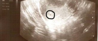
PERSONAL EXPERIENCE. The child is 10 days old; during bathing, the mother notes the blueness of the nasolabial triangle. The child was born full-term, weighing 3500. The mother admitted that she smoked during pregnancy. On examination, a murmur was noted at the apex of the heart. The baby was sent for an ultrasound. As a result, an open oval window of 3.6 mm was revealed. The child has been registered.
When the window closes
In more than half of newborn children, the window closes safely in the first year (usually before they reach three months of age). Somewhat less often, the process turns out to be longer and lasts up to five years. In most recorded cases, the pathology is considered a minor anomaly in the development of the heart. The prevalence of the disease was and remains quite high. According to official statistics, an open oval window is present in 50% of completely healthy children and 10-15% of adults.
Treatment methods
Treatment is not prescribed for children under three years of age whose gap size does not exceed 5 mm. After a year, if the interatrial septum does not close, a preventive ultrasound is performed once a quarter.
Children whose PFO persists for up to five years are subject to more careful monitoring by a cardiologist. If even after this age the size of the gap does not decrease, then they talk about pathology and offer a surgical solution to the problem.
Window sizes for prescribing treatment
The oval window in the heart of a newborn is open 5 mm or less, which happens in most cases; the case does not require either medication or, let alone, surgical treatment. Parents are advised to monitor his condition, not exceed permissible physical activity, and avoid overexertion and nervous stress.
If the window size exceeds 5 mm, the child is registered with a cardiologist, and the baby’s condition is diagnosed at least once every six months. If there are symptoms (shortness of breath, fatigue, discomfort and pain in the heart), especially if they increase, maintenance medications are prescribed.
With large window sizes (more than 7 mm), circulatory disturbances occur, which leads to interruptions in the functioning of the heart and heart failure. In such cases, surgical treatment is indicated. Thanks to modern technologies, the operation is performed quickly, without direct contact with the heart. The recovery period is short, after which the child can lead a full life.
Drug therapy
If a child has experienced ischemia (reduced blood supply to organs) or stroke at least once, then treatment with blood thinning drugs (anticoagulants) is prescribed to prevent thromboembolism. They are used to prevent complications caused by increased blood clotting.
| A drug | Dosage | Action |
| Aspirin | 3-5 mg per kg body weight (daily, divided into 2-3 doses per day). | Disaggregant (antiplatelet drug). |
| Warfarin (Coumadin) | The initial daily dose is 0.2 mg per kg of body weight, if liver function is not impaired. For liver diseases, 0.1 mg per kg of body weight is prescribed. | Used in systemic anticoagulant therapy. Blocks the production of vitamin K in the liver, which is responsible for hemostasis (blood clotting process). |
During treatment with anticoagulants and antiplatelet drugs, constant monitoring of platelet levels in the blood is carried out.
Surgery
To understand whether it is necessary to eliminate the opening of the oval window, the amount of blood thrown into the left atrium and the degree of impact of the deviation on the viability of the body are measured. If there are no symptoms in children, especially in the first year of life, surgical treatment is not performed.
The oval window in the heart of a newborn is open and symptoms are clearly expressed in especially rare cases. As a rule, such children have concomitant lesions. In such cases, surgery is recommended.
Low-traumatic x-ray endovascular occlusion is prescribed, which is performed when the child reaches 6 months. If the condition threatens the baby's life, then the operation is performed urgently.
A catheter is inserted into the femoral artery, through which an occluder is supplied to the heart muscle. This is a special device, similar to a double-sided umbrella, that replaces the undeveloped valve. When the occluder falls into place, it straightens and begins to perform the functions of the interatrial septum, improving the functioning of the atria.
Diagnosis of pathology
A physical examination of a young patient, as well as a study of his medical history, often does not make it possible to accurately determine the presence of an open oval window. They can only accept the possibility that an anomaly is present. To establish an accurate diagnosis, additional examinations are required:
- ECG;
- EchoCG;
- chest x-ray;
- probing of the heart cavities.
If an infant or young child is being examined, transthoracic two-dimensional electrocardiography is most often used for diagnosis. This type of study allows you to see whether the valve is closed or not. Once the presence of a patent foramen ovale is confirmed, the procedure makes it possible to determine its diameter. In addition, using transthoracic electrography, it is easy to obtain a graphical image of the movement of the valve leaflets over time and exclude or confirm the presence of an atrial septal defect.
Doppler EchoCG is an even more accurate procedure that provides information not only graphically, but also in color. The study allows you to clarify the size of the hole in the heart and identify the speed of turbulent blood flow in the area of the foramen ovale, and understand the approximate volume of the shunt.
To identify an open window, serious diagnostics are required
The most capacious, from the point of view of obtaining information, and the most aggressive diagnostic method is cardiac probing. The procedure is performed immediately before surgery in a specialized cardiac surgery hospital. In the case of infants, probing is used extremely rarely.
Possible complications
The presence of an open window in the heart can lead to a number of diverse complications. As the baby grows, his heart muscle begins to grow. The problem is that the valve in this case remains the same size. With increased blood flow in the oval window connector and free penetration of blood from one atrium to another, the load on them increases significantly.
Parents need to be especially attentive and vigilant if an open window in the child’s heart is diagnosed against the background of other cardiac problems that increase pressure in the right atrium. The consequence may not be the most pleasant - such pathologies increase the risk of additional opening of the heart valve towards the left atrium.
Important! Sometimes a pathology such as an open foramen ovale in a child’s heart can even be beneficial for health. Thus, in pulmonary hypertension, moving blood to another atrium reduces pressure and thereby has a positive effect on overall health.
The essence of the diagnosis
Between the right and left atria there is a wall.
There is a gap in it. During the period of intrauterine development of the fetus, this hole is a completely normal and even necessary formation. Its formation begins approximately 3 weeks after conception. During an ultrasound examination, it can be detected as early as 5–6 weeks. Through it there is blood supply between the body of the mother and the fetus.
The process of blood supply to an adult and a fetus has some differences. In an unborn child, the lungs do not participate in the blood supply. This is compensated by the participation of the pregnant woman’s body in ensuring the blood circulation of the fetus.
At the stage of intrauterine development, the ductus Botallus enters the blood circulation. Through it, the blood flow is directed from the heart to the aorta. In addition, the open foramen ovale is directly involved in the process of blood circulation, through which blood is directed from the right to the left atrium.
After a newborn baby takes his first breath of air, his lungs open, which automatically includes them in the blood circulation. As a result of such changes, the Botal duct and LLC cease to be necessary for the viability of the baby.
Over a period of approximately 1 month to a year, the open oval window is gradually closed by a valve, the halves of which grow to the walls of the window. By the end of the child’s first year of life, a septum is formed at the site of the valve, which separates the right and left atria.
In some cases, the final closure of the patent foramen ovale is completed 5–6 years after birth. There are cases when the open oval window of the heart remains uncovered throughout a person’s life.
Doctor Komarovsky's opinion
The famous Russian pediatrician and TV presenter, Dr. Evgeniy Komarovsky, agrees with the fact that almost every baby has a hole in the heart. Its complete closure during the first breath occurs in a very small percentage of babies. In most cases this takes time. In half of the cases, the window is completely overgrown within two years. Closer to the age of five, the anomaly mostly disappears on its own, without causing any harm to the child.
In children under one year old, the hole is more common because their lungs are not yet fully functional. As they open, the pressure inside the right atrium will gradually decrease. As a result, due to the pressure difference, the valve will press against the wall and begin to overgrow.
Dr. Komarovsky especially emphasizes the fact that a hole in the heart septum is not dangerous to life and health. This anomaly is one of the developmental features and in most cases occurs without symptoms and closes without medical help.
If you suspect a problem, don’t delay visiting a doctor.
If a child is diagnosed with this disease, parents should not panic. The risk that the problem will develop into a serious pathology is extremely small. However, until the valve becomes overgrown with connective tissue, it is necessary to periodically examine the baby. This will allow you to keep the situation under control and take the necessary measures in a timely manner if something suddenly goes wrong.
When should the valve close
Immediately after birth, the child inhales for the first time, the lungs “open” and begin their work. A pressure difference occurs, the valve expands and closes the interatrial septum. This happens normally, but not always. In the vast majority of newborns, the gap turns out to be wide, and the valve is too small and cannot completely close the window.
With age, all the baby's organs grow, including the oval window valve. By the age of one year, it reaches the optimal size to close the gap.
There are often cases when the development of blood vessels outstrips the development of connective tissue, which makes up all the valves and sphincters of the body, and the window closes only by 3-5 years. This condition is also considered to be within normal limits, but requires additional monitoring by a pediatrician and pediatric cardiologist.