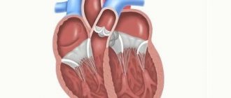
What is the interatrial septum
The right and left atria communicate with each other through an opening (foramen ovale) in the septum. This gap is open only during the intrauterine development of the baby. The supply of oxygen to the child is carried out through the umbilical cord; the pulmonary system does not work, so large quantities of nourishing blood are not required.
As a result, in the process of closed blood circulation in a small circle, a certain volume of blood is discharged from the right atrium through the hole into the left. The window is closed by a valve that works like a door on a spring: opening occurs only towards the left atrium.
However, after the baby is born, changes begin in his body. When the baby inhales air for the first time, the pulmonary system is cleansed of amniotic fluid, filled with oxygen, and blood is supplied through the pulmonary circulation. At this point, the oval window stops working.
In the area of the left atrium, there is an increase in pressure, due to which the valve covering the opening is strongly pressed against the septum between the atria. Thus, the door is no longer able to open, which contributes to its overgrowing.
Norms
The valve in the septum between the atria usually heals between 3 months and 2 years of age. Although even in a 5-year-old child, this phenomenon is still considered the norm. According to statistical data, every second healthy child at the age of 5 years and every fourth at the age of 10 years have a similar feature.
It should be noted that such a defect is not a defect. Experts call this condition MARS (minor anomaly in the development of the heart). With such a deficiency, the cardiac structure differs slightly from anatomical norms, although this does not pose a direct danger to human health.
The hole varies in volume: within 3-19 mm (usually 4.5 mm). Initially, the dimensions of the window are determined by the age of the patient and the size of the organ itself. Whether an operation will be prescribed is decided by the doctor, based not on the size of the hole, but on how much the valve covers it and what the stage of compensation is.
What may be defects in interatrial communication in a newborn?
A defect in the septum between the atria is a congenital heart abnormality (birth defect). If the defect is incomplete, there is a window in the partition; if it is complete, there is no partition at all. This pathology is characterized by the presence of interatrial communication.
The disease is most often detected incidentally during an ultrasound of the heart, since in most patients the disease may not manifest itself for a long time.
Forms and classification
To make it more convenient to understand the condition of a patient with a similar illness, the pathology is classified into 3 types of anomalies in the septum between the atria. The defect can be presented as:
- Open oval window - this phenomenon is observed during the formation of the fetus in the womb. This opening helps perform gas exchange when the pulmonary system is not yet functioning. Subsequently, it is closed by a special valve that tightly adheres to the septum between the atria. Occasionally, this “door” does not take root or the dimensions exactly repeat the outline of the window. In such a situation, even a slight emotional impact provokes the opening of the valve and blood transfusion from the left ventricle to the right.
- Primary anomaly - this condition is characterized by insufficient development of the valves that separate the atria and ventricles, and their size is not sufficient to tightly close the window.
- Secondary defect - in a similar situation in newborns, there is an abnormal development of the hollow superior venous vessel.
Diagnosis of the disease
If any of the above signs are detected, you should consult a pediatrician and, if necessary, a pediatric cardiologist. To identify the presence of abnormal development of the septum between the atria, appropriate diagnostics are prescribed, which will determine whether there are abnormalities in the formation of the heart and begin therapeutic actions.
The most informative thing in such a situation is to conduct the following studies:
- X-ray examination of the sternum area - this technique makes it possible to determine changes in the size of the heart and its elements separately, which may indicate the formation of the described anomaly, and to identify congestion in large venous vessels (they are visualized as enlarged in size);
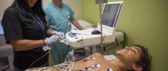
- electrocardiograms - this examination helps determine the congestion of the ventricle and atrium, located in the right half of the heart;
- Ultrasound of the heart (echocardiography) - this technique allows you to detect the presence of an oval window in the septum and changes in the volume of the right ventricle in a larger direction;
- cardiac catheterization - this diagnostic method allows you to identify more oxygenated blood taken from the right atrium as opposed to the left, in addition, you can insert a catheter through the left atrium into the right;
- angiocardiography - this examination allows you to obtain information about whether there is an interatrial septum. For this purpose, contrast is introduced into the bloodstream of one of the atria, thanks to which the resulting film can be used to see how blood moves through the cardiac system.
Peculiarities
VSD is a congenital heart defect (CHD). As a result of the pathology, a hole is formed connecting the lower chambers of the heart: its ventricles. The level of pressure in them is different, which is why when the heart muscle contracts, some blood from the more powerful left side enters the right. As a result, its wall stretches and enlarges, and the blood flow of the pulmonary circle, for which the right ventricle is responsible, is disrupted. Due to increased pressure, the venous vessels are overloaded, spasms and compactions occur.
The left ventricle is responsible for blood flow in the systemic circle, so it is more powerful and has higher pressure. With pathological flow of arterial blood into the right ventricle, the required pressure level decreases. To maintain normal indicators, the ventricle begins to work with greater force, which further increases the load on the right side of the heart and leads to its enlargement.
The amount of blood in the pulmonary circle increases and the right ventricle has to increase pressure to ensure normal flow through the vessels. This is how the reverse process occurs - the pressure in the pulmonary circle now becomes higher and blood flows from the right ventricle to the left. Oxygen-enriched blood is diluted with venous (depleted) blood, and a lack of oxygen occurs in organs and tissues.
This condition is observed with large holes and is accompanied by disturbances in breathing and heart rhythm. Often the diagnosis is made in the first few days of the baby’s life, and doctors begin immediate treatment, prepare for surgery, and, if it is possible to avoid surgery, conduct regular monitoring.
A small VSD may not immediately appear or may not be diagnosed due to mild symptoms. Therefore, it is important to know about possible signs of the presence of this type of CHD in order to take timely measures and treat the child.
Treatment
Currently, several effective techniques can be used to eliminate such an anomaly. The goal of each of them is to reduce hypervolemia in the pulmonary circulation, thereby significantly reducing the load on the myocardium.
If a similar anomaly is detected in a baby, the doctor registers the baby with constant monitoring, since often a small window in the heart septum can close on its own before a certain age. If several holes or combined defects in the heart are detected, surgery is usually prescribed. Let's get acquainted with the main directions in the treatment of this disorder.
Therapeutic
This treatment for abnormal development of the cardiac septum is usually prescribed if the oval window is small. In this case, they rely on the valve’s ability to heal on its own. Although this technique is not applicable if several windows are detected at once, or the hole has reached a significant size.
In addition, small patients who have a small window in the interatrial septum are taken under observation - the majority of children with such an anomaly can be cured without outside intervention by 3 months of age. Although even in this situation it is necessary to regularly visit a doctor and undergo appropriate diagnostics.
Medication
Not a single medication can provoke overgrowth of windows in the cardiac septum. However, the correct use of medications helps to normalize the heart rhythm, stabilize the patient’s well-being, and also helps to quickly eliminate anomalies formed in the interatrial septum.
As a medical treatment for such a cardiac septal defect, the doctor may prescribe the following medications:
- beta blockers;
- Digoxin;
- medications that help reduce blood clotting - anticoagulants that reduce the risk of thrombosis, thereby minimizing the possibility of stroke and heart attack (the most commonly used medication in this group is Aspirin).
Sometimes, if a patient is diagnosed with an atrial septal defect, surgery may be required in addition to medications.
Carrying out the operation
The operation will be performed if the symptoms of the pathology intensify and the heart rate increases. Although surgical intervention is contraindicated for stage 4 cardiac hypertension.
Photo gallery: possible causes of congenital heart disease
Fetal infection
Environmental factor
Bad habits of parents
Genetic predisposition
Taking medications during pregnancy
Maternal diseases
Exposure to toxic substances
Forecast
If you are examined in a timely manner and begin to implement medical recommendations, the prognosis can be quite favorable: the patient’s life expectancy increases significantly, and the possibility of relapse is reduced to a minimum.
In 85-92% of cases, if proper therapy is prescribed, an adult with a similar disease will live for more than one year. Postoperative mortality if the oval window is closed in childhood is minimal. If the operation is performed on an adult patient, the possibility of death of such a patient is slightly higher - it is approximately 2-5%.
If recovery from surgery is managed properly, it will help further reduce the risk and restore the person to health.
Possible complications
A small anomaly (less than 2 mm) against the background of normal well-being of the child does not threaten his life. It is necessary to visit a doctor regularly for examination, since the defect can disappear on its own as the baby grows older.
Windows of significant size can cause the formation of disturbances in cardiac functions, which can occur already in the first days of a baby’s life. If a child is diagnosed with anomalies in the interatrial septum, he does not tolerate colds and pathologies of an infectious nature; often such diseases give complications to the pulmonary system (for example, in the form of pneumonia).
The development of such children proceeds at a slower pace than that of their peers; they have difficulty withstanding physical activity. As you grow older, shortness of breath may appear even in a calm state. In addition, there is a development of problems in some systems in the body due to a lack of oxygen.
Due to abnormal development of the interatrial septum, the following consequences may develop:
- pulmonary hypertension - increased resistance in the vascular system of the lungs, against the background of which an insufficient right ventricle and Eisenmenger syndrome are formed;
- acute disorder of cardiac function;
- endocarditis is an inflammatory process of the inner lining of the heart of infectious etiology;
- thrombosis, risk of stroke;
- disruption of the functioning of valves in the heart, the formation of valvular heart disease.
To minimize the harmful consequences for the condition of the newborn, you need to promptly seek help from a doctor.
Mitral heart defects: types, causes, diagnosis and treatment
Defects are persistent pathological changes in the myocardium that affect its functions.
These are mainly caused by organic disorders of one or more valves (insufficiency), or narrowing (stenosis) of the corresponding openings.
Mitral heart disease (hereinafter referred to as MVC) is a common phenomenon, in most cases (at least 96%) it is a consequence of rheumatic endocarditis.
Why is there a problem?
MPS develops against the background of rheumatic damage to the mitral valve of the heart. With a pathology such as rheumatism, all structural elements of the myocardium are involved in the abnormal process - the endocardium, pericardium, and the vascular system of the muscle. The symptoms and severity of the disease are determined by how severely the valve apparatus is affected.
Mitral defects develop as follows: fibrous tissue that forms on the valve leads to thickening and proliferation of its valves - this, in turn, makes them less mobile and dense. Cicatricial wrinkling (a consequence of the proliferation of fibrous tissue) over time shortens the valve leaflets, resulting in valve insufficiency.
At later stages of development of the pathology, the fused holes become “overgrown” with deposits of calcium salts - this entails disruption of the function, decreased mobility (change in antiphase) of the mitral valve (MV).
Important: adhesion and, as a result, fusion of valve leaflets (commissures) is the cause of mitral stenosis (narrowing). It is noteworthy that several years may pass from the onset of endocarditis to the appearance of primary signs of MV stenosis.
In addition to rheumatic pathologies of the mitral valves (the main cause of the problem), other provoking factors can lead to the appearance of defects.
Thus, according to experts, MPS in some cases are a consequence of the hemodynamic effects to which the functioning valve is exposed.
The commissures are subject to stress, tears appear on them, which, in turn, gradually become overgrown with blood clots. This entails further development of stenosis and progression of the defect.
Congenital MVP (for example, mitral valve prolapse) is a consequence of a violation of the intrauterine development of the child or the result of an infectious disease suffered by the mother. Acquired defects occur against the background of rheumatism or bacterial endocarditis.
In rare cases, the following reasons can lead to stenosis (insufficiency) of the bicuspid valve:
- myxoma (benign tumor formation that occurs in the atrium);
- systemic diseases (scleroderma, lupus);
- valve prolapse with thin, bent, severely altered valves and the threads of connective tissue holding them back;
- cardiac aneurysm;
- myocardial infarction;
- obstructive hypertrophic cardiomyopathy;
- severe forms of hypertension;
- aortic insufficiency;
- Marfan syndrome.
Clinical picture
Rheumatic mitral stenosis is usually diagnosed in women. Mostly, patients consult a doctor with the following complaints:
- shortness of breath (accompanying physical activity of increased intensity);
- chest pain;
- sometimes – rapid heartbeat.
Shortness of breath is a consequence of increased pulmonary congestion against the background of increased release of blood into the pulmonary circle with increased physical activity. Often this symptom is supplemented by a cough (manifests itself while walking), as well as tachycardia (rapid heartbeat); when listening, an altered tone of the mitral valve opening is determined.
Combined mitral disease leaves its mark on the patients' complexion - it acquires a yellowish-pale tint, the lips become bluish. Patients with MPS look asthenic, often with features of physical infantilism. In some cases, the course of the disease is accompanied by a characteristic bright red blush on the cheeks - a consequence of the synthesis of histamine-like substances by the liver.
Valve insufficiency is extremely rarely an independent disease - it is, rather, an intermediate stage in the development of mitral and aortic heart defects (combined). After this, as a rule, stenosis occurs.
Important: with combined defects, serious disorders of the heart are diagnosed, they are extremely difficult, and in most clinical cases they cause rapid decompensation.
When to see a doctor immediately
Moderate MPS can be completely asymptomatic for many years. But at a certain point, patients begin to notice characteristic symptoms:
- severe shortness of breath due to increased physical activity or emotional shock;
- feverish conditions;
- attacks of suffocation;
- dry cough streaked with blood;
- frequent dizziness;
- chest pain, feeling of heaviness in the heart area;
- periodic fainting;
- increasing weakness throughout the body, excessive fatigue, decreased ability to work.
All of the above signs are a reason to urgently contact a cardiologist. Thus, with the development of stroke, heart attack, atrial fibrillation and acute circulatory failure, the patient requires qualified medical care.
How to deal with the problem
At the compensation stage, treatment of MPS requires adherence to an appropriate regimen. This is necessary in order to prevent possible complications that can subsequently lead to heart failure.
Since combined mitral disease is mainly a consequence of rheumatic endocarditis, in case of relapses of the underlying disease, the patient is prescribed complex antirheumatic therapy (carried out exclusively in a hospital setting under the strict supervision of a physician).
Patients with MPS should avoid increased physical activity, stress, that is, all those factors that could lead to the appearance of the main symptoms of cardiac dysfunction - shortness of breath, tachycardia.
For congenital and acquired mitral valve defects, drinking alcoholic beverages, smoking, and hypothermia are prohibited.
Important: drug treatment for combined MVP is aimed not only at combating rheumatism, but also at preventing the development of heart failure.
Drug therapy is also carried out in case of complications of mitral valve insufficiency in children and adults (thromboembolism, atrial fibrillation).
Main drugs:
- Anticoagulants.
- Antiplatelet agents.
- Beta blockers.
Types of operations for MPS:
- Intravascular method. A special catheter is inserted into the femoral artery (or other large vessel), which, under the control of X-ray equipment, is directed to the bicuspid valve. Next, with the help of appropriate instruments, the surgeon widens the valve opening in case of stenosis, or, if we are talking about insufficiency, the patient undergoes mitral valve repair. It is noteworthy that such an operation is also performed in utero, which makes it possible to prevent congenital MPS.
- Open heart surgery (involves temporary circulatory arrest). Using this technique, surgeons can perform plastic surgery of the valve fibrous ring, as well as install a biological implant (artificial prosthesis).
Surgical treatment of mitral stenosis involves commissurotomy (elimination of deflection, separation of fused valve leaflets), valvuloplasty - restoration of their original shape, and prosthetics.
Commissurotomy is a surgical procedure indicated for patients with mitral stenosis. In case of valvular insufficiency, patients undergo surgery to replace the affected valve.
Important: in the later stages of pathology development, surgical intervention is ineffective.
With moderate MPS, women are allowed to give birth to a child. Severe MV defects are a direct contraindication to pregnancy. If surgery was performed on the valve, conception occurs no earlier than a year after the procedure.
Preventive measures
To prevent the development of abnormalities in the heart septum in a newborn, the expectant mother, while carrying a child, should give up any alcoholic beverages or medications that could provoke the development of any heart problems in the baby.
Leading a healthy lifestyle by the expectant mother, as well as psychological and emotional peace, is the key to having a healthy baby.
Watch a video about atrial septal defect: