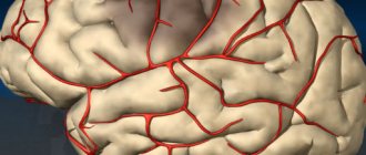
Lenticulostriate angiopathy in newborns - description, causes, consequences
What is dangerous and what can result from lenticulostriate angiopathy in newborns? A qualified doctor will answer this question. There is probably no need to explain to anyone what a huge role the brain, one of the most important organs, plays in the development and normal functioning of a person. Modern medical data indicate an alarming fact - an increase in the number of young children, including newborns, with neurological pathologies of various types associated with the brain. To make an accurate diagnosis, various methods have been developed to study the disease.
When is the brain diagnosed?
Currently, effective methods for studying the brain in children at an early age are widely used in pediatric healthcare practice. Such studies are carried out if there is an assumption of the presence of:
- pathological abnormalities in brain development;
- focal lesions and brain tumors;
- intracranial hemorrhage;
- occlusive hydrocephalus.
In school-age and older children, reasons for brain research, in addition to the above reasons, may be:
- constant unrelenting headaches;
- frequent fainting;
- blood pressure variability.
The studies carried out help to diagnose various diseases of the brain system at a child’s early age. Timely diagnosis is the key to successful treatment and speedy recovery.
Hello, my baby is 6 months old. Based on the results of the ultrasound, he was diagnosed
Hello, my baby is 6 months old. Based on the results of the ultrasound, he was diagnosed with striatal vasculopathy due to cerebral ischemia. Please explain what this means? Does this need to be treated? How? Thank you very much.
The term "ischemia" refers to circulatory problems. Probably, for many, the terrible words “heart attack” and “stroke” have already surfaced from the depths of their memory, but in this case everything is far from so serious. It can be assumed that the events developed as follows: the mother suffered an infection during pregnancy, perhaps the fetus experienced hypoxia, and as a result, blood circulation was disrupted in one of the areas of the baby’s brain, in a small vessel.
If the disorder affects a small area, there is nothing wrong with it, since the body is very smart and cunning, it has a lot of workarounds, plans A, B, C,... U, etc. Therefore, striving for self-healing, our body can fix some problems yourself, find ways to get around the obstacle that has arisen, using additional collaterals (back-up routes).
And on an ultrasound we see the “problem area”. As a rule, calcification subsequently forms at the site of microdamage (this is a focus of calcification, for example, a non-resorbable hematoma can become covered with a capsule over time, that is, the body seems to wall it up so that it does not interfere with its work).
As a rule, if striatal vasculopathy does not manifest itself in any way, it is discovered by chance and is regarded as a finding caused by an unfavorable process suffered in utero. Brain cysts are often treated in a similar way. Often they turn out to be completely harmless and even resolve on their own.
Western scientists, during a mass survey, discovered that brain cysts are more common in those people who do not complain of headaches than in the group of sufferers. It’s just that before, without having the necessary equipment, doctors could not detect foreign formations in the brain. Now they have this opportunity, but it is not always decisive.
So, in the case of striatal vasculopathy, its detection gives us the opportunity to say that the child suffered an unfavorable condition in utero associated with infection or hypoxia, but coped with it, and it will not have any special consequences.
It is necessary to understand that vasculopathy is the body’s reaction to some unfavorable process, therefore there is no specific treatment for striatal vasculopathy. If the problem that caused the vasculopathy led to the emergence of other problems, treatment will be prescribed aimed at eliminating the underlying problem, alleviating symptoms, and means that promote the resorption of hemorrhages.
Retinal angiopathy in children
Symptoms of retinal angiopathy
As already mentioned, the causes of angiopathy are various diseases. Therefore, as such, the symptoms of angiopathy itself are almost impossible to notice on their own. If only with various injuries to the eye and head, a red mesh of blood vessels or small spots will appear on the white of the eye. Otherwise, you can observe the symptoms of the underlying disease.
Based on all of the above, it should be clear that first of all, it is necessary to treat the disease that has caused a change in the condition of the blood vessels in the eye. Once this disease is established, complex therapy is prescribed. The main efforts will be aimed at curing the disease, and along the way, drugs that improve blood microcirculation can be prescribed. The latter, in turn, normalizes the condition of the blood vessels in children's eyes. Although, there is an opinion that only our doctors know about angiopathy. In the rest of the world there is not even a concept of this, and no one is treating it.
Malignant angioendotheliomatosis
is a rare disease characterized by damage to the vessels of the cerebral cortex with the development of rapidly progressive impairment of its function.
It was first described in 1959 by L. Pileger and J. Tappeiner. Morphological examination reveals occlusion of small arteries of the brain and hemorrhagic or ischemic infarctions.
Cells that block the lumen of blood vessels do not have molecules characteristic of endothelial cells (EC) on their membrane, but express antigenic determinants of lymphocytes (CD45, LN-1, LN-2). Some researchers believe that the term “intravascular lymphomatosis” more correctly reflects the essence of this disease.
There are malignant and benign forms of the disease. In the malignant form, damage to the blood vessels of the central nervous system is diffuse, and patients die within the first 6 months from the onset of the disease. In the benign form, isolated damage to certain areas of the brain is noted, and the prognosis is more favorable.
The disease clinically and angiographically resembles primary CNS vasculitis. According to JTLie (1992), who observed 22 patients with CNS vasculitis for 20 years, malignant angioendotheliomatosis occurred in 3 cases.
The onset of the disease is usually acute, in the form of progressive mental disorders, up to the development of dementia. Mental symptoms often precede or develop simultaneously with transient visual disturbances and diffuse headaches. Sometimes angioendotheliomatosis debuts as an acute cerebrovascular accident.
Occasionally, general inflammatory signs are observed - fever, weight loss, arthralgia and myalgia. Some patients experience skin lesions, namely hemorrhagic or non-hemorrhagic subcutaneous formations, plaques, telangiectasias and necrotizing ulcers.
Laboratory abnormalities are nonspecific, but a sharp increase in ESR is characteristic. There are no changes in the cerebrospinal fluid. Cryoglobulins, rheumatoid factor (RF), antinuclear factor (ANF), circulating immune complexes (CIC), and decreased complement concentrations are usually not detected. Angiography of cerebral vessels reveals areas of arterial narrowing and occlusion.
Computed tomography data indicate diffuse atrophy and infarctions of the brain substance. The diagnosis of the disease is rarely made while the patient is alive. The most informative is a brain biopsy with morphological and immunopathological examination.
Preparation and method of conducting the study
Neurosonography does not require preliminary preparation. The baby should not be hungry or thirsty. If the baby falls asleep, there is no need to wake him up; this is even welcome: it is easier to ensure that the head remains still. The results of neurosonography are issued 1-2 minutes after the completion of the ultrasound.
You can take baby milk and a diaper with you to place your newborn baby on the couch. Before the NSG procedure, there is no need to apply creams or ointments to the fontanel area, even if there are indications for this. This worsens the contact of the sensor with the skin and also negatively affects the visualization of the organ being studied.
The procedure is no different from any ultrasound. A newborn or infant is placed on a couch, the place where the skin comes into contact with the sensor is lubricated with a special gel substance, after which the doctor performs neurosonorgraphy.
Causes of cerebrovascular accidents
Against the background of pathology of cerebral circulation or hypoxia (oxygen deficiency), a condition such as lenticulostriate mineralizing angiopathy occurs. There are several reasons for the appearance of such pathological changes:
- congenital pathology of the walls of blood vessels (for example, telangiectasia). Small formations appear on the blood vessels of the brain, which increase in size when filled with blood. As a result of the rupture of such formations, blood penetrates into the brain tissue and hemorrhage occurs;
- calcification of the walls of striatal vessels - there is a process of gradual deposition of calcium salts, which lead to obliteration. Obliteration of the vessel is observed throughout its entire length. When the blood supply to adjacent tissues is disrupted and oxygen starvation occurs;
- intrauterine infection of the fetus - this condition poses a danger to the life and health of the child, as it often leads to disability or death;
- decreased tone of the vascular wall - a disorder of nervous regulation;
- traumatic damage to blood vessels - the presence of hemorrhages in the meninges and brain matter.
Oxygen starvation and intrauterine infections quite often lead to the development of a conditionally pathological condition in a child, which is called lenticulostriate mineralizing angiopathy. In this case, comprehensive monitoring over time is necessary.
Lenticulostriate angiopathy in children is not a disease, but is a feature of the blood supply to the subcortical structures of the brain. In cases where the child does not have developmental deviations due to age, and there is no burden of somatic diseases, treatment in such cases is not required.
Causes
The following reasons can provoke a pathological state of blood flow in a child:
- oxygen starvation in the prenatal period;
- congenital malformations of the vascular system;
- infection of the fetus during pregnancy;
- lack of nutrients and substances;
- prolonged entanglement of the fetus with the umbilical cord;
- decreased tone of vascular formations;
- hypertonicity of the uterus during pregnancy;
- premature aging of placental vessels;
- prolonged stay of the fetus in the mother's reproductive tract.
If there are no signs of a pathological condition, there is no need to treat the baby.
Risk factors
In addition to the underlying chronic disease, the development of angiopathy is facilitated by:
- elderly age;
- individual structural features of the vascular system;
- autoimmune disorders;
- impaired metabolism;
- bad habits;
- overweight and obesity;
- physical inactivity;
- traumatic injuries of the brachiocephalic vessels of the neck;
- excess salt in the diet;
- toxins and frequent contact with harmful substances;
- insufficient intake of vitamins and minerals;
- heredity.
The most common form is considered to be diabetic background vascular angiopathy due to the high incidence of diabetes mellitus. Every year the number of patients increases.
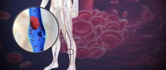
The aorta is the largest artery in the human body, through which blood flows to all parts and systems of the body. As the main vessels branch, they form a complex structure of small capillaries. Changes can also affect the smallest vessels. Such lesions are called microangiopathies. Chronic diseases and other factors can provoke the development of metabolic angiopathy in any area of the vascular bed.
Against the background of diabetes, vascular angiopathy of the lower extremities develops over time. Lack of adequate treatment for angiopathy and diabetes leads to amputation of limbs due to the development of gangrene of the legs. Dystrophic changes in blood vessels occur due to metabolic disorders of glucose metabolism and insulin resistance.
Vascular angiopathy in a diabetic Elevated blood sugar levels destroy the endothelium - the internal lining of blood vessels. These cells play an important role in the functioning of the entire body. They secrete a lot of hormones, prostaglandins and other biological substances for the harmonious functioning of all systems.
Damage to the endothelium leads to impaired blood supply, thrombosis and lack of nutrition and oxygen. Changes can occur in small capillaries (microangiopathy), or affect large vessels, which over time leads to the formation of trophic ulcers and large areas of necrosis. Tissue necrosis is the main factor in the need to amputate part of the damaged limb to save the patient’s life. Adults and especially older people with type 2 diabetes are most prone to this outcome.
Diet and sport
In case of diabetic damage to the blood vessels of the lower extremities, the following foods should be excluded from the diet:
- red meat;
- simple carbohydrates;
- hot and overly spicy seasonings;
- sour, fried and salty dishes.
Despite this, a healthy diet when sick involves eating tasty foods, which include:
- vegetables;
- fish;
- fruits;
- beans;
- whole grains;
- rice;
- buckwheat;
- quinoa;
- dietary meat.
During the treatment period, you should not neglect vitamins, especially vitamins B and D. They play an important role in the healthy state of the nervous system.
Is it necessary to treat lenticulostriate angiopathy?
As is known, the thalamo-striatal region of the brain is responsible for a number of important functional aspects on which the normal functioning, growth and development of the child’s body depends. That is why, after discovering pathological inclusions of calcium salts in a given area, most neurologists suggest the baby’s parents undergo a course of treatment. The main goal of such therapy is to restore adequate blood circulation in the cerebral tissue and eliminate the consequences of ischemia.
What happens if, during the first neurosonographic study, calcifications were discovered in a child aged one month, which affected the striatal zone?
Such young patients are registered with a pediatric neurologist, and at three months of age they undergo a repeat ultrasound examination with monitoring of changes. Sometimes it happens that pathological inclusions resolve on their own; in other cases, calcifications increase in size and provoke an expansion of the ischemic zone.
In the latter case, the child is prescribed drug treatment aimed at correcting disorders, improving trophism and metabolism of cerebral structures.
If there are calcifications along the striatal vessels, the child is recommended to take daily medications to help restore normal blood flow in ischemic tissues. This treatment is carried out for three months, after which its results are assessed. As a rule, young patients take the drugs Cortexin and Pantogam, against which the inclusions either completely disappear or decrease in size.
In cases where the baby is resistant to the therapy and calcifications increase to large sizes, the child’s parents are recommended to consult a pediatric neurosurgeon, who will offer the option of the most correct surgical intervention to eliminate the pathological formations.
Treatment
The striatal zone of the brain is responsible for the development and growth of the child. If there are calcium deposits in this area, the neurologist will suggest a course of therapy to improve blood circulation and prevent possible ischemia. For this, the child is prescribed nootropics and drugs that improve blood circulation.
Therapy is aimed at normalizing trophism and cerebral metabolism. Treatment lasts about 3 months. If there is no therapeutic result from treatment, the baby is prescribed the most gentle surgical intervention.
What is the danger of the disease?
In itself, this disease is not a dangerous condition for the child, but the same cannot be said about its consequences. As is known, the thalamic nuclei are responsible for a number of important functions related to the perception and processing of information that comes from the senses. That is why, with the progression of the pathological process and an increase in the ischemic zone, the following complications are possible:
- complete or partial atrophy of the thalamus;
- child's retardation in psychomotor, mental and physical development;
- the appearance of signs of disorders on the part of the pyramidal and extrapyramidal systems;
- disturbances in the functioning of the organs of perception (except smell);
- the appearance of pathological reflexes in the baby;
- determination during examination and instrumental examination of signs of ischemia of cerebral tissues;
- mental disorders in a child.
Consequences of lenticulostriate pathology
If we take into account the structural features of the thalamus, we can talk about the clinic of its damage due to insufficient blood supply.
The thalamus is the center for processing almost all information coming from the senses (the only exception is the perception of smells). With a lack of blood supply, one or another sense may be impaired, and with significant oxygen starvation, atrophy of the thalamus may occur.
More severe consequences of the development of lenticulostriate vasopathy may be a delay in mental and mental development. In this case, there is a lag of several months, and later even years, in addition, the appearance of symptoms of damage to the pyramidal and extrapyramidal systems, and sensory organs. This will be accompanied by the appearance of pathological reflexes in childhood, as well as clinical manifestations of cerebral ischemia.
Is it possible to prevent the development of the disease?
To minimize the risks of developing lenticulostriate angiopathy in her child, a woman should take care of her health even at the stage of intrauterine development. Prevention of the disease consists of strictly following all prescriptions of the obstetrician-gynecologist:
- rejection of bad habits;
- power control;
- prevention of premature aging of the placenta;
- timely treatment of TORCH infections.
If complications arise during childbirth, after being discharged from the maternity hospital, it is recommended to show them to a pediatric neurologist, who will advise how to avoid the development of a pathological condition associated with the formation of calcifications in the thalamus.
Prevention
In order to prevent striatal angiopathy, it is necessary to follow the following recommendations:
- during pregnancy, stop smoking and drinking alcohol;
- adhere to the instructions of the obstetrician and doctor;
- balance your diet as much as possible;
- prevent premature aging of the placenta;
- treatment and diagnosis of sexually transmitted diseases before pregnancy.
In case of complicated childbirth, additional consultation with a neurologist after discharge from the maternity hospital is advisable.
Causes of the disease
Why can lenticulostriate pathology of the brain develop and what are the prerequisites for its formation? The main reason for the appearance of lenticulostriate calcifications is cerebral ischemia. Normally, while the baby is in the womb, he receives the necessary nutrients and oxygen from the mother's blood and does not lack them. But during childbirth, the following violations may occur:
- Long stay of the child in the maternal birth canal. In this case, the child experiences significant oxygen starvation, which primarily affects the brain, the main oxygen-dependent organ. This leads to the development of ischemic foci in the blood supply zone of the middle cerebral artery and, as a consequence, to calcification of blood vessels and nervous tissue adjacent to the thalamus.
- Birth injury. It is the second most common cause of intracerebral ischemia. Leads to the formation of intracerebral cysts, which, located in the thalamus, can be perceived as calcifications.
In addition to the birth process, the fetus may experience hypoxia during its development.
The main reasons for this are:
- Hypertonicity of the uterus. Contraction of the uterus leads to spasm of the vessels feeding the fetus (extending from the placenta), which causes intrauterine fetal hypoxia to develop, which can lead to premature birth or miscarriage.
- Encircling the fetus with the placenta. The development mechanism is the same, only its cause is a long placenta, which, as a result of fetal movements, can wrap around parts of its body or neck, leading to hypoxia. May be fatal to the fetus.
- Lack of iodine. This microelement is essential for the development of the fetal nervous system. With its deficiency, the neural tube is poorly differentiated, and existing foci of hypoxia are transformed into microcalcifications.
All these conditions are fundamental in the development of lenticulostriate angiopathy.
Features of the disease
This term appeared around the 60-70s, when child neurology was just beginning to develop. However, due to the imperfection of diagnostic procedures and the paucity of equipment, it did not receive specifically substantiated signs and was practically not mentioned in textbooks on pediatrics and neurology. It was given to all children, without exception, in whom, during a neurosonographic study, the smallest changes were identified in the area of the subcortical nuclei, more precisely, the thalamus.
- Eskar eye drops will help eliminate itching and burning;
- feeling of sand in the eyes;
- cloudiness and glare in the field of view;
- redness (including due to pathogenic microorganisms).
If you look at it literally, the term “mineralizing” means that as a result of a pathological process, organized calcifications (deposits of salt ions, mainly calcium) can form in the nervous tissue or in the thalamus itself.
Such formations usually indicate previous cerebral ischemia in these areas. One of the options for the appearance of foci of calcification may be the development of Farah disease (however, this disease is extremely rare).
Angiopathy of cerebral vessels is a prerequisite for the development of this pathology. A feature of the disease is that microcalcifications are formed along these vessels and are localized mainly in the thalamus.
On the one hand, any changes visualized in such small patients must be eliminated. On the other hand, if there are no clinical manifestations in the child (development of sensory disorders or other pathology), then no treatment may be required. But you should definitely schedule a consultation with a pediatric neurologist and conduct a control ultrasound examination of the brain.
Radiation damage to blood vessels
The possibility of developing vascular damage after exposure to radiation on the human body was known already at the end of the 19th century. The changes usually affect medium- or large-caliber arteries. In the areas exposed to irradiation, intimal thickening, mural thrombosis, stenosis and/or vascular occlusion are detected.
The formation of aneurysms and their ruptures also occur. 10 or more years after exposure to ionizing radiation, progressive fibrosis of the vascular wall and perivascular space develops. Early appearance of signs of atherosclerotic vascular lesions was noted.
In patients receiving radiation therapy, vasculopathy should be differentiated from vascular damage due to tumors. In contrast to the latter, with vasculopathy, changes are localized in medium and large arteries, and there are no other manifestations of paraneoplastic syndrome.
Sensations when the blood supply to the brain is disrupted
If normal blood circulation in the brain is disrupted, regardless of the cause, the child experiences characteristic symptoms, such as:
- dizziness, sometimes temporary loss of consciousness;
- frequent headaches;
- memory impairment;
- sleep disturbance (the child sleeps restlessly at night and completely refuses to sleep during the day);
- decreased concentration;
- distracted state.
The child may complain of weakness throughout the body, fatigue and drowsiness. And sometimes the child’s actions manifest aggression, restlessness, causeless irritability and hot temper. Some children with pathological abnormalities of the neurological system have hyperexcitability syndrome.
Without a thorough neurological examination, it is impossible to determine the cause of the disease. In this case, the help of not only qualified doctors, but also the child’s parents is important. They must pay attention to inadequacy in the behavior of their children, analyze their actions and respond in time.
Diagnosis of brain diseases in children
If a child develops characteristic symptoms indicating disturbances in cerebral blood flow, it is necessary to undergo a thorough examination.
There are several methods for diagnosing the state of the vascular system of the brain and identifying circulatory disorders in young children. The main methods include:
- X-ray. Allows you to identify the causes of increased intracranial pressure. Helps give a correct assessment of the condition of sutures and fontanelles in infants;
- CT scan. Such a method is needed to obtain information about the structure of the brain and the interaction of the hemispheres;
- ultrasound (neurosonogram). Provides the opportunity to visualize the structure of the brain substance, its functions and biochemical characteristics. Helps identify vascular diseases in the early stages. A completely safe method, therefore it can be used when examining newborns (as long as the fontanelles are open);
- Dopplerography and duplex scanning. This method allows you to examine and examine the vessels, the condition of the vascular wall, and the uniformity of blood flow through the vessels. Effectively identifies the causes of headaches in children of primary and school age. Helps identify the causes of intracranial pressure disturbances in cerebrovascular accidents. The method is safe, so it can be used during the neonatal period;
- MRI. It is a more informative and safe method, as it allows one to avoid radiation exposure to the body during research. Provides results on the nature of metabolism in various parts of the brain. The method allows you to determine even slight displacement of the brain, detect hemorrhages, heart attacks and brain tumors, and birth injuries. Can be used from the first days of a child’s life;
- EEG. This method provides a graphical representation of electrical impulses from the brain, which makes it possible to judge the child’s convulsive readiness. Provides information regarding the appearance of brain tumors, brain injury, neuroinfections, and diseases of the nervous regulatory system.
All modern methods of neurological research make it possible to quickly recognize changes or deviations in the functioning of brain structures and determine the quality of the condition of blood vessels, even in the absence of characteristic symptoms in a child.
Parents should remember that if their child has any complaints, they should consult a qualified doctor. A special examination and a correctly established diagnosis will help choose effective treatment for vascular pathology in a child.
Hyperexcitability syndrome
Emotional instability, frequent crying and whims, trembling chin, poor sleep and appetite characterize a hyperexcitable child. These children constantly move, change positions, and grab objects. They are interested in the world, but high emotionality prevents them from interacting with it. They are impressionable and easily vulnerable. Hyperexcitability syndrome increases mental exhaustion and fatigue.
If you notice manifestations of hyperexcitability syndrome in a child, do not worry, just visit a doctor. Hyperexcitability can be a manifestation of a natural temperament of the choleric type and an adaptation to changing environmental conditions.
Very rarely, hyperexcitability is a sign of perinatal pathology of the central nervous system. The sooner the child is examined by a specialist, the sooner you will find out the cause of hyperexcitability and eliminate the problem.
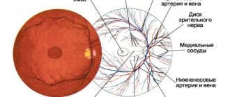
Neonatal retinopathy
One of the most dangerous forms of angiopathy is retinopathy of retinal vessels in newborns. This condition often develops in very premature babies. Retinal capillaries begin to grow in the second half of pregnancy. And only towards the end of the standard gestation period does the full formation of the eye vessels occur, which ensures the baby’s timely and complete development of vision and its high acuity in the future.
In children born as a result of premature birth, the process of normal development of retinal vessels is completed at the time of birth. An underdeveloped vascular system is not able to provide the baby with good vision. The disturbances are aggravated by exposure of the eyes to oxygen. There are cases of complete blindness of a child. The only way out in such a situation is timely diagnosis and surgery using modern equipment.
Extreme prematurity as a particular risk factor
Symptoms
Due to circulatory disorders, the following symptoms are observed:
- headache;
- absent-mindedness;
- decreased concentration;
- loss of consciousness;
- sleep disorders;
- decreased memory performance;
The child exhibits emotional signs of angiopathy:
- prolonged crying;
- increased general fatigue;
- anxiety;
- drowsiness.
To determine the stage of the pathological condition of the blood vessels, it is necessary to seek medical help and undergo diagnostic measures.