One of the problems of the infant's cardiovascular system is a failure of the heart rhythm. Arrhythmia in a newborn, just like in a fetus in the womb, develops under the influence of certain factors and can lead to very serious consequences. What does a heart rhythm disorder look like in a newborn or baby in the womb, how is the pathology treated, and why is this condition dangerous for an infant? We will look into the material below.
What is fetal arrhythmia?
In general, arrhythmia refers to some disruption in the sequence of contractions of the heart muscle. It is worth noting here that the heart of a person (even a very tiny one) beats with a certain rhythm, which is inherent in nature for a healthy and strong body. If the sequence of heart contractions is disrupted, this is already a pathology.
Modern cardiologists can determine the heart rate of the fetus in the womb from the 19th week of its development. In this case, the heart rate is considered normal within the range of 120–160 beats/min. If the rate of contractions of the heart muscle is less than 100 beats/min, the pathology is called bradycardia. A heart rate above 160 is called tachycardia.
Bradycardia in the fetus is most pronounced between 6 and 7 a.m. or between 3 and 7 p.m. During these periods, the baby's activity is noticeably reduced. Tachycardia can best be detected in the fetus between 11 and 15 o'clock in the afternoon or between 19 o'clock in the evening and 1 o'clock at night.
Important:
in most cases, arrhythmia in the fetus during pregnancy is considered normal if the deviations in the frequency of contraction of the heart muscle are only short-term. The pathology is persistent sinus tachycardia (more than 200 contractions per minute), the same bradycardia (less than 100 contractions per minute) or extrasystoles, which occur more than 1 time per 10 healthy and normal heart contractions. Such palpitations require medication adjustment and medical supervision.
Cardiac arrhythmia in newborns: causes and consequences
One of the problems of the infant's cardiovascular system is a failure of the heart rhythm. Arrhythmia in a newborn, just like in a fetus in the womb, develops under the influence of certain factors and can lead to very serious consequences. What does a heart rhythm disorder look like in a newborn or baby in the womb, how is the pathology treated, and why is this condition dangerous for an infant? We will look into the material below.
In general, arrhythmia refers to some disruption in the sequence of contractions of the heart muscle. It is worth noting here that the heart of a person (even a very tiny one) beats with a certain rhythm, which is inherent in nature for a healthy and strong body. If the sequence of heart contractions is disrupted, this is already a pathology.
Modern cardiologists can determine the heart rate of the fetus in the womb from the 19th week of its development. In this case, the heart rate is considered normal within the range of 120–160 beats/min. If the rate of contractions of the heart muscle is less than 100 beats/min, the pathology is called bradycardia. A heart rate above 160 is called tachycardia.
Bradycardia in the fetus is most pronounced between 6 and 7 a.m. or between 3 and 7 p.m. During these periods, the baby's activity is noticeably reduced. Tachycardia can best be detected in the fetus between 11 and 15 o'clock in the afternoon or between 19 o'clock in the evening and 1 o'clock at night.
Important: in most cases, arrhythmia in the fetus during pregnancy is considered normal if the deviations in the frequency of contraction of the heart muscle are only short-term. The pathology is persistent sinus tachycardia (more than 200 contractions per minute), the same bradycardia (less than 100 contractions per minute) or extrasystoles, which occur more than 1 time per 10 healthy and normal heart contractions. Such palpitations require medication adjustment and medical supervision.
The reasons why arrhythmia develops in utero or in a newborn may be different. In particular, in the fetus, disturbances in the contraction of the heart muscle occur for the following reasons:
- Diabetes mellitus in a woman (type 1 insulin dependent).
- Endocrine diseases of pregnant women.
- Lesions of the nervous system in a pregnant woman.
- Pathologies of fetal development due to hypoxia (anemia and anemia in the mother).
- Frequent stress in a pregnant woman.
- Smoking and drinking alcohol.
- Failures in metabolic processes.
- Unbalanced nutrition and lack of electrolytes in the body of the expectant mother (potassium, iodine, manganese, iron).
The causes of arrhythmia in newborns are the following pathologies and conditions:
- Congenital and acquired heart defects.
- Hemorrhage in the area of the cardiac conduction tract.
- Immaturity of the heart muscle due to disturbances in its intrauterine development.
- Intoxication due to tonsillitis and other viral diseases.
- Early birth (prematurity).
- Pathological pregnancy.
- Emotional experiences and stress.
- Anemia.
- Problems with the thyroid gland (hypo- or hyperthyroidism).
For an embryo in the womb and for a newborn baby with an abnormal heart rhythm, their own clinical picture is drawn. The main symptoms and signs in both cases are given below.
Here, the main symptoms of an abnormal heart rhythm are changes in the frequency of contractions of the heart muscle and low/high fetal mobility. The number of heartbeats can be heard both on an ultrasound and simply through the abdominal wall of a pregnant woman using a special stethoscope tube.
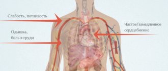
Most often, an abnormal heart rhythm in babies under one year of age hardly manifests itself at all. Pathology is mainly detected by chance during a routine medical examination. If there is a serious deviation in the functioning of the baby’s heart, then the main signs of this condition are:
- paroxysmal shortness of breath and heavy breathing;
- pallor or cyanosis of the nasolabial triangle;
- restless behavior of the baby;
- small weight gain (deviations from the norm);
- visually noticeable pulsation of veins in the neck;
- restless sleep.
Important: if one or more of the listed signs appear, you should immediately contact a pediatrician or a pediatric cardiologist. The sooner the pathology is detected, the greater the chances of a speedy recovery for the baby.
It is possible to diagnose abnormal heart rhythm in utero using two methods:
- Ultrasound in M-mode. In this case, the specialist monitors the movement of the walls of the ventricles and at the same time the movement of the leaflets of the atrioventricular valves. Here the doctor monitors the work of the aortic valve (closing/opening) and at the same time looks at how rhythmically the walls of the atria work. In addition, the doctor studies the speed of blood flow in the heart ventricles.
Arrhythmia in a newborn baby is diagnosed using the following methods:
- ECG (Holter monitoring). In this case, the cardiogram device is worn throughout the day. The data obtained by conducting such a test allows us to track the direct connection between stressful conditions and disruptions in heart rate.
- Ultrasound of the baby's heart (Dopplerography). Makes it possible to track the work of all parts of the heart, including valves, leaflets, atria and ventricles.
Treatment of arrhythmia in the fetus and newborn baby should only be carried out by a pediatric cardiologist. Self-medication is under no circumstances allowed, and even more so, pathology should not be left to chance.
If a serious arrhythmia is diagnosed in the fetus, the expectant mother is prescribed a number of medications that stabilize the functioning of the baby’s heart. In most cases, medications are administered intravenously. In some cases, drugs are prescribed orally. In particular, the following medications are used:
- propranolol, magnesium and lidocaine;
- sotalol and amiodarone (for tachycardia above 200 beats per minute);
- dexamethasone (if fetal myocarditis is suspected).
Important: treatment of the supraventricular form of arrhythmia in the fetus is successful in 90% of cases.
For a newborn, treatment tactics for abnormal heart rhythm are selected strictly individually, depending on the cause of the pathology. In case of serious abnormalities in the heart, the baby should be constantly monitored by a cardiologist and pediatrician.
Surgery is a last resort and is prescribed only if there is a real threat to the baby’s life, or if the selected drug therapy does not produce the expected effect.
Most short-term arrhythmias in newborns or the fetus do not require correction. However, if the pathology manifests itself frequently and remains untreated, the baby may develop the following complications:
If the pathology was detected on time and the diagnosis was made accurately, in 85% of cases the prognosis for the baby is quite favorable.
In general, most types of existing arrhythmias require only constant monitoring of the state of the baby’s heart. Such abnormalities are benign and generally have a favorable prognosis. However, if the baby is diagnosed with atrial fibrillation, paroxysmal or transverse heart block, then the prognosis may be disappointing (in the absence of proper therapy).
In order not to put the health of the unborn baby at risk and to exclude possible heart problems, a pregnant woman needs to adhere to some rules of a healthy lifestyle. These are:
- Frequent walks in the fresh air.
- Timely treatment of any infectious and viral pathologies.
- Undergoing a medical examination before a planned pregnancy.
- A balanced diet with sufficient potassium in foods.
- A calm lifestyle without stress and mental strain.
- Rejection of bad habits.
To prevent a newborn child from suffering from arrhythmia for a long time, it is necessary to take the following preventive measures:
- Neutralize all factors predisposing to arrhythmia.
- Undergo preventive medical examinations in a timely manner.
- Timely follow-up of a child with cardiac pathologies.
- Provide the baby with oxygen baths of sufficient duration and adequate nutrition.
Every mother should understand that the baby’s health comes first. Therefore, you should take care of yourself before the planned pregnancy during the period of gestation. Only high-quality care for the functioning of a woman’s body gives a high chance of conceiving, bearing and giving birth to a baby with a healthy heart.
Heart rhythm disturbances in children of the neonatal period are a fairly common phenomenon that can be caused by both intrauterine negative factors and external stimuli. Arrhythmia in an infant can masquerade as other diseases, and therefore can be diagnosed accidentally during a routine examination.
Parents can understand that a newborn’s heart instability is progressing at home by noticing the following symptoms:
- regular shortness of breath without physical activity (exercises, gymnastics, games);
- fast or slow heart rate;
- visibility of blood pulsation in the vessels of the neck;
- pale skin, cyanosis of the nasolabial triangle;
- the baby gets tired quickly, behaves restlessly and sleeps poorly;
- there is poor weight gain.
Dyspnea in an infant is the main sign of arrhythmia at this age
Parents should understand that the clinical picture may vary individually, since manifestations in children under one year of age and newborns depend on the type of cardiac dysfunction.
Children say! Daughter (3 years old): - Mom, look, sparrows. - Yeah. - And their mother... is a crow.
An abnormal heart rhythm in a child can be detected in the last trimester of pregnancy or immediately after birth. The normal heart rate for a neonatal infant is from 120 to 160 beats/min. The following indicators raise suspicion of arrhythmia:
- normocardia, when the heart rate is from 60 to 120 times;
- bradycardia if heart rate is below 60 beats/min;
- tachycardia, when the heart rate of an infant is more than 100 times.
Arrhythmia in newborns and infants is shown in the photo
In addition, newborns experience atrial fibrillation (this form is characterized by flickering and fluttering of the ventricles and atria), sinus arrhythmia (occurs when the automaticity, excitability and conduction of the heart muscle is impaired), mild or moderate (does not bring much discomfort to the child) and severe, requiring immediate medical care.
Note to moms! Often, heart rhythm disturbances in infants can be a consequence of congenital pathologies of the thyroid gland, hypertension or coronary heart disease. If a child is at risk, he needs a full examination of all organs and systems.
There are plenty of reasons why heart instability occurs in newborns. In most cases, the disease is genetic. Factors influencing the occurrence of heart pathology are divided into cardiac and extracardiac. The first type includes congenital defects: heart failure, atrial and ventricular septal defects, cardiac tumors and injuries of the cardiovascular system.
Extracardiac is considered a disease that is caused by non-cardiac pathologies, i.e. influenza and ARVI, toxoplasmosis, cytomegalovirus, tonsillitis, diphtheria, pneumonia. To avoid these diseases, you should not neglect preventive vaccinations.
ARVI in an infant without proper treatment can develop into arrhythmia
Arrhythmia in newborns can occur due to an unfavorable course of pregnancy, infections in the first and third trimesters, chronic or hidden diseases (such as insulin-dependent diabetes mellitus, as well as tuberculosis in the mother), high or low water. Iron deficiency anemia in the mother and VSD (vegetative-vascular dystonia) also have a bad effect on the heart function of a child after birth.
For parents! Sinus heart pathology in newborns and infants can be a physiological phenomenon and manifest itself as a reaction to hot weather or an emotional outburst.
A slowdown or increase in heart rate in an infant may indicate the presence of a disease, so you should undergo a medical examination to find out the causes of the phenomenon:
- the child is given an ECG in standing, lying and sitting positions;
- assess blood pulsation through the vessels (in children under one year old, the normal heart rate is 140 beats/min);
- X-rays and EEG are taken (for compatibility with central nervous system diseases);
- referred to EchoCG in order to detect organic causes of the disease.
ECG analysis results show arrhythmia in an infant
Treatment of arrhythmia in newborns and infants begins only after confirmation of the diagnosis. Therapy is aimed at eliminating the source that caused the disease: establishing a daily routine, prescribing proper nutrition (balanced); discontinue medications that cause cardiac dysfunction.
Children say! My granddaughter and I are collecting beans. I open the pod and tell her that this is a bean house. She: - So she’s rich. She has her own home!
If the pathology is a consequence of rheumatism and poor blood clotting, they try to get rid of them. When the disease is uncomplicated, it is treated with conservative methods: establishing the electrolyte balance of the myocardium, normalizing cardiac metabolism, and prescribing drugs against arrhythmia in children. Such medications contain magnesium and potassium, procainomide, amiodarone, verapomil, and riboxin.
In more severe cases, when medications have not brought results, surgical intervention is prescribed, in particular, cardiac pacing and implantation are performed, and radiofrequency ablation is possible.
Arrhythmia is not at all a harmless disorder: under its influence, children under one year of age develop heart failure, more pronounced heart rhythm disturbances, myocardial infarction, blood clots can break off, and death is also possible.
Often untreated arrhythmia in infants causes dystonia
Improper functioning of the heart causes pathologies of the brain, since it is poorly supplied with oxygen due to impaired blood flow through the vessels, as a result of which the child experiences lethargy and developmental delays.
We should talk about the prognosis for a child’s recovery after arrhythmia based on the causes of the disease, as well as methods of treating it. The more complex the course of the disease, the risk of complications and relapse increases.
Arrhythmia in an infant can cause a blood clot to break off
To prevent the development of heart rhythm disturbances in infants, infectious diseases should be treated promptly and not delay treatment for sore throat and tonsillitis. During pregnancy, the mother should beware of infections such as toxoplasmosis, hepatitis and others that can cause pathology. If an ailment is detected by ultrasound in the fetus, you should not delay treatment, because the chances of completely getting rid of arrhythmia in utero are quite high.
Watch a video from experienced specialists about childhood arrhythmia.
Heart rhythm disturbances in newborns. As a rule, they are a continuation of arrhythmias in the prenatal period, and their timely detection and treatment largely depend on prenatal screening. In this chapter we will focus only on the most frequent and significant violations encountered in clinical practice.
Contractions of the fetal heart are carried out due to the pacemaker activity of the cells of the sinus node and begin to be recorded from the third week of pregnancy. The normal fetal rhythm is 120 to 160 beats per minute; its variability increases with increasing gestational age. Conventionally, the following gradations of fetal rhythm are distinguished (Ronn E. Tannel M. 2001, as amended):
1. Physiological fetal rhythm (120-160 beats/min).
2. Tendency to bradycardia (101 - 119 beats/min).
3. Bradycardia (less than 100 beats/min for at least 10 s).
4. Tendency to tachycardia (from 161 to 179 beats/min).
5. Tachycardia (more than 180 beats/min for at least 10 s).
When the activity of the sinus node is suppressed or the rate of spontaneous depolarization of the underlying sections of the conduction system increases (due to hypoxia, ischemia, acidosis, hypokalemia, inflammation, etc.), the function of the main pacemaker is assumed by another part of the conduction system of the heart, including myocardial fibers. The result may be the development of various rhythm and conduction disorders (ectopia, microreentry).
Fetal arrhythmias are observed in 1-2% of pregnancies; they can be independent diseases or nonspecific symptoms of other pathological conditions.
Etiology of cardiac arrhythmias in newborns. Heart rhythm and conduction disturbances in the fetus are in most cases associated with congenital anomalies in the development of conductive tracts, but in some cases it is possible to identify other etiological causes.
1. Organic changes in the fetal heart - congenital heart defects (CHD), heart tumors, cardiomyopathies, focal fibromuscular dysplasia of the coronary arteries. Of the heart defects, cardiac arrhythmias most often accompany a common patent atrioventricular canal, corrected transposition of the great arteries, Ebstein's anomaly, and atrial septal defect.
2. External influences on the heart, leading to changes in water-electrolyte (hypokalemia, hypocalcemia) and acid-base balances, hypothermia, disruption of humoral regulation (diabetes mellitus, thyroid dysfunction); the effect of medications taken by the mother (antiarrhythmic drugs, phenothiazides, antidepressants).
3. Systemic diseases of the mother or fetus - connective tissue (systemic lupus erythematosus - SLE), associated with fetopathy, acute inflammatory process (myocarditis, maternal genitourinary tract infection), diseases of the central and autonomic nervous system.
The fetal electrocardiogram can be recorded from the mother's abdominal wall, in rare cases (usually during childbirth) - directly from the fetal head. However, the resulting curves are usually characterized by poorly readable QRS complexes, and the P wave is rarely detected at all. The magnetocardiography method allows you to visualize P and QRS waves almost identical to a conventional ECG, but the cost of the study is unreasonably high. Fetal echocardiography has made fundamental changes in the diagnosis of fetal rhythm disturbances. The heart is examined in M-mode in the apical projection of four chambers; in this case, the slightly inclined plane of the sensor beam passes through the wall of the atrium, the atrioventricular valve and the wall of the ventricle. Analysis of the movement of these structures allows us to determine the presence of atrioventricular block, the nature of tachycardia (sinus, supraventricular or ventricular), and the location of the ectopic contraction. The additional use of Doppler echocardiography makes it possible to accurately calculate the intervals between contractions of the atria and ventricles. This can be achieved in two ways: 1) by analyzing the speed of blood flow through the mitral valve and at the exit from the left ventricle; 2) analyzing blood flows in the superior vena cava and in the ascending aorta [1]. The atrioventricular interval in the first case is measured from the intersection of the “mitral” peaks E and A to the beginning of the ejection wave from the left ventricle (Fig. 9-1). In the second case, the distance from the beginning of the retrograde venous wave A to the beginning of the ejection wave from the left ventricle is measured.
The atrioventricular interval increases with gestational age and with a decrease in heart rate. Its value is on average 35 ms greater than the PR value on the ECG. Measuring the ventriculoatrial (VA) interval allows us to distinguish re-entry type tachycardia (short VA interval) and its other types - sinus tachycardia, ectopic atrial, intraatrial (long VA interval). This may have implications when choosing therapy. In particular, in the first type of tachycardia, digoxin is the drug of choice; in the second type, it must be combined with other antiarrhythmic drugs (flecainide, cordarone).
Doppler echocardiography also makes it possible to evaluate blood flow disturbances in various chambers of the heart, great and umbilical vessels, characterizing the consequences of rhythm disturbances. Unfortunately, there are known limitations for these techniques: maternal obesity, polyhydramnios, uncomfortable fetal position, etc.
There are three main groups of arrhythmias. 1) irregular rhythm (85% of all arrhythmias), 2) tachyarrhythmias (10%), 3) bradyarrhythmias (5%).
In the diagnosis of arrhythmias in newborns, in addition to traditional ECG, it is advisable to use long-term Holter monitoring.
— Return to the table of contents of the section “
Cardiology. "
Table of contents of the topic “Cardiomyopathies in children.”:
In healthy children, the heart rate is 60-90 beats of the heart muscle per minute, and the intervals between them are equal and the child does not feel the work of his heart. Arrhythmia in newborns is a violation of the frequency of contractions. If the beat rate is below 55 beats per minute, this arrhythmia is called bradycardia, but if the beat rate is over 100 beats per minute, it’s tachycardia. The types of arrhythmia include the following heart diseases:
-Sinus arrhythmia, in which the sinus rhythm is irregular.
-Sinus tachycardia, in which there is a frequent regular sinus rhythm.
-Sinus bradycardia. With it, sinus rhythm is rare.
Sinus arrhythmias are diagnosed simply by doing an electrocardiogram.
Arrhythmia in newborns is one of the syndromes of a number of diseases: thyroid disease, hypertension, mitral valve prolapse, coronary heart disease, heart defects. The most common causes of diseases are mainly congenital heart defects, inflammatory diseases, heart rhythm disturbances, and other pathologies.
Some heart rhythm disorders are detected before the baby is born. It is best to carry out diagnosis while the baby is pregnant, this way you can prevent the occurrence of arrhythmia even before the birth of the child. Since a small child will not be able to describe the symptoms of the disease, the first signs are: pale skin, irregular heartbeat, fatigue, weakness, anxiety, shortness of breath, poor sleep and frequent awakenings, increased fatigue. Fainting indicates the severity of the arrhythmia.
Arrhythmia can be diagnosed in the following ways: ECG (both normal and with stress), EchoCG (ultrasound of the heart, if necessary), examination by a cardiologist; in case of complications, you should be monitored at a dispensary. Self-medication is highly undesirable, it can lead to serious consequences!
A child’s heart can be trained by following a daily routine at an early age and spending more time outdoors.
They learned to determine the softness of a steak using x-rays.
Scientists from the Norwegian private research organization SINTEF have created technology to test the quality of raw meat using weak X-rays. A press release of the new technique was published on gemini.no.
A suspended aviation container with an open architecture was created
The American company Northrop Grumman introduced a new aviation hanging container OpenPod for various sensors, created with an open architecture. The weight of the container is 226 kilograms. Thanks to the open architecture, other manufacturers will be able to release their own systems for OpenPod. The container can be installed on F-15 Eagle and F/A-18E/F Super Hornet fighters, A-10 Thunderbolt II attack aircraft, C-130J Super Hercules transport aircraft, as well as various types of helicopters.
Magic Leap has officially announced the creation of a platform for augmented reality developers. You can leave your contacts in the appropriate section on the company’s website. Company representatives announced this at the EmTech Digital conference.
Why is arrhythmia diagnosed in the fetus and newborn?
The reasons why arrhythmia develops in utero or in a newborn may be different. In particular, in the fetus, disturbances in the contraction of the heart muscle occur for the following reasons:
- Diabetes mellitus in a woman (type 1 insulin dependent).
- Endocrine diseases of pregnant women.
- Lesions of the nervous system in a pregnant woman.
- Pathologies of fetal development due to hypoxia (anemia and anemia in the mother).
- Frequent stress in a pregnant woman.
- Smoking and drinking alcohol.
- Failures in metabolic processes.
- Unbalanced nutrition and lack of electrolytes in the body of the expectant mother (potassium, iodine, manganese, iron).
The causes of arrhythmia in newborns are the following pathologies and conditions:
- Congenital and acquired heart defects.
- Hemorrhage in the area of the cardiac conduction tract.
- Immaturity of the heart muscle due to disturbances in its intrauterine development.
- Intoxication due to tonsillitis and other viral diseases.
- Early birth (prematurity).
- Pathological pregnancy.
- Emotional experiences and stress.
- Anemia.
- Problems with the thyroid gland (hypo- or hyperthyroidism).
Impulse conduction blockades in children
As the name suggests, blockades occur when the impulse passes poorly or does not pass at all through any part of the heart muscle. Most often in childhood, the so-called incomplete blockade of the right branch of the Hiss bundle occurs.
Just like mild sinus arrhythmia, it is considered physiological. This arrhythmia is functional in nature and goes away with age, very rarely developing into something more serious.
A more severe version of this arrhythmia is a complete blockade of impulse conduction. Then the wave of excitation, which originated in the sinus node, does not reach the ventricles at all. But the ventricles of the heart are able to generate their own rhythm, only at a too slow pace (about 30 contractions per minute), and therefore the heart does not stop at all.
There may also be pauses in the heart rhythm (usually at night), which will be manifested by loss of consciousness, convulsions, pallor or even bluishness of the skin. Doctors call this phenomenon a Morgagni-Adams-Stokes attack. It usually goes away on its own, but you should definitely consult a doctor or call an ambulance.
Symptoms of arrhythmia
For an embryo in the womb and for a newborn baby with an abnormal heart rhythm, their own clinical picture is drawn. The main symptoms and signs in both cases are given below.
Signs of fetal arrhythmia during pregnancy
Here, the main symptoms of an abnormal heart rhythm are changes in the frequency of contractions of the heart muscle and low/high fetal mobility. The number of heartbeats can be heard both on an ultrasound and simply through the abdominal wall of a pregnant woman using a special stethoscope tube.
Signs of arrhythmia in a newborn baby up to one year old
Most often, an abnormal heart rhythm in babies under one year of age hardly manifests itself at all. Pathology is mainly detected by chance during a routine medical examination. If there is a serious deviation in the functioning of the baby’s heart, then the main signs of this condition are:
- paroxysmal shortness of breath and heavy breathing;
- pallor or cyanosis of the nasolabial triangle;
- restless behavior of the baby;
- small weight gain (deviations from the norm);
- visually noticeable pulsation of veins in the neck;
- restless sleep.
Important:
If one or more of the listed signs appear, you should immediately contact your pediatrician or immediately see a pediatric cardiologist. The sooner the pathology is detected, the greater the chances of a speedy recovery for the baby.
Sinus arrhythmia in children
Sinus arrhythmias differ from other rhythm disorders in that the heart contracts at regular intervals.
There are two types of such arrhythmias:
- Sinus tachycardia is a rapid heartbeat. We can talk about it when the heart rate without physical activity exceeds the age norm by 10 - 20 beats per minute.
- Sinus bradycardia is, on the contrary, a slow heartbeat, that is, when the pulse rate is below the age norm.
For people involved in sports, sinus bradycardia is a common occurrence. A well-trained heart requires fewer contractions to effectively pump blood to the body.
Arrhythmia can exist in three variants:
- A mild form that appears in newborns and infants due to the immaturity of the nervous system, it does not bother the baby in any way.
- Moderate, which occurs in older children (5 - 7 years old), also rarely manifests itself in any way.
- Severe sinus arrhythmia (usually tachycardia), which is characterized by the appearance of paroxysms (attacks) of accelerated heartbeat and has a bad effect on the child’s well-being.
Mild and moderate forms are very common in children and, as a rule, do not require special attention from parents and doctors.
In case of paroxysm of sinus tachycardia, you should definitely consult a specialist.
However, if you called a doctor and are waiting for him, and the heartbeat is very bothering your child, you can resort to the following simple techniques to try to stop this attack:
- Ask your child to close his eyes and apply moderate force to his eyeballs for two to four seconds.
- Have the child tightly cover his nose and mouth with his hands and strain hard, as if trying to exhale.
How is arrhythmia diagnosed in the fetus and newborn?
It is possible to diagnose abnormal heart rhythm in utero using two methods:
- Ultrasound in M-mode. In this case, the specialist monitors the movement of the walls of the ventricles and at the same time the movement of the leaflets of the atrioventricular valves. Here the doctor monitors the work of the aortic valve (closing/opening) and at the same time looks at how rhythmically the walls of the atria work. In addition, the doctor studies the speed of blood flow in the heart ventricles.
Arrhythmia in a newborn baby is diagnosed using the following methods:
- ECG (Holter monitoring). In this case, the cardiogram device is worn throughout the day. The data obtained by conducting such a test allows us to track the direct connection between stressful conditions and disruptions in heart rate.
- Ultrasound of the baby's heart (Dopplerography). Makes it possible to track the work of all parts of the heart, including valves, leaflets, atria and ventricles.
Briefly about heart rhythm and its disturbances
The main function of the heart is to pump blood and distribute it throughout the body.
This requires rhythmic, clearly synchronized contraction of all chambers of the heart: atria and ventricles. The impulse that causes excitation of the myocardium (heart muscle) originates in a cluster of special cells (sinus node), which is located in the right atrium and passes alternately first through the atria and then through the ventricles, and causes their contraction.
In a healthy heart, the sinus node produces impulses at an age-appropriate frequency - sinus rhythm. When the generation of an impulse or its conduction is disrupted, arrhythmia occurs.
So, arrhythmia is a disease that is characterized by various heart rhythm disturbances. And although arrhythmias are quite often diagnosed in children, they are rarely an independent disease and usually serve as a symptom of another, underlying disease.
In childhood, the same variety of rhythm disturbances occurs as in adults.
The most common ones are:
- sinus tachycardia;
- sinus bradycardia;
- respiratory arrhythmia;
- extrasystole;
- atrial fibrillation;
- blockade of impulse conduction.
Treatment of arrhythmia
Treatment of arrhythmia in the fetus and newborn baby should only be carried out by a pediatric cardiologist. Self-medication is under no circumstances allowed, and even more so, pathology should not be left to chance.
Features of treatment of arrhythmia in the fetus
If a serious arrhythmia is diagnosed in the fetus, the expectant mother is prescribed a number of medications that stabilize the functioning of the baby’s heart. In most cases, medications are administered intravenously. In some cases, drugs are prescribed orally. In particular, the following medications are used:
- propranolol, magnesium and lidocaine;
- sotalol and amiodarone (for tachycardia above 200 beats per minute);
- dexamethasone (if fetal myocarditis is suspected).
Important:
treatment of the supraventricular form of arrhythmia in the fetus is successful in 90% of cases.
Features of treatment of arrhythmia in a child under one year old
For a newborn, treatment tactics for abnormal heart rhythm are selected strictly individually, depending on the cause of the pathology. In case of serious abnormalities in the heart, the baby should be constantly monitored by a cardiologist and pediatrician.
Surgery is a last resort and is prescribed only if there is a real threat to the baby’s life, or if the selected drug therapy does not produce the expected effect.
Extrasystole in children
As we remember, in a healthy heart, impulses appear in the sinus node one after another in a certain rhythm. But sometimes an impulse can occur in some other part of the conduction system of the heart. Then, after a normal contraction of the heart, an extraordinary contraction occurs, which is called an extrasystole.
An absolutely healthy person can experience up to 200 extrasystoles per day - this is absolutely normal.
Usually the child does not feel the occurrence of extrasystoles. However, if there are too many of them, they can cause discomfort in the form of a feeling of strange interruptions in the heart. Often, existing extrasystole manifests itself more strongly during physical exertion or after infections.
What complications can there be during the development of arrhythmia, after diagnosis and after recovery?
Most short-term arrhythmias in newborns or the fetus do not require correction. However, if the pathology manifests itself frequently and remains untreated, the baby may develop the following complications:
- heart failure;
- arrhythmogenic cardiomyopathy;
- early disability;
- fibrillation (fluttering of the heart ventricles), leading to death;
- heart failure.
If the pathology was detected on time and the diagnosis was made accurately, in 85% of cases the prognosis for the baby is quite favorable.
Forecast for future health
In general, most types of existing arrhythmias require only constant monitoring of the state of the baby’s heart. Such abnormalities are benign and generally have a favorable prognosis. However, if the baby is diagnosed with atrial fibrillation, paroxysmal or transverse heart block, then the prognosis may be disappointing (in the absence of proper therapy).
Prevention of arrhythmia in the fetus (advice for expectant mothers)
In order not to put the health of the unborn baby at risk and to exclude possible heart problems, a pregnant woman needs to adhere to some rules of a healthy lifestyle. These are:
- Frequent walks in the fresh air.
- Timely treatment of any infectious and viral pathologies.
- Undergoing a medical examination before a planned pregnancy.
- A balanced diet with sufficient potassium in foods.
- A calm lifestyle without stress and mental strain.
- Rejection of bad habits.
Prevention of arrhythmia in a child up to one year old
To prevent a newborn child from suffering from arrhythmia for a long time, it is necessary to take the following preventive measures:
- Neutralize all factors predisposing to arrhythmia.
- Undergo preventive medical examinations in a timely manner.
- Timely follow-up of a child with cardiac pathologies.
- Provide the baby with oxygen baths of sufficient duration and adequate nutrition.
Every mother should understand that the baby’s health comes first. Therefore, you should take care of yourself before the planned pregnancy during the period of gestation. Only high-quality care for the functioning of a woman’s body gives a high chance of conceiving, bearing and giving birth to a baby with a healthy heart.
Respiratory arrhythmia in children
It refers to physiological arrhythmias, that is, it is the norm. The essence of this phenomenon is that during inhalation the heartbeat increases, and during exhalation it slows down. This becomes especially noticeable when breathing deeply; it does not bother the child in any way and such changes should not be alarmed.
Komarovsky E. O.: “This type of arrhythmia is not a diagnosis. It occurs very often, especially in preschool children when listening to heart sounds. There is no reason to panic. The child lives a normal life and can attend all sporting events.”