The role of the artery, its branches, vessels and lymph nodes of the abdominal cavity
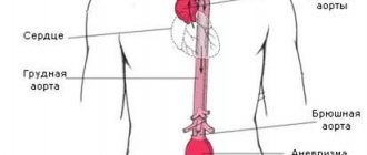
The aorta is the largest arterial vessel in the human body. It arises from the left ventricle of the heart, passes through the chest and abdomen until, at level 4 of the lumbar spine, it divides into two common iliac arteries. In the abdomen, large branches depart from the aorta, which supply blood to all anatomical structures in this part of the body :
- renal arteries;
- abdominal trunk;
- superior, middle and inferior mesenteric arteries;
- splenic artery;
- lumbar arteries;
- suprarenal arteries;
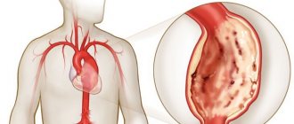
Diseases of the abdominal aorta (detachment, aneurysm, rupture) are considered very dangerous because they are accompanied by large blood losses and disruption of the functioning of key human organs. For a long time, the mortality rate of these pathologies was very high, and only with the advent of high-quality vascular imaging and the development of vascular surgery was it possible to save the lives of such patients.
Lymph nodes of the abdominal cavity and retroperitoneal space are located in various sections, but their greatest concentration is in the thickness of the large peritoneum near the intestine . They play an important physiological role - they are the first barrier to infectious processes that can occur in the abdominal cavity. Here lymphocytes and other cells of the immune system develop and are activated when necessary.
Why is it important to ensure that these anatomical structures are examined? Often the symptoms that occur when they are affected are nonspecific and can completely copy the clinical manifestations of diseases of the digestive system. For example, even for experienced surgeons it is quite difficult to differentiate between appendicitis and lymphadenitis (inflammation of the lymph nodes). The second striking example is that aortic detachment often leads to blockage of the vessels that supply blood to the intestines, which leads to the development of a heart attack and acute digestive disorders.
Ultrasound Dopplerography (USDG)
To study the vessels of the abdominal cavity, a special mode is used - Dopplerography .
It is based on the ability of ultrasonic waves to be reflected from blood at different frequencies depending on its direction and current speed (Doppler effect). Modern ultrasound machines are able to display this difference on the screen using different colors (red and blue), which makes it possible to identify arterial and venous vessels , and also clearly shows their integrity and the presence of areas of blood clot formation . This study is performed immediately after a routine ultrasound of the abdominal organs.
Doppler ultrasound has been used in clinical practice for more than two decades. Its greatest advantage is the ability to quickly diagnose blood flow disorders in the aorta in an outpatient clinic, clinic or emergency room, which significantly saves the time required to transport the patient to the vascular surgery center.
Criteria for damage to the branches of the aorta
In order to diagnose lesions, visualization is used in conventional black and white mode and in color mapping mode. The combination of two modes allows you to examine the geometry and structure of the walls of the vessel, as well as draw conclusions about the degree of vasoconstriction. An important criterion for vessel stenosis is an increase in peak systolic velocity. In this case, it is important to pay attention to the ratio. This refers to the speed in the vessel to the speed of blood movement in the abdominal aorta. Exceeding this value by 3 times may indicate stenosis.
Indications
Ultrasound examination of the abdominal aorta and lymph nodes is necessarily indicated if the patient has the following symptoms:
sharp pain in the abdomen, regardless of location, cutting or burning in nature, the intensity of which does not depend on body position, eating or drinking water, and intensifies with palpation;- nausea, repeated vomiting;
- muscle tension in the anterior or lateral abdominal wall;
- the appearance of impurities of pus or blood in the stool ;
- deformation of the abdomen;
- pronounced pulsation in the midline of the abdomen;
- acute urinary retention;
- pallor of the skin and mucous membranes;
- decreased blood pressure (below 90/60 mmHg), increased heart rate;
- disturbances of consciousness;
- severe general weakness, dizziness and tinnitus in a sitting or lying position.
It is important that ultrasound of the abdominal cavity along with the aorta and lymph nodes, according to national protocols, is indicated for all patients with acute abdominal pain upon admission to hospital.
For what purpose is ultrasound of the abdominal aorta performed?
Abdominal aortography is performed when there is a persistent increase in blood pressure for patients who have reached the age of 40. Other indications:
- the appearance of abdominal pain of unknown etiology (if it is pulsating, this is an even more alarming symptom);
- intermittent claudication and pain in the legs;
- erectile disfunction;
- constant headaches and dizziness;
- ischemia;
- frequent pulsation in the temples and back of the head;
- “spots” in front of the eyes that appear when you turn your head;
- epilepsy;
- reaching 60 years of age;
- long history of smoking;
- suffered a stroke;
- abdominal injuries;
- persistent hypertension and hypotension;
- memory impairment;
- hypertensive crisis.
Also, indications for ultrasound diagnostics are a feeling of heaviness in the abdominal cavity, increased pulsation in it or bloating. Vague symptoms make it difficult to make an accurate diagnosis. In this case, duplex scanning and color mapping are indicated.
Using ultrasound, aortic insufficiency is diagnosed, which is difficult to detect with other research methods.
Preparation
If a patient is admitted to a medical facility with a suspected acute abdomen (a condition that may require immediate surgical intervention), or pathology of the abdominal aorta, then no special preparation is carried out , since delaying time can have a detrimental effect on his condition. In this case, the patient undergoes an ultrasound scan directly in the emergency department, or he is immediately transported to the ultrasound diagnostic room.
The possibility of special preparation is available only for abdominal pain of moderate intensity without disruption of digestive function, when the patient’s general condition remains satisfactory. In this case, it is identical to that for a general examination of the abdominal organs.
Since the study must be carried out on an empty stomach, it is advisable that the last meal be taken no later than 8 hours before the procedure (in children this time may be shorter).
Help If there is increased gas formation on the day of diagnosis, the patient is given sorbents or simethicone preparations (“Espumizan”, “Bobotik”, “Sab Simplex”).
Duplex scanning technique
The procedure is carried out in the first half of the day by a highly specialized specialist - preferably a vascular surgeon. Technique for performing duplex scanning of the abdominal cavity:
- The patient lies on the couch on his back.
- The doctor applies a special contact gel to the anterior abdominal wall and slowly moves the sensor over the skin.
- The equipment monitor displays a picture, on the basis of which the doctor makes a conclusion about the condition of the vessels and the speed of blood flow.
During the procedure, the patient does not experience discomfort or pain. The duration of the study is 15-20 minutes.
Decoding the results
Most often, during examination of the aorta, the following pathologies can be detected:
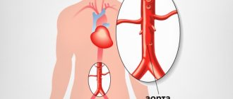
- aneurysm - a protrusion of the aortic wall of various sizes;
- aortic dissection - a rupture of the walls, which leads to the fact that blood gets between its layers (most often epithelial and muscle);
- complete rupture of the wall with internal bleeding;
- atherosclerosis of the aorta;
- formation of parietal thrombi in the aortic lumen;
- calcification (deposition of calcium salts) in the wall;
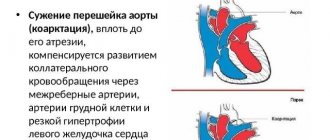
- congenital malformations of the aorta (most often – narrowing of the lumen).
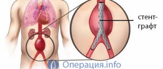
Please note: If a rupture or dissection of the abdominal aorta is diagnosed during an ultrasound, this is an indication for immediate surgical intervention in a vascular surgery center.
During an ultrasound of the abdominal lymph nodes, the following pathologies can be diagnosed:
- lymphadenitis - inflammation of the lymphatic tissue, which can be either infectious or a reaction to damage to the intestines, stomach or other organs of the digestive system;
- metastases of malignant tumors (most often the primary source is in other anatomical structures of the abdominal cavity, urinary or reproductive systems);
- acute or chronic leukemia is a malignant hematological disease that is accompanied by damage to lymphatic tissue and bone marrow.
Standard sizes
During the diagnosis, the doctor measures the transverse dimensions of the aorta and its largest abdominal branches, and compares them with normal values:
| Index | Norm |
| Transverse size of the abdominal aorta under the xiphoid process | Up to 30 mm |
| Transverse size of the abdominal aorta in the area of division into the iliac arteries | Up to 20 mm |
| Diameter of the iliac arteries | Up to 15 mm |
Help Lymph nodes are normally practically not visualized. Their size in an unchanged state should not exceed 2-3 mm.
Other Imaging Techniques
Ultrasound diagnostics is only one of the methods for examining the condition of the abdominal aorta. Its comparison with other visualization methods is shown in the following table:
| Diagnostic method | Description of the technique | Positive sides | Flaws |
| Rheography | Measuring pulse fluctuations in blood vessels using special sensors | · low cost; · availability; · speed of examination | low information content (does not allow diagnosing major aortic diseases) |
| Ultrasound in Doppler mode | Ultrasound method for diagnosing blood vessels using the Doppler effect | · availability; · low cost; · the ability to quickly make a diagnosis; · no contraindications | · with flatulence and constipation, image quality is significantly reduced; · information content strongly depends on the level of qualification of the doctor |
| Color Doppler mapping (CDC) | Ultrasound using the Doppler effect, which allows better visualization of the speed and volume of blood flow | · good visualization of the aorta and its largest branches; · the ability to make a final diagnosis; · no contraindications | · the study is available only in large clinics; · with flatulence and constipation, image quality decreases; · information content strongly depends on the level of qualification of the doctor |
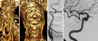
| CT angiography | A radiosensitive contrast agent is injected intravenously, after which a CT scan is performed (a series of X-rays that are processed on a computer and produced in the form of a three-dimensional image) | · very high information content; · the ability to make a final diagnosis; · the study lasts 5-10 minutes; Possibility of simultaneous examination of the whole body or digestive system | · presence of contraindications (renal, heart failure, thyroid disease); Possibility of side effects (allergic reactions, deterioration of kidney function) |
| MRI angiography | A non-toxic contrast agent (based on Gadolinium) is injected intravenously, after which an MRI of the abdominal organs is performed. MSCT angiography can also be used. | · very high information content; · possibility of volumetric visualization of changes in the aorta and its branches (valuable before planned surgical intervention) | · presence of contraindications (installed metal implants, pacemakers, insulin pumps, obesity); Possibility of allergic reactions to contrast; · the procedure lasts more than 30 minutes |
| X-ray contrast angiography | A radiopaque contrast agent is injected intravenously or through a special catheter into the patient intravenously, and then a series of images are taken in real time on an angiograph. | · very high information content (you can find disturbances in blood flow in small branches of the aorta); Possibility of making an accurate diagnosis | · high radiation exposure; · presence of contraindications (renal, heart failure, thyroid disease); Possibility of side effects (allergic reactions, thrombosis, vascular trauma, deterioration of kidney function) |
Important Duplex scanning is not suitable for diagnosing aortic diseases. It allows you to evaluate blood flow in the system of braphiocephalic arteries, which are located inside the skull.
Advantages and disadvantages
The main advantage of this method is its information content and extensive diagnostics. It is worth noting the fact that this procedure does not have any contraindications, which simplifies the research several times. The duration of the procedure can also be called an advantage, because the scanning does not last long. In addition, after the scan, the person is not limited in anything, and can immediately go home.
Price
In Moscow and St. Petersburg, ultrasound examinations of the aorta and lymph nodes are available in almost all clinics. They can be either separate diagnostic services or part of a general examination of the abdominal organs.
In the capital, in public hospitals, the cost of ultrasound of the abdominal aorta and its branches ranges from 750 (City Clinical Hospital No. 1) to 2000 (Clinical) rubles.
In private clinics the price will be 1500-4500 rubles. A separate examination of the lymph nodes of the abdominal cavity costs 550-1200 rubles in Moscow.
In St. Petersburg, the lowest price for ultrasound of the abdominal aorta is in private clinics KallistoMed (740 rubles) and Makhaon (700 rubles), and in most public hospitals it is 900-1700 rubles. Diagnosis of lymph nodes will cost 400-1250 rubles.
In Yekaterinburg and Kazan, the price of an aortic ultrasound is in the range of 500-2000, in Nizhny Novgorod – 800-1850 rubles. Examination of lymph nodes in these cities will cost the patient 400-1050 rubles.