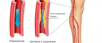
The vascular system delivers blood rich in oxygen and nutrients to all parts of the body. If the important arteries that connect the aorta to the lower extremities are blocked or modified, then a person's health is susceptible to serious disturbances in the blood supply to the legs.
One of the popular methods for diagnosing vascular diseases is ultrasound. In particular, duplex scanning of the arteries of the lower extremities is popular.
What it is
Duplex scanning of the veins of the lower extremities allows you to determine the condition of the vessels with great accuracy and in the shortest possible time, assess the blood flow through them and decide on further actions. Duplex simultaneously combines both conventional ultrasound and Doppler ultrasound, which opens up new possibilities for this diagnostic method and allows an accurate diagnosis in the vast majority of cases.
The technique is based on the peculiarities of the interaction of ultrasound with various tissues and fluids of the body. The waves emitted by the sensor pass differently through formations of different structures and, accordingly, are reflected from it differently.
Duplex scanning is widely used to study blood vessels throughout the body. The name of the method indicates a combination of two diagnostic methods - conventional ultrasound and Doppler studies.
To more easily understand the principle of ultrasound, you can imagine a conversation through a thin wall: the voice will acquire different distortions depending on whether this wall is made of wood, metal, or concrete. Based on the characteristic differences between the resulting ultrasound echo and tissues with different structures, the device analyzes the data received and creates a picture of the organ being examined.
The Doppler phenomenon is based on the fact that ultrasound is reflected differently from liquids flowing in different directions relative to the device’s sensor. Thus, waves reflected from red blood cells floating towards the sensor acquire greater speed in comparison with the same waves reflected from red blood cells floating in the opposite direction relative to the sensor.
Duplex scanning of the veins of the lower extremities Advertising partners
Duplex scanning of the veins of the lower extremities
Many people are concerned about the symptoms of circulatory disorders in the legs. To establish the cause of the pathological condition and diagnose the disease, the doctor prescribes certain examination methods that allow visual examination of the vessels. Few patients know what duplex scanning of the veins of the lower extremities is. But every patient has heard about ultrasound and Doppler sonography. So, it's the same thing. Let's take a closer look at this issue.
When examining a patient, a doctor can only see external changes, but in order to determine the true process of disease development, you need to know how the blood vessels function. This can be done using duplex scanning. Such research is especially relevant when it comes to the need for surgical intervention.
Duplex scanning of the veins of the lower extremities is a modern method of studying blood vessels, provides an accurate clinical picture and greatly facilitates the process of identifying pathology
Duplex scanning of blood vessels is a modern research method, which includes a standard examination of blood vessels and blood flow characteristics, as well as Doppler ultrasound. This diagnostic method provides an accurate clinical picture and greatly facilitates the process of identifying pathology.
In addition, it is very easy to use, can be performed on anyone without age restrictions, and is even allowed during pregnancy.
This method is often used in healthy people who are at risk of developing venous diseases, since it is informative even in the early stages, when the patient is not bothered by any symptoms.
The procedure itself takes about 45 minutes and allows the specialist to assess the condition of the walls of the veins, their valves and the blood flow through them. You can also see the presence of blood clots, the length of the affected area, compactions, and lumen diameter. All this data makes it possible to accurately establish a diagnosis.
To determine pathological phenomena, the following types of vascular scanning are used:
- Doppler ultrasound.
- Ultrasound duplex scanning.
- Color duplex scanning.
The last two methods are used most often in phlebological practice.
Many people are concerned about the symptoms of circulatory disorders in the legs. To establish the cause of the pathological condition and diagnose the disease, the doctor prescribes certain examination methods that allow visual examination of the vessels. Few patients know what duplex scanning of the veins of the lower extremities is. But every patient has heard about ultrasound and Doppler sonography. So, it's the same thing. Let's take a closer look at this issue.
Advantages and disadvantages
Duplex angioscanning of blood vessels involves the use of the properties of ultrasonic waves, so the procedure can be prescribed even to pregnant women, as it is absolutely safe and does not affect the condition of the fetus. Doppler ultrasound is considered an inexpensive technique, so all patients can afford it. There are other advantages:
- high information content;
- non-invasive and no discomfort;
- speed and simplicity;
- no age restrictions or side effects;
- no drugs are used.
This type of diagnosis has no disadvantages, but is still carried out only as prescribed by a doctor.
Duplex angioscanning of blood vessels involves the use of the properties of ultrasonic waves, so the procedure can be prescribed even to pregnant women, as it is absolutely safe and does not affect the condition of the fetus. Doppler ultrasound is considered an inexpensive technique, so all patients can afford it. There are other advantages:
What is determined
The data obtained must be interpreted correctly. Based on the scan results, the following pathological processes can be identified:
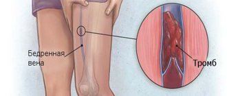
- the formation of blood clots, their size and stage of development, the state of the lumen of the vessel at the site of thrombus formation, the danger of its rupture, the degree of influence on the blood flow and the condition of the soft tissues surrounding the pathology;
- reasons for repeated damage to the vascular network after surgical treatment of varicose veins;
- violation of the functional ability of the venous network, with clarification of the cause and location;
- changes in the valve system of the venous network;
- condition of the vascular wall and possible prognosis for further development of the disease;
- atherosclerosis and endarteritis.
When receiving results, thanks to the availability of any information, everyone can independently assess the initial condition of the veins and arteries.
Types of scanning
In addition to duplex scanning of the veins of the lower extremities, it is possible to conduct research:
- Arteries and veins of the kidneys, as well as the ureters themselves.
- Scanning of brachycephalic vessels (arteries and veins in the head and neck area).
- Scanning of intracranial arteries, sinuses of the dura mater.
- Study of the arteries and veins of the eyeball and, in particular, the retina.
- Examination of the abdominal aorta, inferior vena cava and portal veins.
- Direct duplex scanning of vessels of both lower and upper extremities.
When diagnosing pathologies of the vessels of the lower extremities, it is possible to use different types of ultrasound examination:
- Dopplerography. Allows assessment of blood flow only, without examining the condition of surrounding tissues.
- Duplex scanning. Due to the combination with conventional ultrasound examination, it allows to achieve a comprehensive assessment of the condition both from the blood flow and from the surrounding tissues.
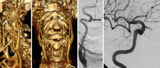
- Triplex scanning. It is the most progressive and expensive type of diagnostics using ultrasound at the moment. Allows you to construct 3D models of the vessels under study.
Preparation and execution
When performing CDS, there is no need for hospitalization; the process itself lasts on average 15 minutes, in rare cases reaching one hour.
Despite the apparent simplicity of the procedure, it is advisable for the patient to know in advance how duplex scanning of veins is performed.
In the treatment room, you will need to expose your hips up to the hip crease. After which the doctor will apply a special gel to the skin and the device’s sensor, which is used in ultrasound diagnostics to reduce friction and improve signal conductivity.
The procedure is performed lying or standing on a stand or couch. In order for the specialist to gain as much information as possible about the state of the vessels and blood flow characteristics, the patient will have to change position a number of times. The doctor may ask you to hold your breath for a while.
To examine the popliteal vessels and veins of the upper leg, the patient should lie on his stomach. To study the main and large superficial ones - lying on your back.
It is important to remember that in late pregnancy it is highly not recommended to lie on your stomach.
Ultrasound and Doppler Doppler - what is the difference?
Vein scanning scheme
Duplex scanning has an optimal ratio of price and quality of the results obtained. Other diagnostic methods are either several times more expensive than this method or do not provide sufficient information.
Conducting conventional ultrasound of tissues of the lower extremities is used quite rarely due to the small amount of data obtained. Such a study can show the presence of a tumor outside the bone, hematoma, ruptures of muscles and tendons, hemorrhages in the joint cavities, but will provide very little information about fractures. All these pathologies are easily determined even without additional diagnostic methods. Moreover, this method is not able to assess the state of blood flow in the vessels.
Ultrasound Dopplerography (USDG) is designed to assess the state of blood flow and filling of veins and arteries. At the same time, it provides information only about the blood flow, but is in no way capable of assessing the condition of the soft tissues and vessel walls. Thus, with ultrasound, the cause of the change in blood flow remains unidentified.
Duplex scanning evaluates both blood flow through the vessels and the condition of soft tissues. This allows you to simultaneously not only identify the cause of the pathology, but also at the same time determine the degree of its severity.
Options for Dopplerography of the legs
Vascular Doppler ultrasound is a way to study blood flow in large and medium-sized veins and arteries of the human body. The essence of the technique is related to the Doppler effect, which implies that ultrasonic waves reflected from moving objects will change frequency in proportion to the speed of movement of these objects.
Dopplerography of the vessels of the lower extremities is divided into 3 main types:
- Doppler ultrasound of the vessels of the lower extremities is an outdated ultrasound diagnostic that is used less and less. This is due to the fact that devices have appeared that allow research to be carried out at a higher level. Previously, this method was widely used to assess the patency of veins and the integrity of valves in various groups of vessels.
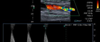
- Duplex scanning of leg vessels (USDS) is a method based on the use of color Doppler echography. Unlike ultrasound of the lower extremities, this method reveals not only the main indicators of hemodynamics, but also structural changes in the walls of blood vessels.
- Ultrasound scanning with color mapping (triplex scanning) is a modern method using pulsed Doppler, which allows you to assess blood flow in any part of the vascular bed in real time and give a color (blue and red shades) designation of its speed. The more saturated the shade, the higher the speed. The advantage of triplex is the ability to simultaneously assess the condition of the vessel in three ultrasound modes, while duplex scanning uses only two modes.
Dysfunction of the valves leads to the appearance of the opposite direction of blood flow in direct and indirect communicating veins
Indications for the study
Duplex scanning of the veins of the lower extremities is always performed if there is a suspicion of a disease of these vessels, or if the diagnosis has already been established in order to monitor its progression and the effectiveness of treatment.
The following symptoms may be a reason for duplex scanning:
- Swelling in the ankles, feet and legs. In this case, you should always remember about the possible presence of heart failure or neoplasms in the pelvis.
- Heaviness in the legs, especially worse in the evening. Often combined with the previous symptom.
- Cramps and involuntary contractions of muscles or individual muscle bundles. It is necessary to exclude possible diseases of the central or peripheral nervous system.
- Pain or discomfort in the legs both after physical activity and at rest.
- Absence of vascular pulsation in the lower extremities.
- Visible expansion of the saphenous veins, the presence of spider veins.
- Signs of tissue malnutrition: pale, cold skin on the legs, the appearance of peeling or trophic ulcers.
You should also periodically carry out preventive duplex scanning of the veins of the lower extremities in the following conditions:
- Already identified varicose veins, thrombophlebitis or other vascular pathology. Diagnosis should be repeated 1–2 times a year or during exacerbation.
- Condition after undergoing surgery for the treatment of diseases of the veins of the lower extremities for the purpose of early detection of complications or relapses - the frequency and timing of the examination are determined on an individual basis.
Conducting research
The procedure is performed starting from the inferior vena cava. This stage requires maximum attention; you need to make sure that there is no blood clot and make sure that the external and internal veins are patency. After this, all veins, as well as superficial vessels, are examined.
Scanning of the arteries of the lower extremities is carried out as follows:
- First of all, the patient should be examined in a horizontal position, and then in a vertical position.
- Next, you need to perform a Valsalva maneuver. By holding your breath while inhaling, the pressure in the abdominal area increases, and in this way a successful test situation is created, which allows you to check the operation of the valves of the veins (deep and superficial).
- Next, you need to conduct a respiratory and cough test. In this case, the patient must inhale as much as possible or simulate a cough. Such actions lead to reverse blood flow if the patient has a pathology of the valve apparatus.
Ultrasound scanning of the leg arteries is performed in two modes (gray and color). In addition, during the study it is necessary to measure the patient’s blood pressure in different parts of the limb. For these purposes, special cuffs are used. The procedure lasts about 40 minutes. If it is carried out by a phlebologist, then treatment can be prescribed immediately after the examination.
How to properly prepare for an examination
Carrying out a duplex study does not require compliance with any special rules. The study requires compliance with standard standards of body care and can be carried out at any age and condition of the patient.
Relative contraindications are:
- The presence of bleeding from veins with a pathological process.
- Extensive trophic ulcers on the lower extremities.
- Infectious skin diseases.
- Severe or terminal condition of the patient.
May be of interest:
|
Decoding
The results are analyzed only by a trained ultrasound physician.
Normal vessel walls are about 2 mm thick, smooth and elastic, there should be no seals or blood clots on them. The lumen is anechoic, with visualization of the valve leaflets. The latter normally function in full, fully ensure the pushing of blood, and close tightly. A color scan of veins should normally show uniform coloration of the blood flow .
The diameter of the vein is greater than that of the artery of the same name, but not more than 2 times.
Deviations from the normal picture allow for a more in-depth study to identify the cause of the pathological process, establish an accurate diagnosis and carry out therapy.
It is important to perform CDS of the veins of the lower extremities horizontally and vertically.
Even if the patient complains of a painful process in one limb, both legs are always scanned for a comparative assessment and diagnosis of changes in the initial stages .
How is the examination carried out?
This diagnostic method is carried out without prior hospitalization or any manipulations. The patient is examined as usual.
The scan is performed while lying on your stomach or back. This allows for maximum convenient and quick examination of the largest number of veins of the lower extremities. It is first necessary to free your legs from clothing to prevent compression of blood vessels. For this reason, you should take off (not roll up!) your pants, shorts, skirt, shoes and socks.
A special gel is applied to the skin to improve contact between the device’s sensor and the skin to obtain more accurate results. At first, the device operates in normal ultrasound mode and only when the necessary vessels are found, it switches to duplex mode. In order to avoid obtaining false results due to positional changes in blood flow in the veins, scanning the same area is carried out several times in different body positions (lying on the back, on the stomach or on the side).
Carrying out additional breathing tests, tests with changes in the position of the limb or the whole body allows a more accurate assessment of the condition of the veins of the lower extremities.
How is ultrasound of veins performed?
Ultrasound scanners consist of a console containing a computer and electronics, a control keyboard and a video display. A sensor is attached to the console and is used for scanning. The sensor is a small device that resembles a microphone attached to the console with a cord. The sensor sends inaudible high-frequency sound waves into the body and then receives reflected echoes from tissues and organs. Depending on the degree of absorption and reflection of sound waves by various tissues, their ultrasound image is formed, which is immediately visible on the display screen.
An ultrasound of the veins is usually performed while lying down, but a standing exam can be used to detect reverse flow in the saphenous veins. The sensor is passed to the places where the venous vessels of interest to the researcher are located. For saphenous veins, linear sensors are more often used; for studying deep veins, arcuate - convex sensors are more suitable. In parallel with the study of the two-dimensional structure of the vein, methods are used to detect blood flow and measure its speed. This is color Doppler blood flow mapping and Doppler ultrasound. During Doppler ultrasound, a sound signal is heard, which reflects the nature of the blood flow in the vessel. Ultrasound of veins usually lasts for 30 minutes and is completely painless for the patient.
Decoding the results
The peak systolic velocity, at which the vessels dilate significantly, is usually 90-140 cm/sec. From here, possible deviations are calculated:
- a value that is in the higher range may indicate stenosis;
- a low value suggests slow flow due to proximal or distal occlusion.
The scanner visualizes the presence of deposits on the walls of the arteries, and the specialist assesses the degree of stenosis. Vasodilation during systole allows for the creation of a reservoir of blood that is present in the arteries during diastole. This volume is enough to supply blood to the extremities; the remainder flows back into the abdominal aorta.
If in the diastole phase the limbs need additional blood volume, then this is indicated by stenosis or occlusion of the iliac arteries. With overlying problems, vortex blood flows or “turbulence” are created in the arteries, which is recorded by an experienced diagnostician.
In the area of stenosis, the speed of blood flow more than doubles. Color Doppler duplex scanning of the vessels of the lower extremities displays the gradation of blood flow in shades.
During occlusion, the velocity decreases as it travels through collateral vessels. Doppler visualizes the cessation of blood flow. There are vessels that are inaccessible to the scanner, but pathologies in them are determined by vortex flows or calculated speed.
Vein lumen assessment
The anatomy of veins is complex and variable, since the vessels are divided into the following types:
- superficial;
- deep;
- perforating
Venous return of blood resists gravity. The pump is the calf muscles on the periphery and the diaphragm, which pulls up the blood like a pump. Valves in the veins allow one-way blood flow. Anatomically, blood from superficial vessels is directed to deep ones, where the muscle pump works more efficiently.
During a duplex of the veins of the lower extremities, the specialist pays attention to valve leakage due to damage or stretching of the vessel. If they fail, blood stagnates in the lower leg, peripheral resistance increases and varicose veins develop
Treatment is usually done surgically. As for the “ligation” of incompetent veins at the places where they flow into deep vessels, if thrombosis develops precisely in them, then elastic bandaging is used to increase venous pressure.
During the procedure, the patient lies on his back with his leg hanging at an angle of 20 degrees from the horizontal to allow the veins to fill with blood. The gel is applied to a probe, which is installed in the groin area near the femoral vein. The probe is advanced to the place where the saphenofemoral junction is located.
The vein is clamped and released to check for regurgitation. If the reverse flow persists for more than a second, then the saphenous vein is faulty.
Sometimes the patient is asked to close his mouth and nose to increase intra-abdominal pressure, then the reverse flow is recorded in the femoral occluded veins.
Similarly, CDS of the veins of the lower extremities reveals the affected perforating veins by pressing the calf muscle. The veins along the back of the thigh are examined in the same way.
When scanning deep veins, the sensor is pressed into the vessel until the blood clot is visualized.
After research
After the examination, you are wiped off the remaining gel from your skin and allowed to get dressed. After the ultrasound examination, no restrictions on normal activities are required. It will be necessary to wait for a written conclusion from a specialist on the results of the study. No complications after ultrasound examination of veins have been noted so far.
An ultrasound diagnostic doctor or phlebologist will analyze the results of the study and draw up a conclusion, which will be the main document on the diagnosis performed. In most cases, ultrasound of the veins is the final diagnostic method for venous diseases, but sometimes additional research methods such as venography are required.
The conclusion on ultrasound of the veins should answer the following questions:
- Are the venous trunks passable?
- What is the diameter of the trunks of the saphenous veins
- Are there thromboses in the superficial and deep veins?
- Do venous valves work and what is the direction of venous blood flow?
- Are there incompetent perforating veins?
Similarities and differences between methods
Ultrasound using Dopplerography and duplex examination also have common features:
- do not cause any discomfort in patients during the procedure;
- safe for health (used during pregnancy);
- do not use invasive methods;
- Ultrasound reflection is used to image vessels.
The similarities end at these four points, because ultrasound and ultrasonic ultrasound scans have a number of differences.
Ultrasound Dopplerography (Dopplerography):
- for diagnosis, the doctor installs the device’s sensor almost blindly, analyzing the location of the vessel, based on the general rules of the anatomical structure of the body;
- allows you to visualize only the valves;
- vein screening can only suggest the possibility of pathology in the form of varicose veins;
- determines only the patency of blood vessels;
- can scan the perforating veins that connect the venous collectors of the deep and superficial systems.
Ultrasound with duplex scanning:
- makes it possible to install the device’s sensor in the required location, since all adjacent tissues are displayed on the monitor;
- establishes the exact cause of vascular obstruction;
- visualizes venous valves;
- examines perforating veins anywhere;
- allows you to determine the cause of recurrence of varicose veins;
- can determine the stage of thrombosis.
All these parameters should be taken into account, since the information content and accuracy of the methods differ significantly.
Features of the technique
CDS of the lower and upper extremities to determine the general condition of the vascular and venous walls allows you to obtain a clearer initial image of the real state of health of the vascular system, to exclude in the early stages any complications of diseases such as varicose veins, thrombophlebitis, thrombosis.
Carrying out diagnostics with a duplex scanning device, a real picture of the condition of the blood vessels is given, which allows one to establish the initial cause of the development of the disease. A duplex scan makes it possible to see the movement of blood flow through the arteries and measure its speed.
In case of damage to the veins and arterial system in the legs, the device will detect pathology with further exclusion of the development of vascular atherosclerosis.
The process of examining vessels and venous walls takes about 45 minutes.
During this time, the specialist will determine how functional the patient’s blood circulation is, show the presence of blood clots, the duration of the affected area, the size of the blood clot, and venous outflow.
During the examination, the specialist sets the level of ultrasound flow with a frequency of 6-12 megahertz, based on the depth of the affected area. Deep vein lesions are examined with a low-frequency probe. All indications for varicose veins are recorded, after which a conclusion is issued.
The procedure has no contraindications, does not cause discomfort, and does not injure tissue during the procedure.
Color duplex scanning is a new modern device that allows you to display a full-scale, clear image of the condition of the blood vessels in the legs in color format. CDS of blood vessels is necessary in emergency situations, when it is necessary to accurately determine the course of the disease with immediate subsequent therapy.
What is ultrasound scanning of the lower extremities and what is its essence?
What is RF face lifting
Ultrasound diagnostics of the venous system of the legs allows you to determine the presence of blockages. Based on the data, the specialist determines whether the patient has varicose veins. If it is present, then the degree of its development is revealed.
Also, based on the indications, it is possible to determine the body’s predisposition to the formation of blood clots in the veins.
The study of the brachiocephalic arteries is the most popular technique. This modern method allows you to study large channels that are responsible for blood flow to the brain.
These include the following arteries:
- subclavian;
- vertebral;
- sleepy.
The areas of their connections are also explored.
The method makes it possible to detect the level of the lumen, determine the presence and position of plaques and blood clots, and examine pathologies and ruptures.
If the indicators are normal, the healthy areas in the resulting image will have a uniform color; there should be no gray areas.