Doppler ultrasound of the vessels of the lower extremities is a conventional two-dimensional ultrasound combined with Doppler ultrasound. It shows, in addition to the structure and position of the vessels, a measurement of the speed of blood flow along the bloodstream. Ultrasound doppler diagnostics is informative for both the examination of arterial and venous blood flow. This diagnosis allows you to successfully identify diseases of the veins and arteries of the legs at the initial stages.
Doppler ultrasound has a number of advantages: safety and painlessness, no special preparation for the study and no need for a recovery period after manipulation. This allows the diagnostic to be used multiple times to monitor dynamic changes during treatment.
What does ultrasound examination of the blood vessels of the legs show?
The abbreviation USDG stands for Doppler ultrasound; the diagnostic principle is based on the Doppler effect. This is the measurement of the reflection of sound waves from moving objects. The information on the monitor of the ultrasound machine after processing the received data shows a two-dimensional color image. It shows the system of blood vessels, as well as places where normal blood flow is obstructed.
Top: Doppler ultrasound showing blood flow in the right femoral artery.
Using ultrasound of the leg vessels, the following veins and arteries can be examined (see table).
| Arteries | Vienna |
| Iliac, femoral, popliteal, tibial, arteries of the foot. In addition to the large arteries listed above, their branches can be assessed. | Femoral, popliteal, greater saphenous, lesser saphenous, anterior tibial, sural, circumflex iliac, dorsal venous arch of the foot, posterior arched vein or vein of Leonardo, perforating veins. |
On the ultrasound monitor, the sonologist can see:
- vessel walls;
- diameter of the vessel lumen;
- shape and location;
- condition of venous valves;
- the speed of blood flow through the vessels, and places where the flow is obstructed;
- determine reflux (backflow of blood);
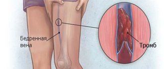
- thrombus and its parameters;
- atherosclerotic plaques;
- arteriovenous malformation (AVM) is an anomaly of vascular development, which is a tangle of intertwined arteries and veins.
Disease prevention
To avoid the occurrence and development of atherosclerosis in the legs, you need to follow simple clinical recommendations. The first of them is, if you have a genetic predisposition, monitor the health of your feet and consult a doctor if alarming symptoms appear. People over 40 years of age are recommended to undergo examination twice a year.
To maintain the composition of the blood and the condition of the walls of blood vessels in good condition, you need to constantly adhere to a diet: give up sweet and fatty foods, strong and carbonated drinks, fast food, pickles and spicy seasonings. You also need to give up smoking and alcoholic beverages. A glass of wine with dinner a week will not hurt, but alcohol abuse will provoke the rapid development of atherosclerotic deposits. You need to include more fresh vegetables and fruits, steamed dishes, nuts and seafood in your diet.
Another important recommendation is to monitor your weight and lead an active lifestyle. Excess weight not only increases the load on the legs, but also causes impaired lipid processes, heart and vascular diseases. Physical inactivity negatively affects blood circulation in the lower extremities, leading to stagnation and decreased mobility of joints and ligaments.
Prevention also includes avoiding tight shoes. Doctors advise walking barefoot on grass, sand, and water in the summer. This is an excellent foot massage that improves the movement of blood and lymph. It is also necessary to avoid stressful situations and work in the evening, and establish a rest and sleep schedule.
Atherosclerosis of blood vessels and arteries of the legs is one of the most common diseases. The danger of the disease lies in its unnoticed development in the first stages: often the symptoms of atherosclerosis are equated with fatigue or the consequences of wearing uncomfortable shoes.
Diagnosis and treatment of atherosclerosis takes place in several stages and depends on the stage of the disease, the presence of concomitant diseases and inflammatory processes, age and gender. At the initial stage, treatment may consist only of lifestyle changes. Drug therapy is prescribed if the vessel is blocked by 50% or more. Surgical methods are resorted to only if the medications do not have a positive effect and the patient’s life is in danger. If you do not follow the doctor's recommendations and instructions, the disease can be fatal. Mortality from leg ischemia in atherosclerosis is 61%.
Indications
Ultrasound scanning of the vessels of the lower extremities is prescribed to patients with the following symptoms:
- swelling of the legs;
- constantly cold feet;
- feeling of numbness in the legs;
- pain and cramps in the calf muscles;
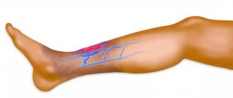
- vascular “networks”, “stars”, protruding veins;
- heaviness in the legs when walking for a long time or that occurs for no reason;
- numbness and changes in sensation in the limbs (for example, goosebumps);
- when determining asymmetry in the patient’s legs (one may be thicker than the other);
- the formation of bruises with minor damage, abrasions that do not heal for a long time.
In addition to complaints, the procedure is prescribed for people suffering from such diseases as:
- thrombosis;
- atherosclerosis;
- thrombophlebitis;
- trophic ulcers;
- arterial aneurysms;
- vasospasm (arterial spasm);
- obliterating endarteritis;
- varicose veins of the legs;
- arteriovenous malformations.
Types and stages of atherosclerosis of the lower extremities
Occlusion of the main arteries of the lower limb by an atherosclerotic plaque causes a characteristic clinical picture depending on the stage of the process.
In practice, doctors use the classification of the process based on the patient’s functional abilities (according to Fontaine-Pokrovsky) , presented in the table.
| Degree of violation | Pain syndrome | Additional signs |
| I | During significant physical activity (walking 1 km or more) | Weakness, fatigue |
| II | A. Pain develops at a distance of 200-1000 m | Development of trophic manifestations: decrease in muscle and fat mass, thickening of nails and skin of the feet |
| B. Intermittent claudication when walking 50-200 m | ||
| III | Pain at rest | To reduce pain, patients lower the affected limb, which becomes purple-bluish in color. Progressive muscle atrophy |
| IV | Stage of destructive changes | The appearance of trophic ulcers, gangrene |
Stages III and IV of disorders are considered “critical ischemia”, which requires emergency medical care, most often surgical.
In addition, the North American classification , which takes into account ankle pressure measurements.
| Degree | Intermittent claudication | Ankle pressure |
| 0 | Asymptomatic | Indicators are within normal limits |
| 1 | Minor | More than 50 mm. rt. Art. after load |
| 2 | Moderate | Intermediate indicators between the 1st and 3rd stages |
| 3 | Expressed | Less than 50 mm. rt. Art. at rest |
| 4 | Pain at rest | Less than 40 mm. rt. Art. to a state of rest |
| 5 | Minimal ulcers, local gangrene, foot ischemia | Up to 60 mm. rt. Art. at rest |
| 6 | Gangrene that rises above the metatarsophalangeal joints | Like in the 5th power |
At 5th and 6th degrees of ischemia, vascular angiography determines the expansion of collaterals and complete occlusion of the main arteries of the foot.
What do legs look like with atherosclerotic lesions at different stages?
Changes in the appearance of the leg are characterized by the level of damage, the degree and duration of the process.
Characteristic signs of disorders at different stages of pathology:
- The phase of functional compensation, which develops as a result of a generalized reaction of the body aimed at improving blood flow. Pallor and coldness of the skin and periodic tingling in the fingers are detected.
- Subcompensation phase, which is characterized by dry skin and decreased elasticity. The affected limb is smaller in volume than the healthy one. Callus (hyperkeratosis) develops on the feet, nails become rough, break, and areas of baldness on the legs are observed. Photo of atherosclerosis of the lower extremities of the 2nd degree:
- The decompensation phase, in which there is pronounced atrophy of muscles and subcutaneous tissue. The skin becomes thinner, minimal trauma leads to the formation of ulcers and cracks. Photo with a trophic ulcer on the inner surface of the left ankle joint.
- The phase of dystrophic changes during which dry gangrene of the foot develops. The terminal stage of atherosclerosis, which requires immediate surgical intervention to prevent infectious and septic consequences.
Diseases that can be found
Dopplerography of the vessels of the lower extremities of the main blood flow allows you to diagnose the following diseases of the veins and arteries:
- Obliterating endarteritis, if the diagnosis is confirmed, urgent treatment is required.
- Obliterating atherosclerosis, the formation of atherosclerotic plaques, the degree of their development, as well as the effect of existing formations on blood flow are assessed.
- Vascular aneurysms occur when pathological stretching of the vessel wall leads to its thinning. The risk of rupture of a damaged vessel during stress and the need for surgical intervention are assessed.
- Varicose veins of the legs. A sedentary lifestyle and excess body weight contribute to the development of the disease. Overfilling of the vascular network leads to dilation of the veins, and subsequently to the formation of blood clots, which pose a danger to life if they rupture.
- Venous insufficiency of the legs is determined by incomplete collapse of the vein; during the contraction phase, part of the blood is thrown back, which leads to the accumulation of blood in the vein cavity and stretching of the vascular wall. This later manifests itself in insufficient nutrition of the tissues of the lower extremities.
- Thrombosis is a complication of varicose veins; when a thrombus is detected, its location, the degree of filling of the vessel, a young or old thrombus and the risk of its rupture are assessed.
In addition to making a diagnosis, ultrasound of the arteries allows you to assess the degree of blood supply to tissues and identify violations of the patency of deep veins.
Main causes of the disease
The presence of atherosclerotic plaques in the arteries of the leg leads to disruption of normal blood flow, which causes unpleasant diseases in this part of the human body. Among the main factors that cause arteriopathy of the lower extremities are the following:
- Addiction to bad habits - frequent smoking increases the risk of developing the disease several times; the lumens of the arteries quickly narrow.
- High blood cholesterol is one of the most common factors causing peripheral arterial disease of the lower extremities.
- Hypertension - increased blood pressure becomes a significant problem, also causing damage to the arteries of the legs.
- Obesity - it has been noticed that people suffering from excess body weight are much more likely to be susceptible to the disease in question.
- Diabetes mellitus is often the cause of arteritis of the lower extremities. This serious illness causes blockage of the blood vessels in the legs.
- Hypodynamia (sedentary lifestyle) - this phenomenon often leads to various undesirable pathologies and contributes to disruption of the circulatory process.
Arteritis of the legs is observed in people who suffer from heart failure. The color of the patient's skin is also one of the factors leading to the development of obliterating atherosclerosis: dark-skinned people are more susceptible to developing the disease.
Preventive Doppler ultrasound of the legs
It is advisable to undergo preventive ultrasound examination of the vessels of the legs once every six months or a year if you are at risk. People at risk include:
- pregnancy;
- smoking;
- overweight;
- diabetes;
- menopause period;
- age over 45 years;
- people with vascular diseases of the legs;
- long-term use of oral contraceptives;
- heavy physical labor (athletes, loaders);
- work requiring constant standing and walking (security guards, waiters, bartenders, salespeople, teachers).
And also look, the more of these factors you have, the greater the reason to conduct preventive ultrasound diagnostics of blood vessels.
Obliterative arterial disease
Obliterating disease of the peripheral arteries is a dangerous and severe chronic pathology characterized by a progressive course. Manifests itself in the form of chronic ischemia of internal organs and limbs. With this disease, there is a disruption in the flow of arterial blood to the lower extremities, this is due to a violation of the elasticity of the blood vessels. Blood circulation does not occur in the required volume, narrowing of the arteries occurs, and sometimes their complete closure.
Risk factors for developing this disease include: high blood pressure, diabetes, smoking, high blood fats, and a sedentary lifestyle.
The first sign of the development of pathology is pain in the lower leg, calf muscle, and buttock. Gradually, the pain begins to intensify, it becomes difficult for the person to move long distances, and eventually he stops walking altogether.
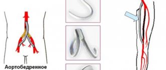
Treatment of pathology is aimed at restoring natural blood circulation in the affected area. As a rule, anti-inflammatory drugs are prescribed; in later stages, surgery is prescribed, the purpose of which is to restore impaired blood flow.
If gangrene develops, amputation of the limb will be required.
Preparation
No special preparation is required for ultrasound examination of the vessels of the lower extremities. But your feet must be clean. If you have thick hair on your legs, of course, it is advisable to shave them to make the procedure easier.
On the day of diagnosis:
- Do not drink alcohol, coffee, strong tea, or energy drinks.
- Do not carry out physical activities such as running, lifting weights, or training.
- Do not smoke 2 hours before the session.
If the diagnosis is carried out in a government institution and is free of charge, then you need to take a diaper to lay on the couch, and a towel to wipe off the gel applied by the doctor. Take the referral with you, as well as the results of previous examinations, if any, to assess changes and dynamics.
Prevention
Narrowing of the peripheral arteries of the extremities causes a lot of trouble, so prevention of the disease is extremely important. To protect yourself from leg arteritis, you should change your lifestyle. Quitting smoking, active physical activity, and healthy eating will help reduce the risk of pathology.
It is important to control cholesterol and blood sugar levels; blood pressure should not exceed the norm. Changes in the structure of the limbs should be alarming. People suffering from obesity should take daily walks and try to reduce excess body weight.
How to do an ultrasound scan of the vessels of the lower extremities
The procedure can be performed with the patient lying or standing. Naturally, you need to free your legs from clothing, then the doctor applies a special gel and moves the ultrasound sensor over the skin of the legs in the examination area.
The ultrasound examination procedure is performed in a lying or standing position.
Before starting the procedure, the doctor conducts various tests to determine the area with possible violations, in which visualization is carried out with the greatest attention. Doppler ultrasound reveals a fundamentally altered type of blood flow and allows the doctor to determine the amount of treatment required.
The following veins are accessible to the Doppler method:
- lower hollow;
- thigh vein;
- subcutaneous large and small;
- ileal;
- deep shins;
- popliteal;
- fibula;
- and sural veins.
The diagnostician sees the speed of filling and strength of the veins with blood in real time in the form of noise and graphic effects.
If necessary, the doctor will ask you to perform simple actions, such as getting off the couch or holding your leg up. This is necessary to assess the behavior of the vascular network in a changed position and evaluate the work of the veins under load.
The ultrasound procedure of the lower extremities takes from 10 to 15 minutes of pure research time, maybe longer. During this period, the sonologist will fully assess the functionality of the veins and identify foci of pathology. At the end of the procedure, the patient is given a study protocol, which must be shown to the attending physician. Some indicators can be deciphered and you can understand on your own what is written there, but you don’t need to draw your own conclusions.
Severe narrowing of the arteries of the lower extremities
When the arteries are severely narrowed due to lipid plaques or are completely blocked (peripheral artery thrombosis), pain in the legs appears even during a period of rest. The legs may appear completely normal, but the toes are pale in color, sometimes with a bluish tint. They are usually cold to the touch and have weak or absent impulses.
In the most severe cases of oxygen deficiency, tissue necrosis (death) begins. The lower part of the leg (ankle) becomes covered with trophic ulcers; in the most advanced cases, gangrene develops, but such a complication is rare.
Decoding
Let's decipher what indicators should be normal in the protocol, and what these or other terms mean.
- The veins are passable, not dilated, the walls are not thickened. The thickness of the vascular wall normally ranges from 0.9 to 1.1 mm.
- Compressive - they have not lost their normal tone, they can shrink.
- The lumens of the arteries are not narrowed.
- The valves are healthy, there are no refluxes - the valves are completely closed and there is no reverse blood flow.
- There are no pathological refluxes when performing compression tests and the Valsalva maneuver - blood does not flow in reverse direction when performing tests.
- The movement of blood flow in each individual vessel in different locations (velocity has its own normal values at each site and cannot be measured in only 1 place). Normally, the speed of blood flow in the femoral artery is approximately equal to 100 cm/s, in the arteries of the leg - 50 cm/s.
- No intraluminal structures were identified—there were no atherosclerotic plaques or thrombi.
- There are no pathological connections between the vessels - the norm.
- The blood flow is phasic - it means faster when you exhale and slower when you inhale, which is normal.
Diagnosis of the disease
The doctor interviews the patient, measures blood pressure, asks about bad habits and lifestyle. After which he feels the pulse in the artery, in the damaged area.
For an accurate diagnosis, the doctor prescribes special tests to determine whether the arteries of the extremities are affected or not. One way to study peripheral arteries is to measure blood pressure in the leg and arm and compare the results. This will allow us to make an assumption about the development or absence of vascular pathology. In some cases, the doctor prescribes an ultrasound of the lower extremities to examine the peripheral arteries; this will provide complete information about the blood circulation in the affected area.
If the doctor still has doubts after the procedures performed, he prescribes angiography (X-ray examination of blood vessels) and tomography (examination of the condition and structure). If there is a suspicion that the patient has a late stage of development of the disease, he is prescribed radiography.